Comparison of the Effectiveness of Phosphorus 32 Application and 10
Mg/Cc Triamcinolone Acetonide Intralesional Injection on Keloid
Abstract Objective: To analyze the effectiveness of phosphorus 32 (32P) application
compared to 10 mg/cc triamcinolone acetonide (TA) intralesional injection in keloid.
Methods: A single blind (evaluator blind) randomized clinical trial on 52 keloids was conducted during the period of May – August 2014 in Tumor and Skin Surgery Clinic, Department of Dermatovenereology and Department of Nuclear Medicine and Molecular Imaging, Dr. Hasan Sadikin General Hospital Bandung.
Results: Lesions were divided into two groups with each group consisted of 26 lesions. Group A was treated with 32P application whereas group B was
treated with 10 mg/cc TA intralesional injection. Flattening of the lesion of more than 50% was higher in the group treated with 32P application (76.9%)
compared to the group that was treated with 10 mg/cc TA intralesional injection (57.7%) on the 8th week, but the difference was not statistically
significant (p>0.05).
Conclusions: Based on the statistical analysis, the application of 32P was as
effective as the intralesional injection of 10 mg/cc TA for keloid lesions.
Keywords:Flattening, keloid, phosphorus 32, triamcinolone acetonide
IJIHS.2016;4(1):26–31
Introduction
Keloid is a skin disorder caused by an dermal collagen overgrowth , as a response of abnormal wound healing in a predisposed individuals.1,2
These fibrous growths result from a connective tissue response to trauma, burns, surgery, or inflammation, and seems occasionally occur spontaneously.3,4 Psychosocial problems often
occur due to cosmetic issues and contracture as complications from keloid.2
Keloids may occur at any age, but tend to develop at the age of 10–30 years old.3 The
keloid incidence of between 4.5% and 16% has been reported in a predominately black
and hispanic population with no difference found between man and woman.3,4
In general, the clinical manifestations of keloids include nodules or demarcated plaques with regular or irregular shape, pink, purple, or hyperpigmented with a shiny surface.3 Keloids
tend to occur in a high skin tension area, such as shoulder, sternum, mandible, arms, and upper back and may be painful, hyperesthetic, or pruritic.3,4
Keloids occur due to the imbalance between collagen production and degradation, resulting in excessive collagen deposition.2 Fibroblast
abnormal activities, increased growth factor level, decrease in the metalloproteinase level, decreased apoptotic activity, increased level of plasminogen activator inhibitor 1 (PAI-1), and tissue hypoxia are the pathogenesis of keloids. However, the exact mechanism of keloid still remains unclear.3 It has been recently shown
Hani Indrayati,1 Jono Hadi Agusni,2 Asmaja Soedarwoto,2 Achmad Hussein S. Kartamihardja3
1Faculty of Medicine, Universitas Padjadjaran-Dr. Hasan Sadikin General Hospital
2Department of Dermatology and Venereology, Faculty of Medicine, Universitas Padjadjaran-Dr. Hasan Sadikin General Hospital
3Department of Nuclear Medicine and Molecular Imaging, Faculty of Medicine, Universitas Padjadjaran-Dr. Hasan Sadikn General Hospital
Correspondence:
Hani Indrayati, Faculty of Medicine, Universitas Padjadjaran-Dr. Hasan Sadikin General Hospital Jl. Pasteur No. 38, Bandung, Indonesia
e-mail: [email protected] Received:
December 29, 2015
Revised:
February 27, 2016
that transforming growth factor- β (TGF- β) is the key factor in the pathogenesis of keloids, because this growth factor has the ability to stimulate fibroblasts proliferation, stimulate collagen and fibronectin productions, as well as inhibiting collagen degradation.5,6 The activity
of TGF- β is normally turned off when wound reparation process is complete but in keloids the production and activity of TGF- β remains, resulting in fibrosis. Basic fibroblast growth factor (bFGF) inhibits collagen production and stimulates collagen degradation.1
A real challenge is faced in the therapy of keloid since no treatment is highly effective for this condition to date. There are several therapies for the treatment of keloid, such as laser, silicone gel, excision, interferon, corticosteroid intralesional injection, radiation, cryotherapy, and 5-fluorouracil injection, or combination.4,7 Corticosteroid injections are
the first choice therapy for keloid with TA as the most commonly used corticosteroid. The concentration have arbitrarily varied from 10 to 40 mg/cc. Corticosteroids can only soften and flatten the scars to some extent with symptomatic relief.4 TA intralesional injection
has an inhibitory effect on TGF-β production and induces the synthesis of bFGF in dermal fibroblasts. In one study, intralesional injection of TA in keloids, given twice a week for two months, has a good outcome in the form of flattening of the lesions of more than 50% in 40% patients. The use of corticosteroids has been associated with unacceptable side effects, such as atrophy, telangiectasia, ulceration, hyperpigmentation, hypopigmentation, and hypertrichosis. Pain associated with the injection and the need for repeated injections can cause discomfort and reduce compliance of the patients. This therapy cannot be used in large or multiple keloids due to the pain and the maximum dose limit of corticosteroids.8
Application of 32P has been reported to have
a good outcome in the treatment of keloids.7,9 32P is a radionuclide that emits β particles
continuously, resulting in death of fibroblasts and inhibit the proliferation of fibroblasts.7,10
This is easy to use, noninvasive, painless, as well as convenient for the patients.11,12 This
application can cause mild side effects, such as hypopigmentation, edema, erosion, erythema, ulceration, dermatitis, and hyperpigmentation. However, these side effects commonly subside spontaneously in two weeks.11
The objective of this study was to compare the effectiveness of 32P application to TA
intralesional injection as the main therapy of keloids.
Methods
This study was an eight week, single blind (evaluator blind) randomized clinical trial on 52 keloids that was performed at the Tumor and Skin Surgery Clinic of the Department of Dermatovenereology and the Department of Nuclear Medicine and Molecular Imaging, Dr. Hasan Sadikin General Hospital Bandung. Informed consent was obtained from all participants.
The patients must be over 14 years old. All lesions had to be a minimum of 0.5 cm and maximum of 1 cm in height, and the size does not exceed 5x5 cm. Lesions were randomized into two study groups. In group A (application of 32P), all lesions were treated with 32P patch
which was then removed after three days of application. In group B (Intralesional injection of 10 mg/cc TA), lesions were treated once every two weeks with intralesional injection of 10 mg/cc TA (dosage 0.1 cc every 1 cm2)
for a total of four treatments. The application of 32P was conducted at the Department of
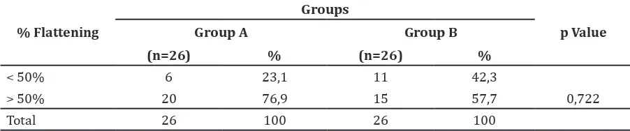
Table 1 Comparison of Effectiveness of Therapy between Two Groups
% Flattening
Total 26 100 26 100
Note: p value is obtained from Kolmogorov Smirnov test between 2 groups
Nuclear Medicine and Molecular Imaging, Dr. Hasan Sadikin General Hospital Bandung.
The study was approved by the Ethical Committee of the Faculty of Medicine, Universitas Padjadjaran-Dr. Hasan Sadikin General Hospital, Bandung.
The lesions were evaluated every two weeks in both groups. To determine the greatest height of the lession, a vernier caliper was used. The percentage of flattening was defined as the percentage of height reduction after treatment compared to the baseline height. Flattening of the lesion of more than 50% considered as a result of an effective treatment.
Results
Of the 15 subjects (56 lesions), 14 completed the full 8 week follow-up period. One subject with four lesions failed to complete the study, hence, the total number of lesions was 52. The
lesions were divided into two groups with 26 lesions in each group. Group A was treated with
32P application whereas group B was treated
with 10 mg/cc TA intralesional injection. At baseline, there was no difference found in the incidence between man and woman, duration of lesions, predilections, symptoms (pain and pruritic), history of previous therapy, and precipitating factors. Of the 14 subjects, 28.58% were in 14–19 years old group. Most of the lesions (55.76%) were <2 years old and the most predilection site was on the shoulder (27%). Pruritus was the most common symptom (75%) with most of the lesions (51.92%) already received previous treatments. Acne vulgaris (73.07%) was the most common precipitating factor found in the sample. Two subjects (14.29%) had a keloid history in the family.
The comparison of the effectiveness between 32P application (Group A) and 10
mg/cc TA intralesional injection (Group B) on keloid were presented (Table 1). A flattening Table 2 Comparison of Flattening of the Lesion Every Two Weeks of Follow-up between Two Groups
Follow-up
Flattening
Zm-w p Value
Group A (n=26) Median (Range)
Group B (n=26) Median (Range)
K1 Week 2 0.19 (0.03–0.58) 0.19 (-0.07–0.49) 0.832 0.493
K2 Week 4 0.32 (0.04–0,66) 0.29 (0.03–0.55) 0.693 0.722
K3 Week 6 0.35 (0.06–0.66) 0.32 (-0.05–0.63) 0.832 0.493
K4 Week 8 0.36 (0.18–0.75) 0.36 (0.03–0.63) 1.109 0.171
Note: p value is obtained from Mann Whitney test
K1: 1st follow up; K2: 2nd follow up; K3: 3rd follow up; K4: 4th follow up
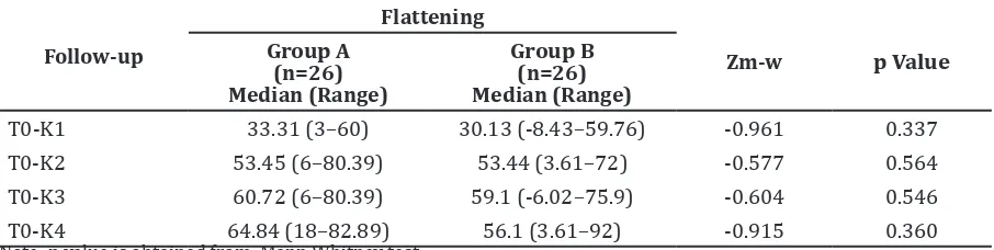
Table 3 Comparison of Flattening (%) in Every Follow-up to Initial Height between Two Groups
Follow-up
Flattening
Zm-w p Value
Group A (n=26) Median (Range)
Group B (n=26) Median (Range)
T0-K1 33.31 (3–60) 30.13 (-8.43–59.76) -0.961 0.337
T0-K2 53.45 (6–80.39) 53.44 (3.61–72) -0.577 0.564
T0-K3 60.72 (6–80.39) 59.1 (-6.02–75.9) -0.604 0.546
T0-K4 64.84 (18–82.89) 56.1 (3.61–92) -0.915 0.360
Note: p value is obtained from Mann Whitney test
Hani Indrayati,Jono Hadi Agusni, et al.
of the lesion of more than 50% was found in 20 lesions (76.9%) in group A, compared to 15 lesions (57.7%) in group. However, this difference was not statistically significant. The comparison of lesion flattenning between group A dan B every two weeks of follow up is shown (Table 2). The mean flattening of lesions of more than 50% in both group was achieved in the second follow-up (Table 3). Group A showed more flattening of the lesion in the second follow-up compared to group B, albeit not statistically significant.
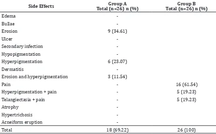
The side effects of each group were listed (Table 4). During the study, no severe side effects were observed in both groups. In group A, 34.61% of the subjects reported some degree of erosion and 23.07% experienced hyperpigmentation. No ulcers and pain were reported by all of the subjects in group A. Nearly all injections were painful (61.54%) in group B, but no erosions and ulcers were seen.
Discussion
This study shows that there were no difference in the incidence of keloid between
men (50%) and women (50%). This study has the same results with the previous study conducted by Davis et al.13 which revealed the
same incidence of keloid between men and women. Effects due to old wounds, other skin diseases, postoperation, and acne vulgaris are the most common reasons that the subjects seek for medical treatment.13
Keloid may occur at any age, but it tends to develop at the age 10–30 years old.3 Patient’s
age also influences the precipitating factor. In this study, most of subjects were in 14–19 years old group, which is the group with the highest prevalence of acne vulgaris according to Shen et al.14 This correlates with the
finding that acne vulgaris is the most frequent precipitating factor in this study.
In this study, shoulder was the most common predilection site of keloid (26.9%). This is similar to the result of a study by Kontochristopolous.6 Skin tension is the most
important factor in the occurrence of keloid. Wound may cause disappearance of some skin tissue and lead to increase in skin tension for closing the wound.4 Skin on the shoulder,
chest, and upper back are prone to have increased skin tension because of respiratory Table 4 Side Effects in Group A and Group B
Side Effects Total (n=26) n (%)Group A Total (n=26) n (%)Group B
Edema
-Bullae
-Erosion 9 (34.61)
Ulcer
-Secondary infection
-Hypopigmentation
-Hyperpigmentation 6 (23.07)
Dermatitis
-Erosion and hyperpigmentation 3 (11.54)
Pain - 16 (61.54)
Hyperpigmentation + pain - 5 (19.23)
Telangiectasia + pain - 5 (19.23)
Atrophy
-Hypertrichosis
-Acneiform eruption
motions. Increased skin tension can stimulate fibroblasts to produce collagen through the use of growth factors and other cytokines.15
Repeated motions in high skin tension areas can cause continuous stimulation of collagen production by fibroblast, which then leads to compression on the dermis area. This leads to disruption of the blood vessel and decreased oxygen tension that will cause higher TGF-β1 activities.16
In this study, acne vulgaris was the most commonly found precipitating factor of keloid (73.07%). Chronic inflammation as seen in acne vulgaris can be a precipitating factors in the occurrence of keloid. Propionibacterium acnes can stimulate the polymorphonuclear cells to produce proinflammatory cytokine such as IL-1 and TNF-α, which increases VEGF expression.17
32P is a radionuclide that emits β particles
which can penetrate well into the tissue with a minimum exposure to the surrounding environment.9,11,12 32P is suspected to cause
direct disruption of fibroblasts, p53 activation, and increased expression of the apoptosis genes.18 Radiation of β particles can cause
ionization of DNA molecules inside the nucleus, induce breaking of double stranded DNA, causing mitotic death.19 The disruption
of double stranded DNA will activate ataxia telangiectasia mutated (ATM), causing p53 to become an active form, and eventually results in apoptosis.10 This process causes a decrease
in number of fibroblasts, collagen synthesis.7
Vivante et al.9 conducted a study in six keloid
lesions applied with 32P with most of the
lesions had experience flattening of more than 50% in a week and become 70% in the fourth week. One lesion had complete remission after 2 months of therapy. In Vivante’s study, the author did not show the percentage of effectiveness; however, a different result is seen in this study.9 Majority of the lesions in this
study had flattening of more than 50% in the second follow-up (fourth weeks of therapy). This difference is probably caused by variations
in the duration of radiation (range from 20–59 hours) in Vivante’s study and also by the fact that some of the lesions had received previous treatment using intralesional injection of TA or 5-fluorouracil. Another study conducted by Yan et al.7 revealed 69% of effectiveness in 151
keloid lesions using 32P application in 72–96
hours and when combined with CO2 laser. The radionuclide was applied a month after the laser.7 Yan’s study result was similar to this
study (76.9%), despite the fact that in this study did not combine the therapy with laser.
This study revealed that erosion and hyperpigmentation were the most frequent side effects in 32P application group, 34.61%
and 23.07% respectively. This condition is due to inflammation processes following radiation and it can develop shortly or 2 weeks after the application and can heal in two weeks with or without topical treatment.9 There was no
pain reported after the application of 32P in all
subjects in group A. In TA injection group, we found hyperpigmentation, telangiectasia, and pain as side effects. All subjects complained about the pain caused by the injection.
Flattening of the lesion of more than 50% is characterized as a result of an effective treatment. In comparison with the TA injection as the first choice therapy for keloid, the application of 32P seems to be more effective
with a few side effects. Flattening of the lesion of more than 50% was found in 20 lesions (76.9%) in group A compared to 15 lesions (57.7%) in group B but this difference is not statistically significant. The study concluded that the application of 32P is as effective as
the intralesional injection of 10 mg/cc TA in keloid lesion treatment. This is based on the fact that the differences between the two types of therapy are not statistically significant. To our knowledge, this is the first randomized clinical trial to compare these two treatments for keloid. Although the difference is not statistically significant, the 32P application is
shown to be easy to use, noninvasive, painless, and convenient for the patients.11,12
References
1. Jfri A, Rajeh N, Karkashan E. A case of multiple spontaneous keloid scars. Case Rep Dermatol. 2015;7(2):156–60.
2. Olabanji JK, Onayemi O, Olasode OA, Lawal OAR. Keloids: an old problem still searching for a solution. Surg Pract. 2005;9(1):2–7.
3. Shaffer JJ, Taylor SC, Cook-Bolden F. Keloidal scars: a review with a critical look at therapeutic options. J Am Acad Dermatol. 2002;46(Suppl 2):S63–97.
Hani Indrayati,Jono Hadi Agusni, et al.
1999;25(9):631–8.
5. Bouzari N, Davis SC, Nouri K. Laser treatment of keloids and hypertrophic scars. Int J Dermatol. 2007;46(1):80–8.
6. Kontochristopoulos G, Stefanaki C, Panagiotopoulos A, Stefanaki K, Argyrakos T,
Petridis A, et al. Intralesionals 5-fluorouracils
in the treatment of keloids: an open clinical and histopathologic study. J Am Acad Dermatol. 2005;52(3):474–9.
7. Yan D, Zhao B, Yang H, Zhu B, Wang J. A combination of nonoperative treatment modalities used for treatment of keloids. Dermatol Ther. 2014;27(1):48–51.
8. Roques C. Teot L. The use of corticosteroids to treat keloids: a review. Int J Low Extrem Wounds. 2008;7(3):137–45.
9. Vivante H, Salguiero MJ, Ughetti R, Nicolini J, Zubillaga M. 32P-patch contact brachyradiotherapy in the management of recalcitrant keloids and hypertrophic
scars. Indian J Dermatol Venereol Leprol.
2007;73(5):336–9.
10. Salguiero MJ, Duran H, Palmieri M, Pirchio
R, Medina V, et al. Bioevaluation of 32P patch
designed for the treatment of skin diseases. Nucl Med Biol. 2008;35(2):233–7.
11. Pandley U, Sarma HD, Ingle AD, Kullolli BS,
Samuel G, Venkatesh M. Radioactive skin
bandages incorporating 32P for treatment of
superficial tumors. Cancer Biother Radipharm.
2006;21(3):257–62.
12. Saxena SK, Kumar Y, Pandey U, Shinde SN,
Muthe KP, Venkantesh M, et al. A facile, viable
approach toward the preparation of 32P patches for the treatment of skin cancer. Cancer Biother Radipharm. 2011;26(5):665–70.
13. Davis SA, Feldman SR, McMichael AJ. Management of keloids in the United States, 1990-2009: an analysis of the national ambulatory medical care survey. Dermatol Surg. 2013;39(7):988–98.
14. Shen Y, Wang T, Zhou C, Wang X, Ding X, Tian S, et al. Prevalence of acne vulgaris in Chinese adolescents and adults: A community based study of 17,345 subjects in six cities. Acta Derm
Venereol. 2012;92(1):40–4.
15. Agha R, Ogawa R, Pietramaggiori G, Orgill DP. A review of the role of mechanical forces in cutaneous wound healing. J Surg Res. 2011;171(2):700–8.
16. Bux S, Madaree A. Involvement of upper torso
stress amplification, tissue compression and
distortion in the pathogenesis of keloids. Med Hypoth. 2012;78(3):356–63.
17. Mahdavian Delavary B, van der Veer WM, van Egmond M, Niessen FB, Beelen RH. Macrophages in skin injury and repair. Immunobiology. 2011;216(7):753–62.
18. Kalanxhi E, Dahle J. Transcriptional responses
in irradiated and bystander fibroblasts after low dose α-particle radiation. Int J Radiat Biol.
2012; 88(10):713–9.