The journal homepage www.jpacr.ub.ac.id p-ISSN : 2302 – 4690 | e-ISSN : 2541 – 0733
Analysis and Prediction of Some Histone-derived
Antimicrobial Peptides from Toads Duttaphrynus melanostictus
and Phyrinoidis asper
Muhammad Dailami,1* I Made Artika,1,3 Mirza Dikari Kusrini2
1
Department of Biochemistry, Bogor Agricultural University, Dramaga Campus, Bogor 16680, Indonesia 2
Department of Forest Resources Conservation and Ecotourism, Faculty of Forestry, Bogor Agricultural University, Dramaga Campus, Bogor 16680, Indonesia
3
Eijkman Institute for Molecular Biology, Jalan Diponegoro 69, Jakarta 10430, Indonesia
*
Corresponding email: [email protected]; [email protected] [email protected];[email protected]
Received 28 January 2016; Revised 27 April 2016; Accepted 29 April 2016
ABSTRACT
Antimicrobial peptides in skin secretions of toads are promising methods to combat a wide spectrum of bacteria. Histone H2A is a type of DNA-binding protein that acts as a precursor for several antimicrobial peptides. In toads (family Bufonidae) buforin I and buforin II are examples of antimicrobial peptides that derived from histone H2A. This study investigated the genetic diversity and phylogenetic analysis and in silico prediction of antimicrobial peptides derived from histone H2A of Duttaphrynus melanostictus and
Phyrinoidis asper, which were collected from Bogor Agricultural University’s campus area.
A new set of primers (Buf_fwd and Buf_rev) were designed by using PrimerBLAST, to amplify 393 nucleotides of the histone H2A gene that codes 131 amino acids. Haplotype diversity of both species are very low. Phylogenetic analysis shows the sample D. melanostictus and P. asper are separated to each other in two different clades. Several short predicted peptides from histone H2A show a potential as an antimicrobial peptides based on in silico prediction. Psychochemical characteristics and 3D structure of potent antimicrobial peptides are described.
Keywords: Antimicrobial Peptides, Histone H2A, Phylogenetic, In silico prediction, Toads
INTRODUCTION
Bacterial antibiotic resistance is a global problem today. Song [1] reported there is an increase of the resistance rate of various bacteria pathogens to conventional antibiotics, especially in the Asian Region. This is caused by misuses of antibiotics in the clinical and the animal husbandry sectors. Therefore, there is a need for a new generation of antibiotics that can combat resistant bacteria and which do not cause further resistance to become a critical issue. Antimicrobial peptides (AMPs) are short peptides that can act as an alternative for conventional antibiotics, because AMPs can combat most resistant bacteria [2] and natural resistance of bacteria to AMPs is very rare [3].
that produces many AMPs is anuran, the amphibians. Anurans secrete a skin secretion that contains AMPs to protect themselves from microbial pathogens. Xu and Lai [5] reviewed all antimicrobial peptides of amphibians (1900 AMPs) that had been published up to 2013 and classified them into 100 families of peptides. However, in the family Bufonidae, few researchers have reported the presence of AMPs. Several antimicrobial peptides that have been reported from this genus are buforin I, buforin II, maximin and alyteserin-1a [6]. In addition, Garg et al [7] reported the potential of skin secretions of D. melanostictus from India as an antibacterial agent, and Utami et al [8] reported the antifungal activities from this species.
Buforin I and II are examples of AMPs that isolated from B. gargarizans. This buforin has an identical amino acid sequence with histone H2A, it is an AMPs that derived from histone H2A by endo-peptic cleavage. Kawasaki and Iwamuro [9] summarized the potential of histone as a precursor for antimicrobial peptides from various organisms. These facts, show that exploration and identification of AMPs derived from histones needs further study. On the other hand, exploration and identification AMPs by conventional methods are long and tedious. As a cutting edge solution, the screening and identification potential AMPs derived from histone using an in silico approach is a promising solution with many advantages. Several related studies on the exploration and identification of AMPs with the in silico approach have been undertaken for histones of microbes [10] and fish [11, 12]. In this study, we designed a pairs of primer that can amplify the histone H2A gene of D. melanostictus and P. asper. We report the nucleotide sequences of histone H2A from D. melanostictus and the phylogenetic relationship with previous reported histone H2A. In addition, we presented the potential AMPs that can be predicted from histone H2A based on the in silico approach.
EXPERIMENT
Chemicals and instrumentation
Chemicals used for this research were: Alcohol 96% (Novapharin), DNA Isolation Kit (Qiagen), Go Taq Green PCR Mix (Promega), Oligonucleotides Primers (IDT), TBE Buffer (Promega), Agarose (1st Base), Molecular Grade Water (GBioscience). Several consumable plastics that were used: micropipette tips (Axygen), Microtube 1.5 mL (Eppendorf), PCR Tube 0.2 mL (Corning).
Instrumentation applied in this research were: Micropipettes (Eppendorf), Micro-centrifuge (Eppendorf Centrifuge 5424), PCR Thermal Cycler (MJ Mini Biorad PTC1148), Horizontal Electrophoresis tank (Thermoscientific Owl Easycast B1A), Microwave (Sanken), Electrophoresis Power Supplay (Enduro Labnet 300V), Gel doc (Biorad).
Sample collection
Live samples of D. melanostictus and P. asper were collected from Bogor Agricultural University campus area. Toe clips were collected and preserved in alcohol 96% as the DNA sources. The live samples were released to their habitat.
DNA isolation and PCR amplification
with Go Taq green (Promega) in 50 µL reaction volume. The PCR conditions involved an initial denaturation at 95oC for 5 minutes, followed by 40 cycles of 94oC for 1 minute, 50oC for 1 minute, 72oC for 1 minutes and a final extension at 72oC for 10 minutes. The amplicons were analyzed by electrophoresis in 1.5% agarose gel in TBE buffer, stained with gel red, and visualized using Gel Doc. The PCR products were purified and sequenced at 1st Base, Malaysia.
Phylogenetic Analysis
Forward and reverse sequences were proof read, trimmed and assembled using software MEGA 5 [15]. The sequences were aligned by Clustall W and the amino acid sequences were deduced with standard genetic code provided by MEGA 5 [15]. The homology search of nucleotide was done by the Basic Local Search Alignment Tools (BLAST) algorithm of National Center for Biotechnology Information (NCBI) [16]. Phylogenetic tree was constructed with Maximum Likelihood and 1000 replication of bootstrap was performed during the tree construction. Best substitution model was chosen by model test.
Antimicrobial Peptides Prediction
In silico prediction of potent AMPs was conducted based on the methods used by Yoo et
al [17] with a slight modification. Amino acid sequences of histone H2A were submitted to CAMP server [18] and each fragmented into 20 amino acid segments. All fragments were predicted with 4 algorithm provided by the server. Fragments, that were categorized as AMPs
were further analyzed for their isoelectric point (8≤PI≤12), molecular weight and positive charge with EMBOSS PEPSTATs [19]. In vitro and in vivo aggregation levels of peptides
were predicted with TANGO (AGG ≤ 500, 0 ≤ HELIX ≤ 25, 25 ≤ BETA ≤ 100) [20] and
AGGRESCAN (-40 ≤ Na4vSS ≤ 60) [21], respectively. The possibility the candidate AMPs were degraded in the in vivo system was predicted based on the cleavage site of chymotrypsin using a peptide cutter [22] and PEST motif with EMBOSS EPESTFIND [19]. 3D structure of potent AMPs generated with PEPstr server [23] then visualized with VMD 1.9.1 [24].
RESULT AND DISCUSSION
Primer Design and Amplicon of histone H2A
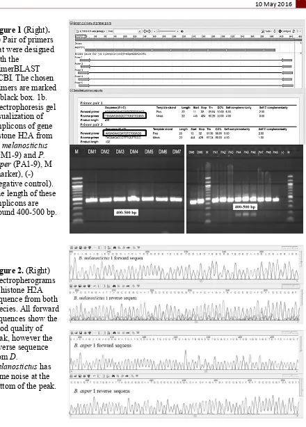
A pairs of primer, forward Buf_Fwd (5’-AAGAGAACGATGTCTGGACG-3’) and
reverse Buf_rev2 (5’-TTAGAAGAGCCTTTGGTTCGGG-3’) were designed and chosen
Figure 1 (Right).
(a) Pair of primers that were designed with the
primerBLAST NCBI. The chosen primers are marked in black box. 1b. Electrophoresis gel visualization of amplicons of gene histone H2A from
D. melanostictus
(DM1-9) and P.
Asper (PA1-9), M
(marker), (-) (negative control). The length of these amplicons are around 400-500 bp.
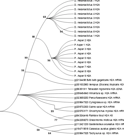
Figure 2. (Right) Electropherograms of histone H2A sequence from both species. All forward sequences show the good quality of peak, however the reverse sequence from D.
melanostictus has some noise at the bottom of the peak.
identity 96% in common with two others sequences (U70133.1 and AF255740.1). There is no sequence of histone H2A of D. melanostictus and P. asper already reported in GenBank. This appears to be first record of histone H2A from D. melanostictus and P. asper from Indonesia. The sequences from D. melanostictus and P. apser have low genetic diversity. From the 393 bp nucleotides, there are only three haplotypes from all 18 individuals (Table 1). Compared to each group of samples, there is one polymorphic site in the D. melanostictus
groups, which is in nucleotides position 124. This is caused by substitution from G to C, which is a transversion (Tv) mutation. On the other hand, there is no a polymorphic site in P. asper groups. This result shows, that histone H2A of D. melanostictus has a higher haplotype diversity compared to P. asper. This is in contrast with the Cytochrome oxidase 1 data [Unpublished data] that shows a higher haplotype diversity on P. asper groups than on D.
melanostictus groups (from same samples).
Table 1. Haplotype groups of D. melanostictus and P. asper
No Haplotype Sample
1 Haplotype 1 D. melanostictus 1, 2, 3, 4, 6, 7, 9
2 Haplotype 2 D. melanostictus 5, 8
3 Haplotype 3 P. asper 1, 2, 3, 4, 5, 6, 7, 8, 9
Figure 3. Nucleotide and amino acid sequences of histone H2A of D. melanostictus and P.
asper. The nucleotides which differ among the 3 haplotype are shown with grey shading, and
the amino acid change is in bold and underlined font.
in amino acid positions 42, 126 and 127, that caused by substitution of the nucleotide in the first codon position. Based on redundancy of the genetic code, every mutation in the second position of a codon will give an amino acid change. Moreover, most of mutations in the third position of the codon will not change the amino acid, and some mutations in the first codon position also do not give amino acid changes.
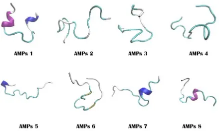
Figure 4. Phylogenetic tree from nucleotide sequences of histone H2A of D. melanostictus
and P. asper with several comparison sequences from NCBI.
D. melanostictus 7 H2A
gi|2104498 Bufo bufo gagarizans H2A mRNA
tested with a model test and chosen based on the Bayesian Invariance Criterion (BIC) value [25]. Tamura 3 parameter with gamma distribution (T93+G) was chosen as the best model with the lowest BIC value compared with other models. A maximum likelihood tree was constructed and tested using bootstrap methods with 1000 replications. The result was provided in Figure 4.
Our phylogenetic tree shows both groups of samples are separated into two different clades with support of bootstrap 80%. This clade is strongly separated from other anuran species which are B. gargarizans and Xenopus tropicaliss with 99% and 56% confident, respectively. In addition, inside of the D. melanostictus clade, samples 5 and 8 that have the same haplotype are separated from other samples that have a different haplotype. On the other hand, all sample of P. asper are together in one big clade. This because the P. asper
sample have the same haplotype.
Prediction of Potent Antimicrobial Peptides Derived from Histone H2A
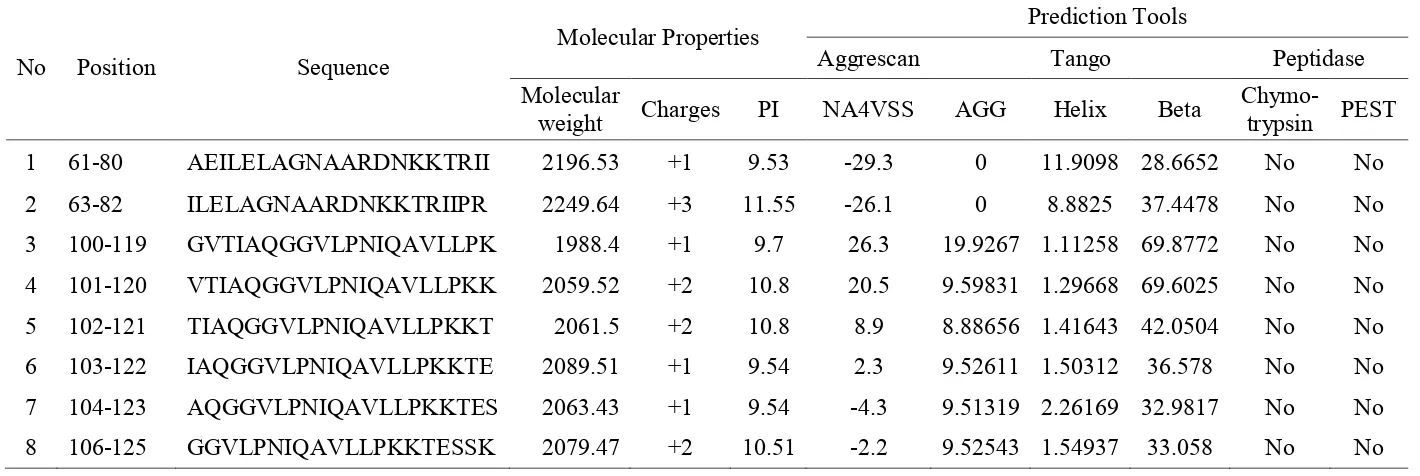
Prediction of fragment peptides that have potential as antimicrobial peptides (AMPs) was conducted by submitting the histone H2A amino acid sequence to collection of antimicrobial peptides release 2 (CAMP R2) server [18]. A total 130 amino acids were used as a template and resulted in 111 fragment peptides with 20 amino acids each. The four algorithms provided by the server are search vector machine (SVM), random forest (RF), Artificial Neural Network (ANN) and Discriminant analysis (DA), were used to calculate and give a prediction score for all peptides. A total 42 peptides considered as candidate AMPs based on its fourth algorithm. Peptides that are predicted Non-AMPs, based on one algorithm, will not be used for further analysis. From the 42 peptides, 13 peptides were chosen based on their positive charge and their isoelectric point (8-12). Peptides with positive charges will easily attach to the bacterial cell wall, which are negatively charged [26].
The aggregation of peptides in solution and in the outer membrane of bacteria, were predicted using software Tango (solution) [20] and Aggrescan (Outer membrane) [21]. Peptides with decrease aggregation in solution but which increase in the bacteria cell membrane is an ideal characteristic for AMPs. By this analysis, only 8 peptides (Table 2) were left for further analysis. In vivo stability based on the possibility that the peptides may be degraded by any peptidase enzymes is an important issue. An endopeptidase that can
reduce the activities of AMPs is α-chymotrypsin. Also, PEST motif can be elevated the possibility of the peptides being degraded. This cleavage site in 8 potent peptides was searched by Peptide cutter [22] and EMBOSS EPESTFIND [19]. All 8 potent peptides did not have any of these peptidase cleavage sites. This shows that the peptides will be stable in
Figure 5. 3D structure of potent AMPs derived from histone H2A of D. melanostictus and P. asper
CONCLUSION
A pair of primers was designed and successfully amplified the histone H2A of both D.
melanostictus and P. asper. The phylogenetic tree based on histone H2A sequence shows all
samples of D. melanostictus and P. asper are separated onto 2 big clades. Three haplotype of these samples are separated in a phylogenetic tree. A total of 8 potent AMPs are successfully predicted based on in silico prediction. We recommend the synthesis these 8 AMPs and the bioassay of their antimicrobial activities in vitro.
ACKNOWLEDGEMENT
Thanks to Himakova IPB students for the sample collection. Thanks to Dr. Dodi Safari for the laboratory facilities in Laboratory of Moleculer Bacteriology, Eijkman Institute for Molecular Biology, Jakarta, Indonesia. Also thanks to M. Majid Khoery and Wisnu Tafroji for helping on the laboratory work.
REFERENCES
[1] Song JH., Antimicrobial Resistance Control in Asia, Monitoring, in Carlet J, AMR Control 2015 overcoming Global Antimicrobial Resistance, Surveillance and National Plans, 2015, TIM PROBART Publisher, United Kingdom.
[8] Utami E., Kusumorini A., Anggadiredja K., Barlian A., Proceed 3rd ICMNS, 2010. [9] Kawasaki H., Iwamuro S., Infect Disord Drug Targets, 2008, 8, 195-205.
[10] Abo Almaali, H.M., Int J Recent Sci Res, 2014, 5(3), 649-655.
[11] Shatyan N., Philip R., Chaithanya ER., Kumar PRA., Sanjeevan VN., Singh ISB., ISRN Mol Biol, 2012, 1-6.
[12] Chaithanya ER., Philip R., Sathyan N., Kumar PRA., ISRN Mol Biol, 2013, 1-7.
[13] Ye J., Coulouris G., Zaretskaya I., Cutcutache I., Rozen S., Madden T., BMC Bioinformatics, 2012, 13(134), 1-11.
[14] Kim HS., Park CB., Kim MS., Kim SC., Biochem Biophys Res Comm, 1996, 229(1814), 381–387.
[15] Tamura K., Stecher G., Peterson D., Filipski A., Kumar S., Mol Biol Evol, 2011, 28, 2731-2739.
[16] Camacho C., Coulouris G., Avagyan V., Ma N., Papadopoulos J., Bealer K., Madden TL., BMC Bioinformatics, 2009, 10(421), 1-9.
[17] Yoo WG., Lee MR., Yun T., Kwon., Kim DW., Bioinformation, 2015, 11(1), 017-020. [18] Waghu F.H., Gopi L., Barai R.S., Ramteke P., Nizami B., Idicula-Thomas S., Nucleic
Acids Res, 2013, 42(1), D1154- D1158.
[19] Rice P., Longden I., Bleasby A., Trends Genet, 2000, 16(6), 276-277.
[20] Fernndez-Escamilla AM., Rousseau F., Schymkowitz J., Serrano L., Nat Biotech. 2004, 22(10), 1302-1306.
[21] Conchilo-Solé O., de-Groot NS., Avilés FX., Vendrell J., Daura X., Ventura S., BMC Bioinformatics, 2007, 8(65), 1-16.
[22] Gasteigr E., Hoogland C., Gattiker A., Duvaud S., Wilkins MR., Appel RD., Bairoch A., Protein Identification and Analysis Tools on the ExPASy Server, in The Proteomics Protocols Handbook, Humana Press. New Jersey, 2005, 571-607.
[23] Kaur H., Garg A., Raghava GPS., Protein Pept. Lett., 2007, 14(7), 626-630. [24] Humphrey W., Dalke A., Schulten K., J Mol Graphics, 1996, 14(1): 33-38.
[25] Nei M., Kumar S., Molecular evolution and phylogenetics, 2000, Oxford University Press, New York, 147-164.
Table 2. Potent peptides as AMPs and their molecular properties
No Position Sequence
Molecular Properties Prediction Tools
Aggrescan Tango Peptidase
Molecular
weight Charges PI NA4VSS AGG Helix Beta
Chymo-trypsin PEST
1 61-80 AEILELAGNAARDNKKTRII 2196.53 +1 9.53 -29.3 0 11.9098 28.6652 No No
2 63-82 ILELAGNAARDNKKTRIIPR 2249.64 +3 11.55 -26.1 0 8.8825 37.4478 No No
3 100-119 GVTIAQGGVLPNIQAVLLPK 1988.4 +1 9.7 26.3 19.9267 1.11258 69.8772 No No
4 101-120 VTIAQGGVLPNIQAVLLPKK 2059.52 +2 10.8 20.5 9.59831 1.29668 69.6025 No No
5 102-121 TIAQGGVLPNIQAVLLPKKT 2061.5 +2 10.8 8.9 8.88656 1.41643 42.0504 No No
6 103-122 IAQGGVLPNIQAVLLPKKTE 2089.51 +1 9.54 2.3 9.52611 1.50312 36.578 No No
7 104-123 AQGGVLPNIQAVLLPKKTES 2063.43 +1 9.54 -4.3 9.51319 2.26169 32.9817 No No