INTRODUCTION
G
lass ionomer cements (GIC) have undergone manymodifications and improvements since their initial
development by Wilson-Kent.1Resin-modified glass
ionomer cements (RMGICs) are a class of hybrid materials created by modifying GICs with the addition of monomer components, typically 2-hydroxyethyl methacrylate
(HEMA) and an associated initiator system.2RMGICs are
said to overcome the problems of low mechanical strength and moisture sensitivity associated with GICs while retain-ing their advantages, includand moisture sensitivity associated with GICs while retain-ing adhesion to tooth structure and base metals, biocompatibility, anticariogenicity associ-ated with fluoride release and thermal compatibility with tooth enamel resulting from similar coefficients of thermal expansion.3-5
Two types of polymerization are evident in RMGICs: a faster setting promoted by light activation, and a slower acid-base chemical reaction initiated by mixing the powder and liquid components and continuing up to 24 h after mix-ing.6
The exact mechanism by which RMGIC bonds to dentine is not well known. A chemical reaction allows bonding to dental hard tissue to be established through a reaction between the carboxylic components of the cement and the
calcium present in the enamel and dentine substrates.7 In
addition to ionic bonding, RMGICs adhere to dentine by
micro-mechanical retention.8,9 Self-adhesiveness of
RMG-
ICs may also be attributed to ionic bonding to the hydroxya-patite around collagen.10
RMGICs are known to adhere successfully to enamel; however, contraction forces applied to RMGICs during light irridation may lead to bond failure between RMGIC and
The Effects of Surface Pretreatment on the Microleakage of
Resin-Modified Glass-Ionomer Cement Restorations
Bayrak S * / Sen Tunc E ** / Tuloglu N ***
Objective:To evaluate the effect of different surface pretreatments on the marginal microleakage of Vitremer restorations. Study Design:Class V cavities were prepared on the labial and lingual surfaces of extracted human third molar teeth. Cavities were randomly distributed into six groups according to surface treatment, as follows: Group NC (negative control): no surface treatment; Group VP (positive control): Vitremer Primer; Group PA: 32% phosphoric acid; Group PAA: 20% polyacrylic acid; Group PLP: self-etch adhe-sive (Prompt L Pop); Group PB: etch&rinse adheadhe-sive (Prime&Bond NT). All cavities were restored with Vit-remer. Teeth were thermocycled, stained with 0.5% basic fuchsin dye and sectioned. Microleakage values were quantitatively assessed by linear measurement of dye penetration using image-analyzing software. Dif-ferences between occlusal and gingival microleakage values within groups were evaluated using paired t-tests, and differences among groups were analysed using one-way analysis of variance (ANOVA) and Tukey tests. Results:There were statistically significant differences between the occlusal and gingival microleak-age values for all groups (p<0.05). Microleakmicroleak-age differed significantly among surface pretreatment groups (p<0.001). The negative control and PLP groups showed similar microleakage values, but were signifi-cantly higher than other groups for both margins. Although there were no statistically significant differences between positive control and PA, PAA, PB groups, microleakage values of positive control group were smaller than all other experimental groups except for PB group. Conclusions:Vitremer restorations require surface pretreatment to prevent excessive microleakage. Pretreatment with etch&rinse adhesives and Vitremer Primer may reduce microleakage of Vitremer restorations.
Keywords: Adhesives, Conditioners, Marginal leakage, Resin-modified glass ionomer cement.
J Clin Pediatr Dent 36(3): 279–284, 2012
* Sule Bayrak, DDS, PhD, Assistant Professor, Department of Pediatric Dentistry, Faculty of Dentistry, Ondokuz Mayıs University, Samsun, Turkey.
** Emine Sen Tunc, DDS, PhD, Associate Professor, Department of Pediatric Dentistry, Faculty of Dentistry, Ondokuz Mayıs University, Samsun, Turkey.
*** Nuray Tuloglu, DDS, Research Assistant, Department of Pediatric Dentistry, Faculty of Dentistry, Ondokuz Mayıs University, Samsun, Turkey.
Send all correspondence to: Sule Bayrak, Department of Pediatric Dentistry, Faculty of Dentistry, University of Ondokuz Mayıs, 55139, Kurupelit, Samsun, Turkey
Tel: +90 362 3121919/3365 Fax: +903624576032
dentine.11 The prevalence of cohesive failures of RMGIC
restorations to dentine has suggested that the smear layer may interfere with the adhesion mechanism; therefore, sur-
face pretreatment to remove the smear layer has been sug-gested as a means of improving bonding.12-15 Studies have
examined the affects of numerous commercially available
pretreatment agents, such as polyacrylic acid,15-19phosphoric
acid,15,17-19hydrogen peroxide,15maleic acid,15citric acid17 and
EDTA.16,20
Given the presence of resin monomers within the RMGIC matrix, recent studies have examined whether or not pretreatment with resin adhesive agents could increase the bonding ability of RMGIC to dental tissue.11,16,18,19 It is
also possible that surface conditioning of the bonding sub-strate with self-etch adhesives, most of which are composed of aqueous mixtures of acidic monomers and which were
developed to simplify bonding procedures,21 could help
improve bond strength between RMGIC and dentine. This study investigated two hypotheses: (i) pretreatment with a self-etch adhesive reduces the microleakage of Vitremer restorations; and (ii) different surface pretreatments perform differently in terms of preventing microleakage.
MATERIALS AND METHOD
Thirty caries-free extracted human third molars were selected and stored in thymol 0.1% saline solution for 1 week following extraction. Residual tissue and calculus were removed with a scalpel and/or scaling instruments, and
teeth were examined under a stereomicroscope for enamel and dentine defects.
Class V cavities were prepared on the buccal and lingual surfaces of each tooth, with the occlusal margins in the enamel and the cervical margins located 1.5 mm apical to the cemento-enamel junction. A cylindrical bur (KG Sorensen Ind Com Ltda, Barueri, SP, Brazil) in a high-speed handpiece and air/water cooling was used, with the bur changed after every five preparations. Standardized cavity dimensions were produced using a template 4.0 mm in width and 3.0 mm in height. Cavity depths were approximately 2
mm, as measured using a periodontal probe.22Preparations
did not include intentional bevels or undercuts for retention. Following cavity preparation, specimens were randomly assigned to one of six groups (n=10) for different surface treatments, as follows:
Group NC: No conditioning (negative control)
Group VP: Vitremer primer (positive control)
Group PA: 32% phosphoric acid
Group PAA:20% polyacrylic acid
Group PLP:Self-etch adhesive (Prompt-L-Pop)
Group PB:Etch&rinse adhesive (Prime&Bond NT).
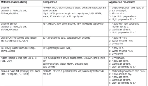
Details of the materials and application procedures used in surface pretreatment are given in Table 1. Following sur- face treatment, all cavities were restored using Vitremer. Vit-remer powder and liquid were dispensed at 2.5:1 by weight,
Material (manufacturer) Composition Application Procedures
Vitremer
(3M Dental Products Co, St.Paul,MN,USA)
Powder: fluoro-aluminosilicate glass, potassium persulphate, ascorbic acid
Liquid: 50% polycarboxylic acid copolymer, 20% HEMA, water, 13% carboxylic acid copolymer
1. Dispense powder and liquid at 2. 5:1 by weight.
2. Mix for 45 s.
3. Inject into preparations. 4. Light polymerize 20 s.* Vitremer primer
(3M Dental Products Co, St.Paul,MN,USA)
46% HEMA, 39% ethyl alcohol, 15% vitrebond copolymer 1. Apply with light scrubbing motion for 30 s.
2. Gentle air stream. 3. Light polymerize 20 s.*
UNI-ETCH Phosphoric acid (Bisco, Inc. Schaumburg IL, USA)
32% phosphoric acid, benzalkonium chloride 1. Apply for 15 s. 2. Water rinse for 15 s. 3. Dry gently.
GC Cavity conditioner (GC Corp.,
Tokyo, Japan) 20% polyacrylic acid, AlCl3
1. Apply 10 s. 2. Water rinse for 15 s. 3. Dry gently.
Adper Prompt L Pop (3M ESPE, ST Paul, USA)
Red cushion: Methacrylic phosphates, BisGMA, photo-initia-tor
Yellow cushion: Water, HEMA, polyalkenoic acid polymer
1. Dry surface. 2. Apply adhesive. 3. Gentle air stream. 4. Light polymerize 10 s.* Prime & Bond NT (Dentsply Ind. Com.
Ltda, Petropolis, RJ, Brazil)
BisGMA, PENTA-P, photoinitiator, ethylamine hydrofluoride, acetone
1. Etch with phosphoric acid 15 s. 2. Rinse and blot dry.
3. Apply adhesive. 4. Gentle air stream. 5. Light polymerize 10 s.*
AlCl3, aluminum chloride; BisGMA, bis-phenol A diglycidylmethacrylate; HEMA, hydroxyl ethyl methacrylate
* Elipar Freelight II, 3M ESPE, USA; light intensity: 1000mW/cm2.
loaded into a Centrix syringe, and injected into the prepara- tion. Then the excess material removed by covering the sur-face with a polyester strip and glass slab and applying slight pressure. Restorations were polymerized, Vitremer finishing gloss was applied to the unfinished restorations and light cured, specimens were then finished with aluminum oxide discs (Sof-Lex, 3 M ESPE, USA), and a second coat of fin-ishing gloss was applied and cured.
Restored specimens were stored for 24 h in deionized water at 37±1°C and then thermocyled for 1.500 cycles
between 5°C and 55°C with a dwell time of 10 s and a trans-fer time of 30 s between baths.23Following thermocycling,
root apices were sealed with sticky wax, and the entire tooth surface was coated with nail varnish up to 1 mm around the circumference of the restoration margins.
Microleakage was evaluated according to dye penetra-tion. Teeth were soaked in 0.5% basic fuchsin dye for 24 h, removed and rinsed under running water for 5 minutes. The roots were cut from the crown, which was sectioned mesio-distally and buccal-lingually using a slow-speed saw.
Images of the material-tooth interface at the cavity mar- gin were captured at x30 magnification using a stereomicro-scope (Nikon SMZ-1500, Osaka, Japan) and stored in a digital format. Microleakage, seen as a pink line at the RMGIC-enamel interface, was measured in millimeters using Photoshop software (Adobe Photoshop CS4 expanded, Adobe Systems, USA). Measurements were made by a single operator (NT) who was blinded to the treatment procedure. Both occlusal and gingival measurements were
taken for each section, for a total of 240 measurements (4 measurements x 30 restorations). For each tooth, the highest measurements were recorded for both occlusal and gingival margins.
Means and standard deviations were calculated from the microleakage data. The difference between the mean occlusal and gingival microleakage measurement in each group was evaluated using paired t-tests (p<0.05). Differ-ences between surface treatment groups for occlusal and gingival margins were separately analyzed by using one-way analysis of variance (ANOVA) and Tukey multiple range tests (p<0.001).
RESULTS
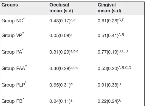
Mean occlusal and gingival microleakage measurements for each of the six different surface treatments are shown in Table 2.
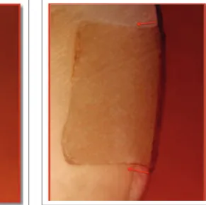
Mean occlusal and gingival microleakage values did not exceed half the cavity depth in any of the groups (Figures 1, 2). Occlusal microleakage measurements were smaller than gingival microleakage measurements in all groups, and these differences were statistically significant in all groups (p<0.05) (Table 2).
ANOVA revealed statistically significant differences among the surface treatment groups (p<0.001) (Table 2). The negative control and PLP groups showed similar microleakage values, but were significantly higher than other groups for both margins. Although there were no statistically significant differences between positive control
Figure 1. Representative stereomicroscopic image of a tooth that without dye penetration in PB group. Original magnification ×30.
and PA, PAA, PB groups, microleakage values of positive control group were smaller than all other experimental groups except for PB group.
DISCUSSION
Resin-modified glass ionomer materials have been widely used and are recommended for restoring cervical lesions that include enamel and dentin margins. RMGICs are specifi-cally recommended for the caries-prone patient because of the possible cariostatic effect of fluoride. Despite recent improvements in RMGIC adhesion to dentin, RMGIC bond strengths are still not comparable to those of dentine bond-ing adhesives.11 Recent studies have shown that the use of
dentin bonding adhesives in conjunction with RMGICs can
improve RMGIC bond strength;11,16however, there is little
known about the effects of self-etch adhesives on RMGIC
bond strengths.11The present study examined the effects of
different surface pretreatments, including a self-etch adhe-sive, on the marginal microleakage of RMGIC restorations. One essential factor in the longevity of a restoration is the marginal sealing ability of the restorative material. Dimen-sional changes and lack of adaptation of the restoration to the cavity walls can lead to marginal leakage, i.e., fluid and molecular movement and ingress of bacteria or bacterial nutrients. Long-term sealing capacity is required to prevent microleakage and its sequelae – including staining, postop-erative sensitivity, pulpal irritation and recurrent caries.23
Many techniques involving dyes have been used to assess
microleakage both in vivoand in vitro.24Dye penetration is a
technique to determine the loss of adhesion.25Other
meth- ods, such as the silver nitrate staining technique, or penetra-tion of radioactive substances, are not commonly used because they are time-consuming and the handling of these
materials is difficult.26 Although marginal leakage studies
usually rely on qualitative criteria,15,25,27-29 some studies
per-form linear measurements of dye penetration at the
tooth-restoration interface to provide quantitative information.30
The present study used linear measurements in order to pro-vide an objective evaluation.
Bonding to the tooth substrate in the area of the cemento-enamel junction continues to remain problematic for clini-cians. In the present study, none of the treatment groups were able to completely resist microleakage. Microleakage at the gingival margin was statistically higher than at the occlusal margin for all test groups (p<0.05). This finding is in line with previous in vitrostudies.27,29
The application of surface treatment prior to RMGIC restorations is controversial. Some authors consider RMG-ICs to be adhesives in their own right by virtue of their
HEMA content, making surface conditioning unnecessary.31
However, it is important to note that HEMA, while strongly
hydrophilic, constitutes only 5% of the cement content.32In
the present study, microleakage in the non-conditioned neg-ative control group was higher than in all other groups except the PLP group. This result is in agreement with other
of the polymer and dentin.7-9Acid conditioning of the tooth
surface creates irregularities or pores in the substrate sur-face, thus facilitating material tag formation. However, in view of the role ionic exchange plays in bonding, care must be taken to ensure that the full mineral content of dentine
and enamel is maintained.27The use of strong acids removes
inorganic components, exposing a microporous scaffold of collagen fibrils and depleting the calcium and phosphate ions required for the ion-exchange mechanism to function; the resulting bond is primarily micro-mechanical and may
be easily broken.14 For this reason, some authors
recom-mended not using phosphoric acid, which is stronger than polyacrylic acid, to treat dentin before RMGIC
restora-tions.14,19However, Yilmaz et al15
found no significant dif-ference between phosphoric acid and polyacrylic acid pretreatment. Our results are in line with these latter find-ings. Furthermore, the PA and PAA groups in our study exhibited less microleakage than the NC group, although the differences were not statistically significant for both margins.
The manufacturers of Vitremer recommend the applica- tion of Vitremer primer to cavities prior to placement of Vit-remer restorative materials. A previous study on permanent dentin found that in line with the manufacturer’s claims, Vit-remer primer was able to modify the smear layer to permit a
closer interaction of the RMGIC and the dentin surface.33In
the present study, the positive control group (VP group) had lower microleakage values than the PA and PAA groups; however, the differences between them were not statistically
Groups Occlusal
mean (s.d)
Gingival mean (s.d)
Group NC* 0.48(0.17)c,d 0.81(0.28)C,D
Group VP* 0.05(0.08)a 0.51(0.41)A,B
Group PA* 0.31(0.29)a,b,c 0.77(0.19)B,C,D
Group PAA* 0.30(0.28)a,b,c 0.53(0.20)A,B,C,D
Group PLP* 0.65(0.31)d 0.91(0.38)D
Group PB* 0.04(0.11)a 0.22(0.24)A
“*”indicate statistically significant differences between occlusal and gingival measurements within groups (p<0.05).
*Differences in superscript letters indicate statistically significant differences in one column (p<0.001).
significant (p>0.001). Similarly, Marquezan et al 33
sug- gested that applying a HEMA-rich primer instead of condi-tioning dentin with polyacrylic acid may compromise the formation of the absorption layer and the continuation of the acid-base reaction taking place within the RMGIC at the dentin interface.
In the present study, the application of an etch&rinse adhesive (group PB) to cavity surfaces prior to the place-ment of Vitremer reduced the microleakage to levels lower
than those of the other materials tested. Pereira et al11found
that the use of adhesive systems associated with RMGICs exhibited the higher bond strength to dentin. It is likely that a properly applied adhesive system is capable of penetrating the demineralized dentin matrix to form a hybrid layer, opti-mizing the mechanical interlocking between the restorative material and substrate and thus reducing microleakage values.
Surface pretreatment with a self-etch adhesive could rep-resent an interesting alternative to other pretreatment agents used in pediatric dentistry because it would reduce the num-ber of steps and eliminate the need for rinsing. However, the present study, found self-etch adhesives to be significantly less effective in preventing microleakage than the other materials tested. This result is in line with many studies that report self-etch adhesives to have less effective bonding properties than etch&rinse adhesives.30,34,35 Several factors
associated with self-etch adhesives can result in the inade-quate formation of a hybrid layer and, consequently, microleakage at the tooth-restoration margins that would
decrease long-term bonding effectiveness.36Included among
these factors are the incomplete alteration and/or removal of smear layer components due to composition (pH, osmolar-ity) and strength of the acidic primer and inadequate resin film thickness, requiring multiple layering techniques and changes in the monomer-water ratio, resulting in phase separations.37-41 microleakage, levels of which were higher at the gingival margin than the occlusal margin.
(2) Surface pretreatment may reduce the microleakage
of Vitremer restorations.
(3) Surface pretreatment with an etch & rinse adhesive and Vitremer Primer resulted in less microleakage than other pretreatment agents. An etch&rinse adhe-sive could be used for surface pretreatment. (4) Surface pretreatment with a self-etch adhesive
resulted in more microleakage than other pretreat-ment agents.
REFERENCES
1. Wilson AD, Kent BE. A new translucent cement for dentistry: the glass ionomer cement. Br Dent J, 132: 133–135, 1972.
2. McLean JW, Nicholson JW, Wilson AD. Proposed nomenclature for glass-ionomer cements and related materials. Quintessence Int, 25: 587–589, 1994. and microstructures of glass-ionomer cements. Dent Mater, 16: 129–138, 2000. 9. Abdalla AI. Morphological interface between hybrid ionomers and
dentin with and without smear-layer removal. J Oral Rehabil, 9: 808–814, 2000.
10. Coutinho E, Yoshida Y, Inoue S, Fukuda R, Snauwaert J, Nakayama Y. Gel phase formation at resin-modified glassionomer/ tooth interfaces. J Dent Res, 86: 656–661, 2007.
11. Pereira PN, Yamada T, Inokoshi S, Burrow MF, Sano H, Tagami J. Adhesion of resin-modified glass ionomer cements using resin bonding systems. J Dent, 26: 479–485, 1998.
12. Pereira PN, Yamada T, Tei R, Tagami J. Bond strength and interface micromorphology of an improved resin-modified glass ionomer cement. Am J Dent, 10: 128–132, 1997.
13. Inoue S, Van Meerbeek B, Abe Y, Yoshida Y, Lambrechts P, Vanherle G, Sano H. Effect of remaining dentin thickness and the use of condi-tioner on micro-tensile bond strength of a glass-ionomer adhesive. Dent Mater, 17: 445–455, 2001.
14. Yap AU, Tan AC, Goh AT, Goh DC, Chin KC. Effect of surface treat-ment and ceYap AU, Tan AC, Goh AT, Goh DC, Chin KC. Effect of surface treat-ment maturation on the bond strength of resin-modified glass ionomers to dentin. Oper Dent, 28: 728–733, 2003.
15. Yilmaz Y, Gurbuz T, Kocogullari ME. The influence of various condi-tioner agents on the interdiffusion zone and microleakage of a glass ionomer cement with a high viscosity in primary teeth. Oper Dent, 30: 105–112, 2005.
16. Nakanuma K, Hayakawa T, Tomita T, Yamazaki M. Effect of the appli-cation of dentin primers and a dentin bonding agent on the adhesion between the resin-modified glass-ionomer cement and dentin. Dent Mater, 14: 281–286, 1998.
17. Yamamoto K, Kojima H, Tsutsumi T, Oguchi H. Effects of tooth-con-ditioning agents on bond strength of a resin-modified glass-ionomer sealant to enamel. J Dent, 31: 13–18, 2003.
18. Setien VJ, Armstrong SR, Wefel JS. Interfacial fracture toughness between resin-modified glass ionomer and dentin using three different surface treatments. Dent Mater, 21: 498–504, 2005.
19. Wang L, Sakai VT, Kawai ES, Buzalaf MA, Atta MT. Effect of adhe-sive systems associated with resin-modified glass ionomer cements. J Oral Rehabil, 33: 110–116, 2006.
20. Fagundes TC, Toledano M, Navarro MF, Osorio R. Resistance to degradation of resin-modified glass-ionomer cements dentine bonds. J Dent, 37: 342–347, 2009.
21. Tay FR, Sano H, Carvalho R, Pashley EL, Pashley DH. An ultrastruc-tural study of the influence of acidity of self-etching primers and smear layer thickness on bonding to intact dentin. J Adhes Dent, 2: 83–98, 2000.
23. Delmé KI, Deman PJ, De Bruyne MA, De Moor RJ. Microleakage of four different restorative glass ionomer formulations in class V cavi-ties: Er:YAG laser versus conventional preparation. Photomed Laser Surg, 26: 541–549, 2008.
23. Kidd EA. Microleakage: a review. J Dent, 4: 199–206, 1976. 24. Taylor MJ, Lynch E. Microleakage. J Dent, 20: 3–10, 1992.
25. Sidhu SK. A comparative analysis of techniques of restoring cervical lesions. Quintessence Int, 24: 553–559, 1993.
26. Iwami Y, Yamamoto H, Ebisu S. A new electrical method for detecting marginal leakage of in vitro resin restorations. J Dent, 28: 241–247, 2000.
27. Yap AU, Mok BY. Reinforced glass-ionomer cements: the influence of conditioners on marginal leakage. J Oral Rehabil, 24: 477–481, 1997. 28. Ceballos L, Osorio R, Toledano M, Marshall GW. Microleakage of
composite restorations after acid or Er-YAG laser cavity treatments. Dent Mater, 17: 340–346, 2001.
29. Hallett KB, Garcia-Godoy F. Microleakage of resin-modified glass ionomer cement restorations: an in vitro study. Dent Mater, 9: 306–311, 1993.
30. Swanson TK, Feigal RJ, Tantbirojn D, Hodges JS. Effect of adhesive systems and bevel on enamel margin integrity in primary and perma-nent teeth. Pediatr Dent, 30: 134–140, 2008.
31. McLean JW. Clinical applications of glass-ionomer cements. Oper Dent, Suppl 5: 184–190, 1992.
32. Mount GJ. Glass ionomers: a review of their current status. Oper Dent, 24: 115–124, 1999.
33. Marquezan M, Fagundes TC, Toledano M, Navarro MF, Osorio R. Dif-ferential bonds degradation of two resin-modified glass-ionomer cements in primary and permanent teeth. J Dent, 37: 857–864, 2009.
34. Ceballos L, Camejo DG, Victoria Fuentes M, Osorio R, Toledano M, Carvalho RM, Pashley DH. Microtensile bond strength of total-etch and self-etching adhesives to caries-affected dentine. J Dent, 31: 469–477, 2003.
35. Aguilar-Mendoza JA, Rosales-Leal JI, Rodríguez-Valverde MA, González-López S, Cabrerizo-Vílchez MA. Wettability and bonding of self-etching dental adhesives. Influence of the smear layer. Dent Mater, 24: 994–1000, 2008.
36. Owens BM, Johnson WW. Effect of single step adhesives on the mar-ginal permeability of Class V resin composites. Oper Dent, 32: 67–72, 2007.
37. Van Landuyt KL, De Munck J, Snauwaert J, Coutinho E, Poitevin A, Yoshida Y, Inoue S, Peumans M, Suzuki K, Lambrechts P, Van Meer- beek B. Monomer- solvent phase separation in one-step self-etch adhe-sives. J Dent Res, 84: 183–188, 2005.
38. Reis A, Albuquerque M, Pegoraro M, Mattei G, Bauer JR, Grande RH, Klein-Junior CA, Baumhardt-Neto R, Loguercio AD. Can the durabil-ity of one-step self-etch adhesives be improved by double application or by an extra layer of hydrophobic resin? J Dent, 36: 309–315, 2008. 39. Cadenaro M, Antoniolli F, Sauro S, Tay FR, Di Lenarda R, Prati C, Bia- sotto M, Contardo L, Breschi L. Degree of conversion and permeabil-ity of dental adhesives. Eur J Oral Sci, 113: 525–530, 2005.
40. De Munck J, Van Landuyt K, Peumans M, Poitevin A, Lambrechts P, Braem M, Van Meerbeek B. A critical review of the durability of adhe-sion to tooth tissue: methods and results. J Dent Res, 84: 118–132, 2005.