Menara Perkebunan 2012 80(2), 57-67
Two-dimensional gel electrophoresis and immunoblotting for
detection of antigenic proteins from natural rubber latex
Gel elektroforesis 2-dimensi dan imunobloting untuk deteksi protein antigen dari lateks karet alam Siswanto*)
Indonesian Biotechnology Research Institute for Estate Crops, Jl Taman Kencana No. 1. Bogor, 16151-Indonesia Diterima tanggal 10 April 2012/Disetujui tanggal 10 Agustus 2012
Abstrak
Lateks karet alam banyak digunakan untuk produksi peralatan medis, industri dan rumah tangga. Reaksi alergi yang disebabkan oleh protein asal lateks telah banyak dilaporkan terutama berkaitan dengan penggunaan sarung tangan asal karet alam. Namun tidak semua jenis protein dari karet alam bisa menyebabkan alergi. Penelitian ini dilakukan untuk mendeteksi jenis protein antigenik yang berasal dari karet alam menggunakan teknik imunobloting. Protein diekstrak dari tiga fraksi sentrifugasi lateks (serum B sebagai fraksi dasar, serum C atau serum sitosolik sebagai fase tengah dan partikel karet sebagai fase atas) dan tujuh jenis sarung tangan komersial, kemudian dipisahkan berdasarkan berat molekulnya melalui Gel elektroforesis 1-D (SDS PAGE) dan 2-D. Selanjutnya untuk deteksi protein antigenik secara immuno-chemi-luminescense dilakukan imunobloting menggunakan IgG antibodi poliklonal anti protein lateks dari kelinci putih New Zealand dan diwarnai dengan Sypro Ruby protein blot fluorescence. Hasil imunobloting menunjukkan bahwa tidak semua protein serum C dan serum B bersifat antigenik. Terdapat lebih dari 14 spot protein antigenik yang terdeteksi dari serum C pada posisi pI antara pH 4,2 s/d pH 6,8, serta lebih dari 16 spot protein antigenik yang terdeteksi pada serum B dengan pI antara pH 5,5 s/d pH 7,0. Protein yang bersifat antigenik dalam serum C a.l: dengan BM 43 kDa diduga Hev b 7,01, BM 22 kDa adalah Hev b 3 dan BM 15 kDa adalah Hev b 8. Sedangkan protein antigenik dalam serum B dengan BM 42 kDa diduga adalah Hev b 10, dan BM 39 kDa adalah Hev b 2. Protein yang bersifat antigenik pada sarung tangan yang terdeteksi dengan IgG kelinci anti serum-C antara lain Hev b 5 dengan BM 14 kDa, Hev b 1 (BM 10 kDa), Hev b 6.03 (BM 18 dan 19 kDa), dan Hev b9 (BM 55 kDa). Sedangkan yang terdeteksi dengan antibodi IgG anti serum B antara lain Hev b 6.02 dengan BM 7 kDa, serta Hev b 10 (BM 46 kDa) dan Hev b9 (BM 55 kDa).
[Kata kunci: Elektroforesis 2-D, lateks-karet alam, sarung-tangan lateks, imunobloting, IgG anti serum B, IgG anti serum C.]
Abstract
Natural rubber latex is widely used for the production of medical, industrial and household devices. Allergic reactions caused by latex proteins have been reported primarily related with the use of natural rubber gloves. However, not all types of proteins from natural rubber can cause allergies. This study was conducted to detect the type of antigenic proteins derived from natural rubber with immunobloting techniques. Proteins were extracted from three fractions of latex centrifugation (B-serum as bottom fractions, C-(B-serum or cytosolic (B-serum as middle phase and rubber particles as upper phase) and seven types of commercial gloves, then separated by molecular weight through 1-D gel electrophoresis (SDS PAGE) and 2-D. Detection of antigenic protein by immuno-chemiluminescense, was performed by immu-noblotting using polyclonal antibody IgG anti-protein latex from New Zealand white rabbits and stained with Sypro Ruby protein blot fluorescence. Immunobloting results indicate that not all proteins from C-serum and B-serum were antigenic. More than 14 antigenic protein spots were detected in the samples of C-serum at pI between pH 4.2 to pH 6.8, and more than 16 antigenic protein spots were detected in the samples of B-serum at pI between pH 5.5 to pH 7.0. Antigenic proteins detected in C-serum were MW 43 kDa suspected as Hev b 7:01, MW 22 kDa was Hev b 3 and MW 15 kDa was Hev b 8. While the antigenic protein detected in B-serum with MW 42 kDa was suspected as Hev b 10, and protein with MW 39 kDa was Hev b 2. Antigenic proteins detected on the rubber gloves with rabbit IgG anti-C serum were Hev b 5 with MW 14 kDa, Hev b 1 (MW 10 kDa), Hev b 6:03 (MW 18 and 19 kDa), and Hev b9 (MW 55 kDa). Whereas antigenic protein of rubber gloves detected with IgG anti-B serum were Hev b 6:02 with MW 7 kDa, and Hev b 10 (46 kDa) and Hev b9 (MW 55 kDa).
[Keywords: 2-D electrophoresis, natural-rubber latex, gloves, immunoblotting, IgG anti-B serum, IgG anti-C serum.]
Introduction
Indonesia is the second largest natural rubber producer after Thailand, which supplies appro-ximately 25% of total world natural rubber. The majority (85%) of rubber plantations in Indonesia is a rubber smallholders plantations involving more than tens million of workers. Natural rubber products are made from latex of Hevea brasi-liensis tree. The crude latex, usually collected in ammoniated solution to prevent microbial growth, contains cellular proteins, lipids and amino acids (Altrich, 2012, Wang et al., 2010). Downstream industries that manufacture latex gloves (medical, household, industrial), balloons, condoms are produced by many small and medium enterprises (SMEs). Therefore, low levels of protein labelling in latex products will have broad impact on the economy, industry and trade.
Industrial manufactures of latex dipping products such as medical gloves, sphygmomano-meters, urine tubes, condoms, balloons, etc. have been faced on the difficult situation to fulfill the international market demand concerning the labeling of lower protein contents. The FDA recommended limit for water extractable protein is 1200 µg per glove or about 150 μg protein/g rubber. In our previous results, the application of protease could reduce the protein level to 68% in medical glove and 93% in latex. Other alternative treatment during the production process to the task of removing latex protein include: chlorination, polymer coating, fumed silica addition, improved leaching protocol and gamma irradiation (Siswanto et al., 2002, Utama et al., 2001).
The raw material of latex and the processing technique in the factories must be improved to produce the hypoallergenic rubber products and meet a demand of the FDA regulation. One possibility is the treatment of protease or irradiation technique to produce the DPNR (Deproteinised Natural Rubber) as raw material for rubber products. The treatment of protease is also possible to be involved during the processing of rubber products to diminish the protein content. FDA (Food and Drug Administration) reported that among 1118 anaphylactic reactions to latex, 15 patients was dead because of mucosal contact from enema cuff exposure (Slater et al., 1996). Our study also demonstrate that 5.8% healthcare workers in five hospital in Jakarta (n = 600) who frequently use rubber products, were allergic to latex. (Sundaru et al., 2002).
The use of examination gloves was dramatically increase, probably caused by increasing consump-tion of gloves to prevent the transmission of disease, like HIV (human immunodeficiency virus) or hepatitis B virus. FDA is requiring all medical medical and surgical gloves (12 brand names) from local industry have protein content varied from 300 to 4500 g/g glove which are much higher than the upper limit of FDA standard (150 µg/g) or ASTM (American Society for Testing and Materials) standard (50 µg/g).
The objective of the study was to analyze the one-dimensional (one-DE) or two-dimensional (2-DE) gel electrophoresis and immunoblotting of latex proteins with polyclonal antibodies of rabbit IgG directed against C-serum and B-serum latex. Those polypeptides which bound to the Rabbit IgG against C-serum or B-serum are classified as antigenic latex proteins. Those polypeptides which bound to the Human IgE from latex-allergic healthcare workers were classified as allergenic latex proteins.
Material & Methods
Samples preparation
Latex fractionation was carried out using a high speed centrifugation, while proteins of gloves were extracted in phosphate buffer saline (PBS) pH 7.2 at 370C for three hours. Fresh latex from AVROS 2037 clone or mix clones was centrifuged at 25.000 g for 45 min, at 4oC in a high-speed SORVAL RC 5B. The main fractions obtained are composed of rubber particles (upper phase), cytosolic serum or C-serum (middle phase) and lutoids (bottom fractions) (Wititsuwannakul et al., 2008, Wang et al., 2010). A lutoid pellet was gently grinned in small volume of Tris-HCl 0.1M buffer pH 7.2 containing 1 mM MgCl2, 10% glycerol, 1 mM EDTA, 1 mM -mercaptoethanol, and subsequently centrifuged to obtain fluid of B-serum. The rubber particles and the gloves were extracted in the same way to obtain the extractable proteins. The glove sample is a mix from seven brands medical glove founded in free market
Menara Perkebunan 2012 80(2), 57-67
around Bogor, Indonesia. Cut pieces of gloves or rubber particles were extracted in PBS pH 7.2 1:4 (w/v) at 370C for three hours. Extracts were clarified by centrifugation at 10.000 rpm, 40C for 15 min to obtain the clear serum. Latex concentrate from mix clones was provided by PTPN VIII, West Java, Indonesia.
Determination of protein content
The protein content of the samples was determined with Bradford reagent kits (Biorad Cat. No. 500-0006). A series of standards (in duplicate) was prepared which contain: 0, 10, 20, 30, 40, 60, 80 g of BSA. In 96 microwell plate ELISA, pipetted 50 L of sample or BSA standard, then add with 200 L of Bradford reagent (diluted 10x). The samples were incubated at 40C for 15 min was read at the absorbance 595 nm in ELISA reader.
Immunization and IgG polyclonal antibody production in rabbit against C-serum or B-serum latex
The antigen for the preparation of antiserum was the total protein from latex B-serum, or C-serum of latex mix clones. Polyclonal antibodies were produced by immunizing the New Zealand white rabbits subcutaneously with 100 μg of protein emulsified in Freund’s complete adjuvant followed by four booster for four to five times at two week intervals. The immune serum was collected, added with 0.02% sodium azide as preservative and stored at -300C until used.
One-dimensional gel electrophoresis (one-DE)
Latex proteins were separated by mono-dimensional acrylamide SDS-PAGE using 12% (w/v) separating gels, pH 8.8 and 4% (w/v) stacking gel pH 6.8. B-serum or C-serum latex proteins were diluted 1: 5 (v/v), whereas glove proteins were diluted 1:1 in a sample buffer (50 mM Tris HCl, pH 6.8, 2% SDS, 10% glycerol, 0.0025% bromophenol blue and 2% β -mercaptoethanol).
Two-dimensional gel electrophoresis (2-DE)
The isoelectric focusing was performed on immobilized pH-gradient (IPG)-strips and the second dimension was run on standard SDS-PAGE
according Amersham Biosciences protocols. Protein samples were diluted or dissolved in rehydration buffer (8 M urea, 2.0% CHAPS, Pharmalyte pH 3-10 without bromophenol blue 2% (v/v)).
Protein gel staining Coomassie blue staining
For Coomassie blue gel staining, gels were stained directly in the Coomassie blue R-250 solution. The solution was prepared by dilution of 0.25 g of Coomassie Brilliant Blue R-250 in 90 mL of methanol:H2O (1:1 v/v) and 10 mL glacial acetic
acid. The gels were destained in the methanol/ acetic acid solution.
Silver staining
After electrophoresis, the gel slab was fixed in 40% ethanol, 10% acetic acid, 50% ddH2O for 1h or
in 5% ethanol, 5% acetic acid, 90% ddH2 for
overnight. The gel then was sensitized in 0.5 M Na-acetate and 1% glutaraldehyde for 30 min, and was then rinsed with deionized water for 2x5 min. The gel resensitized again with 0.05% (w/v) napthalene disulphonic acid in H2O for 30 min, then rinsed
with four changes of deionized water for 5 min each. After rinsing, the gel was submerged in 0.8% (w/v) of silver nitrate diluted in silver reagent stock solution (50 mL ammonia 25%, 850 mL H2O, 80
mL NaOH 1N) for 30 min. The gel was rinsed twice with deionized water for 5 min each, and then developed in 0.1% (v/v) formaldehyde, 0.01% w/v) Na-citrate solution for 1 to 5 min.
Staining PVDF membranes with sypro ruby protein blot stain
After electroblotting proteins to a PVDF membrane, the membrane was allowed to dry completely, then submerged in 7% acetic acid, 10% methanol for 15 minutes. After washing in deionized water four times for five minutes each, the membrane was incubated in SYPRO Ruby protein blot stain solution (Biorad 170-3127) for 15 minutes. The membrane was washed in deionized water two to three times for one minute each to remove excess stain. The membrane may be monitored periodically using UV epi-illumination to determine the level of background fluorescence.
Immunoblotting for detection of antigenic proteins from latex
Following electrophoresis, the gels was equilibrated in transfer buffer pH 9.2 (48 mM Tris, 39 mM glycine, 20% methanol) for 15 min. The gel then was assembly into the sandwich on nitro-cellulose membrane (Schleicher and Schuell) or PVDF membrane (Immobilon-P transfer mem-brane, Millipore) as described in the instruct-tion manual, then electroblotted in Trans-blot Semi-dry electrophoretic transfer cell (Biorad). The transfer condition was set at 300 V, 400 mA for one hour. Detection chemiluminescence of antigenic proteins from latex was performed with Supersignal west dura extended duration substrate, according to the manufacture instruction of Pierce-Perbio in LAS-1000.
Results and Discussion
The present work describes the profile of antigenic proteins of C-serum, B-serum and rubber particle samples from fresh latex, latex con-centrated and extracts of gloves with immuno-chemiluminescense detection after one-dimensional or two-dimensional gel electrophoresis. Fresh rubber latex is the cytoplasm of laticiferous cells of Hevea brasiliensis trees containing about 30% of rubber, which could be separated by high speed centrifugation into three fractions namely rubber particles (white upper layer), an aqueous layer (C-serum) and bottom fraction (B-(C-serum). Latex concentrate, raw material for latex dipping products such as gloves and balloons, is prepared by protein concentration in the fresh latex sample : B- serum are about 14.5 – 19.2 mg/mL, C-serum are 7616 ± 280 µg/mL, respectively. Proteins content in latex concentrated is 11.1 mg/mL and extracts of in seven medical gloves brands (made by Indonesian
factories) is 0.376 mg/mL. In all of the samples, the blue color will develop after addition of Bradford reagent, except for concentrated latex, the solution is in green color that may be caused by the pH of solution which relatively basic (pH 10) and will attributed in the overestimated detection. In spite of such conditions, many proteins remain on the surface of the gloves, even after leaching to remove excess chemicals and proteins. In the latex glove industry, the total extractable proteins of final product are used as an indicator of their potential allergenicity. Extract of seven glove brands used in this experiment contains 376 g protein/mL. It means that the average of protein content of gloves was about 1.5 mg/g of dry rubber since the glove was extracted with PBS at dilution 1:4 (w/v).
In our previous results, the protein content of medical and surgical gloves (12 brand names) from Indonesian factories has showed considerable variation from 284 to 4528 µg/g rubber which is much higher than maximal limit of FDA standard of 1200 µg protein/glove or about 150 µg/g rubber. Moreover ASTM recommended much lower at 50 g protein/g. A relationship between extract-able protein levels in latex gloves and the risk of allergic reaction or sensitization has been demons-trated in several studies, so, the risk for latex sensitization and/or allergic reactions can be reduced by minimizing the amount of extractable proteins (SCMPMD, 2000).
Western blot in one-dimensional gel electro-phoresis for antigenic determinants
Menara Perkebunan 2012 80(2), 57-67
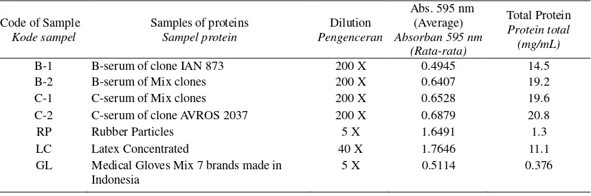
Table 1. Quantity of proteins in the different samples of natural rubber latex and gloves. Tabel 1. Kuantitas protein dari beberapa sampel lateks karet alam dan sarung tangan.
Code of Sample
GL Medical Gloves Mix 7 brands made in Indonesia
5 X 0.5114 0.376
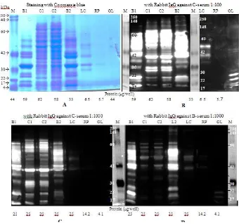
product manual suggested to use the dilution of 2nd antibody at 1 : 10,000, trying to use this antibody more diluted (1:5000) for allergenic protein detection result no signal (data not shown), however after reincubated with the concentration of 1: 1000, we found a good signal of chemi-Figure 1A and after immunoblotting for antigenic detection with rabbit IgG against C-serum or IgG against B-serum are shown in Figure 1B, C and D. Molecular weight standards are shown in lane M. exhibited more than one hundreds polypeptides with molecular weights ranging from 4 kD to 100 kD. Although the protein concentration is not differ significantly than B-serum sample (B1), the ammoniated latex concentrated sample has a more diffuse polypeptide staining with only four faintly protein bands at molecular weight 30, 34, 38 and 50 kDa. Because of the low quantity of proteins (less than 10 g/well) in rubber particles and glove extracts, coomassie blue staining did not able to detect any proteins bands from both samples.
The figure 2B and 2C shows that immunogenic proteins profiles recognized by rabbit IgG against C-serum at dilution 1:100 and 1: 1000 was
relatively identical except the intensity of bands are more fines if the dilution of antibody at 1: 1000 with the results that we can see clear separation of more than 15 bands especially at molecular weight up 40 to 60 kDa in C-serum samples (C1 & C2). However for latex concentrated and glove samples the antigenic proteins only could be detected clearly if the antibody used at 1: 100 dilution. In latex concentrated sample (LC), among four distinct protein bands detected in coomassie blue staining only the polypeptides at 38 and 50 kDa were strongly recognized by this antibody. While this antibody also strongly recognize proteins of glove sample at 10, 14, 16 and 20 kDa and some faintly detected at 38 to 42 kDa which are not able to be detected in coomassie blue staining (Figure 1B). As shown in Figure 1C, the rabbit IgG against C-serum strongly recognized the homologous antigen (C-serum samples) about 15 polypeptides mainly at 14, 20, 28-30, 36, 38, 46-60 kDa. However, this antibody also cross reacted with the heterologous antigen (B-serum samples) and strongly recognizes the polypeptides at 14, 20, 30, 36, 38, 42, 46 – 50 and 60 kDa. Interestingly, some major bands at 42 kDa and 46-56 kDa of B-serum protein was strongly recognized by this antibody whereas these bands were detected very faintly in homologous antigen of C-serum sample.
Figure 1. Coomassie blue staining and immunoblotting of latex proteins with polyclonal antibodies against latex C-serum & B-serum. Proteins from B-serum (B1, B2), C-serum (C1, C2), Latex concentrated (LC), Rubber particles (RP) and Glove (GL). were separated by SDS-PAGE on a 12% polyacrylamide gel. M: Molecular weight protein marker
Gambar 1. Pewarnaan coomassie blue dan imunobloting protein lateks dengan antibodi poliklonal anti serum-C dan serum-B lateks. Protein dari serum-B (B1, B2), serum-C (C1, C2), lateks pekat (LC), partikel karet (RP) dan sarung tangan (GL) dipisahkan melalui SDS-PAGE pada gel polyacrylamide 12%. M: Marka berat molekular protein
polypeptide at 26 kDa in both B-serum samples which only be recognized by rabbit IgG against B-serum but not by rabbit IgG against C-B-serum. The cross reaction of this antibody with antigen heterolog of C-serum samples (C1 & C2) was also observed but less intense at MW > 40 kDa (42, 46, 50, 56 and 58 kDa) with some diffuse staining at low molecular weight polypeptides.
If we compare the profile of total proteins revealed by coomassie blue staining (Figure 1A) with antigenic binding pattern as shown in Figure 1C and Figure 1D, it seems that proteins from B-serum (B1 & B2) are more immunogenic than from C-serum samples (C1 & C2). The proteins dominant of B-serum detected in coomassie blue staining have also a strong signal if immunoblotted with either the rabbit IgG against B-serum or C-serum. Difference in the titers of antisera also results in the differences reactivity obviously noted
if the samples at lower proteins concentrations such as in the latex concentrated, rubber particles and gloves. Natural rubber latex from the Hevea brasiliensis trees contains more than 250 poly-peptides, at least 60 of which demonstrate IgE binding properties. At least 11 different Hevea proteins are known to elicit IgE antibody and these may be subcategorized into four families based on their known biological functions (Hamilton, 2002).
Two-dimensional gel electrophoresis and immunoblotting for …….(Siswanto)
gel electrophoresis, a smeared pattern, indicative of degraded protein was observed. Nevertheless, strong antigenic reactions can be elicited by the proteins/epitopes remaining on the surface of the latex glove and weak reactions in latex concentrate. This result demonstrates that immunoblot assay using rabbit IgG are more sensitive than coomassie blue or silver nitrate staining.
Western blot in two-dimensional gel electro-phoresis for antigenic determinants
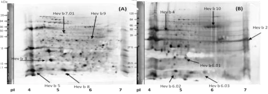
Comparison of silver-stained 2-DE gel of C-serum and B-C-serum samples with an immuno-bloting prepared simultaneously for detection of antigenic proteins was shown in Figure 2 and 3. The latex C-serum 2-DE map was very complex, distributed from pH 4 to 7 and exhibited about a hundred distinct polypeptides from low to high molecular weight (Figure 2 A). Whereas the protein distribution of B-serum samples was accumulated nearly the pH acid (between pH 4-5) (Figure 2 B). However using this IPG strip at pH 4-7, in both samples the horizontal streaking and a smear spots were observed with proteins located at the both side of the membrane in the pH 4 and pH 7. This may be caused by the accumulation of proteins at lower than pH 4 or higher than pH 7 that could not be separated by the strip. For the other experience using the pH strip at a broad range (pH 3-10) the horizontal streaking and a smear spots
was not observed. The resolution of the 2-DE was high and some predictable allergenic proteins in C-serum or B-C-serum that have been reported by WHO-IUIS (International Union of Immunological Societies) and Hamilton, (2002) was indicated by the arrow with the trivial name. The predicted allergenic proteins found in C-serum sample are Hev b.3 at 23 kDa(pH 4.3), Hev b5 at 16 kDa (pH 3.9), Hev b7.01 at 46 kDa (pH 4.8) and Hev b 9 at 48 kDa (pH 5.9). And the predicted allergenic proteins in latex B-serum sample are Hev b2 at 36 kDa (pH 9.8), Hev b4 at 50-57 kDa (pH 4.5), Hev b 6.01 at 20 kDa (pH 5.3-5.6), Hev b 6.02 at 4.7 kDa (pH 4.7 – 4.9), Hev b 6.03 at 14 kDa (pH 6.0 – 7.4), and Hev b 10 at 45 kDa (pH 4.3-6.3). Wang et al., (2010) have reported the Borax/PVPP/Phenol (BPP) protocol to develop an efficient method for protein preparation from different latex subcellular fractions and constructed high-resolution 2-DE maps. They obtained proteins from both total latex and C-serum fraction generate more than one thousand protein spots and several hundreds of protein spots from rubber particles as well as lutoid fraction and its membranes on the Coomassie Brilliant Blue stained 2-DE gels. Hevea latex lectin-like protein (HLL), had MW 17 kDa and a pI value of 7.2, prepared from the bottom (lutoid) fraction and rubber layer of centrifuged fresh latex, leading to the formation of rubber coagulum necessary for a latex coagulation was demonstrated by Wititsuwannakul et al. (2008).
Figure 2. Two-DE of proteins from C-serum (A) and B-serum (B) of latex H. brasiliensis. In the first dimension, proteins were loaded on a 7 cm IPG strip with a linear gradient of pH 4–7, and then a 12% SDS polyacrylamide gel was used for second dimension separation. Proteins were visualized by silver staining. The predicted allergenic proteins reported by IUIS were indicated by arrow.
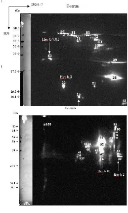
Figure 3 shows the immunobloting profile of C-serum and B-C-serum samples each with the antibody homologous i.e., rabbit IgG anti C-serum or B-serum successively. A number of antigenic proteins were detected in both C-serum and B-serum samples. In C-serum sample, the antigenic proteins are more than 14 spots which are distributed at a broad range of approximately at pH 4.2 to 6.8 from low to high molecular weight (Figure 3 A), whereas in B-serum more than 16 spots of antigenic proteins were observed in the region of pH 5.5 to 7.0 with the molecular weight up to 30 kDa until 100 kDa (Figure 3 B). According to the molecular weight and pI (isoelectric point), an immunoreactive spot in C-serum sample at 43 kDa probably is Hev b 7.01 (patatin homolog, rubber biosynthesis inhibitor), at 22 kDa is Hev b 3 (SRPP= Small rubber particle protein) and at 15 kDa is Hev b 8 (Profilin). Moreover, an immunoreactive spot in B-serum sample at 42 kDa probably is Hev b 10 (Mn-superoxide dismutase), and at 39 kDa is Hev b 2 ( -1,3-glucanase).
In comparison of this immunobloting with the data of protein total of 2-DE presented in Figure 2, we can see clearly that not all of the proteins of C-serum or B-C-serum are antigenic. This remark is not easily to see if the immunobloting was prepared in one-dimensional gel electro-phoresis. It must be noted that in silver staining most of the proteins of B-serum are acidic however most of the antigenic proteins was accumulated close to the pH neutral. Figure 4 shows a comparison of latex proteins resolved in 2-DE prepared in parallel then staining in silver nitrate or blotting against rabbit IgG anti C-serum or B-serum using IPG strips at pH 4.0–7.0. In silver staining, the glove sample consisted of more than 40 distinct polypeptides at low to high molecular weight. The most spots were distributed in the range of pH 5 to 7, and some of vertical streaking and smear spots of proteins were found in the acidic end (at pH 4.0) and at pH 7.0 (Figure 4 A).
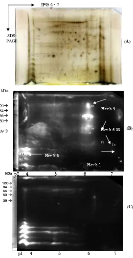
Immunoblot using rabbit IgG against C-serum shows that some strong antigenic proteins was found at around pH 4 at 7, 10, 12 kDa and 14 kDa at in the range of pH 6.5 to 7.0 at 10, 18, 19, 33, 37, 55 and 88 kDa (Figure 4 B). The antigenic protein which also predicted as allergenic proteins are Hev b 5 (Acidic protein) at 14 kDa, Hev b 1 (REF= Rubber elongation factor) at 10 kDa, Hev b 6.03 (Prohevein) at 18, 19 kDa, and Hev b9 (Enolase) at 55 kDa. Immunobloting using rabbit IgG against B-serum shows that the majority of antigenic proteins was found at around pH 4 at molecular weight less
than 7 kDa with some horizontal streaking from pH 4 to 6, and probably one of which identified as Hev b 6.02 (Prohevein). A clear spot at 46 kDa was probably represented Hev b 10 (Mn-superoxide dismutase) (Figure 4 C). In the latex, prohevein is an 18.5 kDa precursor molecule, cleaves post-translationally to produce a 4.7 kDa N-terminal cleavage product, hevein, and a C-terminus peptide of 13.3 kDa.
Prohevein, hevein and the prohevein C-terminus are also named Hev b 6.01, Hev b 6.02 and Hev b 6.03 respectively according to the allergen nomenclature of the International Union of Immunological Societies (Yeang et al., 2007). Hevein is a heat-stable chitin-binding protein in natural rubber latex, constitutes 40% to 70% of the proteins in the aqueous content of lutoids, the B-serum, , is located in the lutoids of natural rubber latex (Yeang et al., 2007).
Two-dimensional gel electrophoresis and immunoblotting for …….(Siswanto)
Figure 3. Immunoblotting of C-serum & B-serum latex proteins with the antibody homologous, i.e.,Rabbit IgG against C-serum or B-serum at dilution 1:1000. Proteins quantity of C-serum or B-serum are 100 g/ gel.
Gambar 3. Imunobloting protein dari serum-C & serum-B lateks masing-masing dengan antibodi homolog yaitu antibodi IgG kelinci anti serum-C atau serum-B pada pengenceran 1:1000. Kuantitas protein serum-C& serum-B adalah 100 g/gel
Since the ASTM (American Society for Testing and Materials) has introduced an IgG-ELISA inhibition assay (ASTM D6499-00) or LEAP (Latex Elisa for Antigenic Proteins), these
antibodies i.e., rabbit IgG raised against C-serum in combination with rabbit IgG raised against B-serum can also be used for reagent detection kits to measure the antigenic proteins in latex products.
Figure 4. 2-D gel electrophoresis of natural rubber glove after silver staining (A) and immunodetection using Rabbit IgG against C-serum (B) or Rabbit IgG against B-serum (C). Proteins quantity is 50 g/gel and Rabbit IgG diluted 1: 1000
Gambar 4. Gel elektroforesis 2-D dari sampel sarung tangan karet alam setelah pewarnaan silver (A) dan imunodeteksi menggunakan IgG kelinci anti serum-C (B) atau IgG kelinci anti serum-B (C). Kuantitas protein adalah 50
g/gel dan IgG kelinci diencerkan 1: 1000
(A)
37
19
12
1
pI 4 5 6 7 (C) 88
55
33
18
10
14
10 7
Hev b 9
Hev b 6.03
Hev b 5
Hev b 1
pI 4 5 6 7
(B)
46
28
7 92
5
4 3
3
Hev b Hev b 10
IPG 4 - 7
SDS-PAGE
kDa
120 84 66 50
39
kDa
120
84
66
50
39
kDa
120
84
66
50
39
Two-dimensional gel electrophoresis and immunoblotting for …….(Siswanto)
Conclusion
Detection of antigenic proteins from natural rubber and latex gloves can be done using rabbit IgG anti-C serum and rabbit IgG anti-B serum. However, detection using rabbit IgG anti-C serum was more effective because it could detect antigenic proteins in a wider pH range. Antigenic proteins detected in the C-serum were more than 14 spots at the position of pI between pH 4.2 to pH 6.8, while that detected from the B-serum were more than 16 spots at the position of pI between a pH of 5.5 to pH 7.0. Immunobloting results on 2-D gel electrophoresis showed that the proteins that were antigenic on gloves were Hev b 6:02 (Prohevein) with MW 7 kDa, Hev b 1 (REF = Rubber elongation factor) MW10 kDa, Hev b5 (Acidic protein ) with MW 14 kDa, Hev b 6.03 (Prohevein) MW 18 and 19 kDa, and Hev b 10 (Mn-superoxide dismutase) in MW and Hev b9 46 kDa (Enolase) MW 55 kDa.
Acknowledgements
This research was supported by DAAD grant in continuation with the 1st DAAD-Fraunhofer Technopreneur Awards in Life Sciences”. I would like to express my sincere thanks to Professor Herwig Brunner, Director of Fraunhofer IGB, the Director of DAAD for the fellowship. I am very grateful and my warmest thanks to Dr. Steffen Rupp, Dr. Kai Sohn, Dr. Anke Burger-Kentischer, Georg Geiger Dipl.Ing., and Doris Finkelmeier for their friendly supports and advices concerning this laboratory work.
References
Altrich ML (2012). Diagnosis of Latex Allergy. The alert Newsletter, American Latex Allergy Association, October 2012, 6 p.
Beezhold DH, GL Sussman, DA Kostyal & NS Chang (1994). Identification of a 46-kD latex protein allergen in health care workers. Clin Exp Immunol 98, 408-413.
Hamilton RG (2002). Diagnosis of natural rubber latex allergy. Methods 27, 22–31.
SCMPMD (2000). Opinion on Natural Rubber Latex Allergy. European commission. Scientific Committee on Medicinal Products and Medical Devices, European Commission Health & Consumer Protection Directorate-General. [Taken from: http://www.europa. eu.int/comm/food/fs/sc/ scmp/ out 31 _en.pdf: 34 pp.]
Siswanto, H Sundaru, T Haryono & Suharyanto (2002). Allergen on natural rubber gloves. Dalam: Konggres III KBI dan Seminar Bioteknologi, PPAU Bioteknologi ITB, Bandung, 10 –11 September 2002
Slater JE, T Vedvick T, A Arthur-Smith, DE Trybul & RGO Kekwick (1996). Identification, cloning, and sequence of a major allergen (Hev b 5) from natural rubber latex (Hevea brasiliensis). American Soc Biochem & Mol Biol 271(41), 25394-25399.
Sundaru H, Siswanto, Karjadi, TH, Suharyanto & L Parede (2002). Perakitan kit diagnostik protein alergen dengan antibodi IgE manusia untuk kontrol mutu dari produk barang jadi lateks dalam negeri. Laporan Hasil Penelitian. Jakarta, Badan Litbang Pertanian, PPTP/P2: 40p.
Utama M, HM Halik, Siswanto, Y Syamsu, S Herwinarni, Suharyanto & B Handoko (2001).
Trial production of low protein content, and free Nitrosamine of prevucanized natural rubber latex in factory scale by Gamma irradiation technique. Atom Indonesia 7, 10-16.
Wang X., X Lu, R Ma., C Wu., A Gio, M Peng & W Tian (2010). A method for protein extraction from different subcellular fractions of laticifer latex in Hevea brasiliensis compatible with 2-DE and MS. Proteome Sci, 8 - 35.
Wititsuwannakul R, P Pasitkul, K Kanokwiroon, D Wititsuwannakul (2008). A role for a Hevea latex lectin-like protein in mediating rubber particle aggregation and latex coagulation. Phytochem 69, 339–347.
Yeang HY, RG Hamilton, DI Bernstein, SAM Arif, K-S Chow, Y-H Loke, M Raulf-Heimsoth, S Wagner, H Breiteneder & RE Biagini (2006). Allergen concentration in natural rubber latex. Clinical Experim Allergy, 36, 1078 – 1086.
Yeang HY, SAM Arif, YH Loke, NP Chew, SM Mohsin (2007). Electrophoretic characterisation of Hevein. J Rubb Res 10(4), 235–244.