www.elsevier.com / locate / bres
Research report
Recurring episodes of spreading depression are spontaneously elicited
by an intracerebral hemorrhage in the swine
a ,
*
a a a,bSheila Mun-Bryce
, Arika C. Wilkerson , Nicholas Papuashvili , Yoshio C. Okada
a
Department of Neurology, University of New Mexico School of Medicine, Albuquerque, NM 87131, USA
b
Department of Neuroscience, University of New Mexico School of Medicine, Albuquerque, NM 87131, USA
Accepted 3 October 2000
Abstract
Intracranial bleeding damages the surrounding tissue in a complex fashion that involves contamination by blood-borne products and loss of ionic homeostasis. We used electrophysiological techniques to examine the functional changes in the developing intracerebral bleed and in surrounding regions using an in vivo swine model. Intracerebral hemorrhage (ICH) was induced by collagenase injection into the primary somatosensory cortex (SI). Somatic evoked potential (SEP) elicited by electrical stimulation of the contralateral snout as well as changes in DC-coupled potential were monitored in the SI from the time of collagenase injection in order to measure the effects of ICH. The SEP decreased in amplitude within minutes of the intracerebral injection. Its short-latency component was abolished within the first hour after collagenase injection without any sign of recovery for the duration of the experiment. As the SEP started decreasing in amplitude, we observed spontaneous, recurring episodes of cortical spreading depression (SD) as early as 20 min post-injection. The timing of SDs in SI is consistent with our interpretation that SDs were initially generated at multiple sites adjacent to the lesion core and propagated into the surrounding area. With time, SD became less frequent near the injection site, shifting to more distant electrodes in the surrounding area. Our results indicate that ICH leads to the reduction in SEP amplitude and induces spontaneous episodes of SD. Loss of ionic homeostasis is most likely the physiological basis for the SEP change and for the induction of SD. Recurring SD spontaneously generated in experimental ICH needs further study in humans with ICH. 2001 Elsevier Science B.V. All rights reserved.
Theme: Disorders of the nervous system
Topic: Ischemia
Keywords: Spreading depression; Somatosensory evoked potential; Electroencephalography; Collagenase; Intracerebral hemorrhage
1. Introduction model to the large gyrencephalic brain of the swine. The
swine brain resembles the human brain by having a large Intracerebral hemorrhage (ICH) occurs in 15–20% of volume and a well-developed pattern of convolutions in stroke patients and in brain injury [8,26]. We have initiated the cortex. Collagenase was injected into the rostrum area a series of studies to characterize the functional changes of the primary somatosensory cortex (SI). The swine snout produced by collagenase-induced ICH since the patho- is somatotopically represented over the large R1 area physiology of hemorrhagic stroke is still poorly understood which is doughnut shaped with the sulcus naris in the in spite of its clinical importance. The temporal pro- middle of the coronal gyrus [5,20]. This somatotopic gression of acute ICH has been characterized in the rat organization enables us to study the effect of collagenase-using the collagenase model of hemorrhagic stroke induced hemorrhage on cortical neurons by recording the [16,23,24]. somatic evoked potential (SEP) from the area of the cortex In order to gain insights into the sequela of ICH in the receiving direct thalamocortical projections from the human brain, we adapted the collagenase-induced ICH stimulated location of the snout before, during and after
collagenase injection into the projection area.
In a preliminary study [17], we observed cortical
*Corresponding author. Tel.: 11-505-265-1711, ext. 4889; fax: 1
1-spreading depression (SD) while monitoring the
DC-po-505-260-0165.
E-mail address: [email protected] (S. Mun-Bryce). tential changes in the hemorrhagic core as a function of
time after collagenase injection. We now characterize the effectiveness of anesthesia was monitored continuously development of SD, which is distinguished by large with ECG and EEG.
changes in the extracellular concentrations of major ions ˆ
[12]. Originally described by Leao [14], SD is generated 2.2. Recording procedure when the extracellular potassium concentration increases
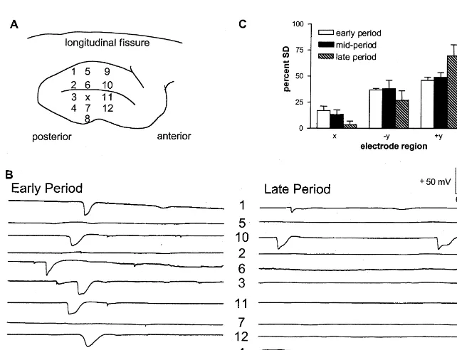
from the resting level of about 3.5 mM to above 10 mM Electrical potential recordings were made with either a [12]. SD is described by temporary loss of neuronal 6-channel array (n54) or 12-channel array (n54) of glass activity in the cortical area undergoing this phenomenon micropipettes (tip diameter 5–10 mm, filled with 2 M with a slow propagation of the affected area from the NaCl). A Ag–AgCl electrode wire contacted the NaCl initiation site toward the surrounding cortical areas [11,13]. fluid and was connected with the amplifier. The glass SD has been associated with epilepsy [25], stroke [18], electrodes were positioned between plexiglass plates and migraine [13] and traumatic brain injury [19]. We therefore held secure with plastic screws. The six electrodes in the hypothesized that SD should be generated initially in the 6-channel array were positioned linearly 1 mm apart. The core of the hemorrhagic injury and propagate to the 12-channel array was comprised of four electrodes surrounding areas. As the hematoma develops and ionic positioned 2 mm apart along three columns (separated by 5 homeostasis is lost in the core area, adjacent tissue loses mm between electrodes 1–4 and 5–8 and 7 mm between the ability to spontaneously generate SD, and SD ceases. electrodes 5–8 and 9–12). Electrode 9 was not functioning The dysfunctional cortical area should gradually move in all the experiments while electrode 8 did not work outward from the ICH core, generating SD farther from the during one of four experiments. Thus these cases were ICH core over time. excluded in the statistical analysis.
After stereotaxically implanting the recording electrode array 3 mm ventral to the cortex, a layer of 4% agarose 2. Materials and methods (type I-A, Sigma Chemical) was poured over the cortical surface to minimize cortical movement. A Ag–AgCl 2.1. Surgical procedure reference electrode was placed in the epidural space of the frontal brain region. Baseline level of spontaneous activity The animal protocol for the present study was approved and evoked responses in the intact SI cortex were collected by the University of New Mexico School of Medicine for 20–30 min prior to collagenase injection. An area of Animal Research Committee. All of the experiments were the snout contralateral to the recorded SI cortex was conducted in the Magnetophysiology Laboratory, Veterans electrically stimulated (2 mA, 100 ms) at 0.5 Hz using a Affairs Medical Center, Albuquerque, NM. bipolar pair of electrodes. The evoked responses Juvenile farm swine, weighing 4–12 kg, 3–5 weeks old, (bandpass50.1 Hz to 1 KHz) were averaged over 50 were initially anesthetized with sodium thiopental, 25–30 epochs. The stimulation site was moved until the short mg / kg intrathoracically. Sodium thiopental was used since latency component of the SEP was maximized in the SD can be elicited under a barbiturate anesthesia [1,2,11] recording electrodes adjacent to the collagenase injection but it is suppressed by anesthetics such as ketamine, which pipette.
blocks N-methyl-D-aspartate (NMDA) receptors and pre- In one group of animals (n58), 625 U of collagenase vents SDs [9]. Following 1% lidocaine application the (type XI, Sigma Chemical) in 5 ml sterile saline15 ml trachea was intubated for mechanical respiration of room heparin (10,000 U / ml) was delivered using an 80-mm air, and the femoral artery was cannulated to monitor outer diameter glass pipette over 16 min with a microinfu-physiological blood gases and for continuous isotonic fluid sion pump (Harvard Instruments). Five microliters of infusion. The cephalic vein was cannulated in the upper saline were placed in the tip of the injection pipette forelimb and the anesthesia was then administered at a rate prefacing the collagenase injectate to insure that baseline of 9 mg / kg / h i.v. during the remaining surgery. The data were not influenced by collagenase leaking into the animal was then placed in a head-holder. The scalp area brain tissue environment. Heparin was added to the carrier, was infiltrated with 1% lidocaine so that an incision could following the protocol of Del Bigio et al. [6]. The be made to expose the dorsal surface of the skull. The SI occurrence of SD was monitored continuously from the cortical surface was exposed bilaterally by removing the time of collagenase injection for up to 6 h with a DC-bone and dura. The animal was then transported into a coupled recording of the potential within the SI. SEPs magnetically and electrically shielded room [21]. A warm within the SI cortex were monitored periodically using the water pad was used to maintain body temperature at 378C. same electrodes.
In the control group (n53), animals received a 5-ml electrode sites did not indicate a uniform propagation from saline injection and electrical potential recordings were a single focus, but rather suggested that SD appeared to monitored using the 12-channel electrode array. No signifi- have originated from multiple foci adjacent to the injection cant differences in SEP recordings were apparent follow- site. The SDs tended to propagate in both directions during ing intracerebral saline injection as compared to baseline the early period. Approximately 90 min after collagenase signals and no SD was detected (data not shown). injection (late period), SDs became less frequent or At the conclusion of the experiment the animal was disappeared altogether near the injection site, while SDs euthanized by thiopental overdose and the brain was fixed were still seen at more distant electrodes (Fig. 3B, right). in formalin for histological assessment. Statistical analysis Electrodes 3 and 4, adjacent to the lesion core, showed a of SEP data was conducted using a two-tailed paired t-test, decrease in SD episodes during the late period as com-and the two-way analysis of variance test for the SD data. pared to the early period, whereas SDs continued to occur
in electrodes 1 and 6 during the late period.
These key temporal and spatial characteristics were
3. Results quantified by examining the SDs in all the recordings
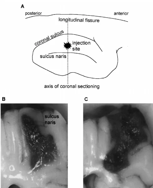
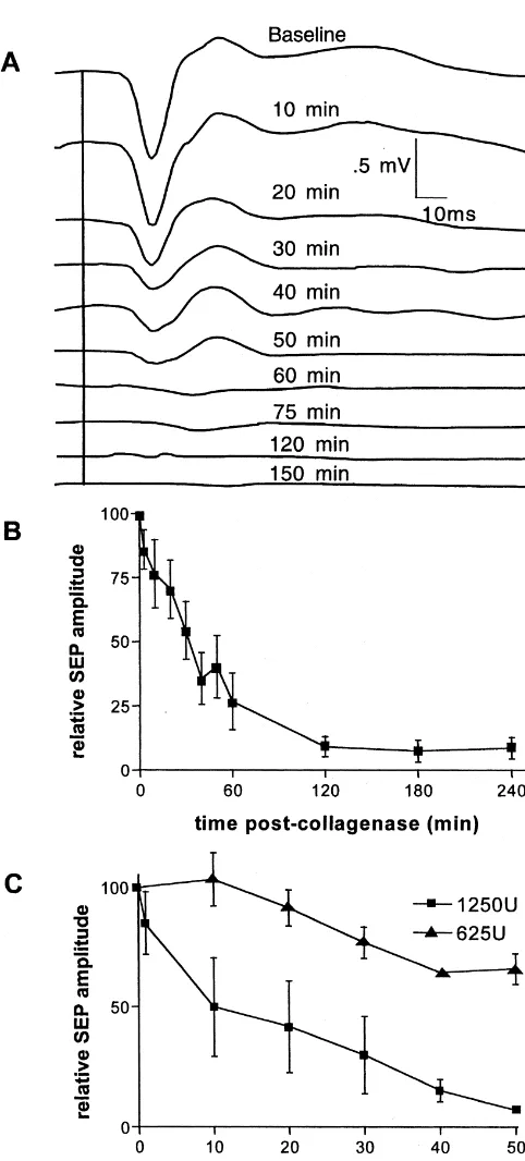
across four animals. Fig. 3C shows the percentage of SDs 3.1. Effects of collagenase on SEP seen at each electrode site relative to the total number of SDs seen at all the electrode sites during that particular Histological examination of the affected cerebral hemi- period: early, mid- and late. Of the 58 total SDs observed sphere revealed that an intracerebral injection of collagen- in all animals during the early period, 19 SDs (33%) were ase produced a hemorrhagic mass primarily in white matter recorded in electrodes 3 and 4 (1 mm away from the of the SI cortex. Fig. 1 displays a representative example injection site). Twenty SDs (34%) were recorded in of the hemorrhage 5.5 h after an injection of 625 U of electrodes 2 and 5 (2 mm away from the injection site) and collagenase (Fig. 1B) and another 6 h after an injection of 19 SDs (33%) were detected in electrodes 1 and 6 (3 mm 1250 U of collagenase (Fig. 1C). These two coronal away from the injection site). Sixty-three SDs were sections were obtained along the plane indicated in the observed during the mid-period: 14 SDs (22%) at 1 mm, illustration of the dorsal view of the cortex (Fig. 1A). The 24 SDs (38%) at 2 mm, and 25 SDs (40%) recorded at 3 extent of the injury was larger for injections of higher mm away from the injection site. During the late period, concentrations of collagenase. 37 total SDs were recorded in the four animals: five SDs SEPs elicited by electrical stimulation of the snout (14%) at 1 mm, 15 SDs (40%) at 2 mm, and 17 SDs decreased in amplitude within minutes after the start of (46%) at 3 mm from the site of injection.
1250 U of collagenase injection into the SI projection area Maximal duration of each period was 24 min in the of the snout (Fig. 2A). These SEPs were recorded with early period, 42 min in the mid-period, and 74 min in the electrode 2 whose location in the 6-electrode array is late period. During the early period SD was seen equally depicted in Fig. 3A. The negative polarity of the SEP often at all electrode sites, indicating that SD was being short-latency component was due to the fact that the generated near the injection site and propagating to outer electrode was positioned 3 mm below the cortical surface. electrode locations. The proportion of SD detected near the In all collagenase-injected animals, the SEP was nearly injection site decreased relative to those at the periphery as abolished by 60 min after the start of collagenase injection time progressed. This interaction between the time periods (Fig. 2B). A significantly more rapid drop in the amplitude and electrode distance from the injection site was statisti-of the SEP short-latency component was measured during cally significant (F53.82, df54 / 27, P,0.01).
the site of collagenase injection, while electrodes positioned medial to the sulcus naris continued to detect substantial SD episodes during the late period.
These features illustrated in Fig. 4B were quantified by an analysis of the entire data set across four animals (Fig. 4C). As in Fig. 3C, the graph in Fig. 4C indicates the percentage of SDs detected in a particular group of electrodes related to SDs recorded at all electrode sites for each period. Of the 110 total SDs observed in all animals during the early period, 22 SDs (20%) were recorded in region x (electrodes 3 and 11) which were the electrodes in the anterior / posterior plane of the injection site. Forty-one SDs (37%) were recorded in the 2y region, which includes all other electrodes lateral to the sulcus naris (electrodes 4, 7, 8, 12), and 47 SDs (43%) were recorded in the 1y region, which included all electrodes medial to the sulcus naris (electrodes 1, 2, 5, 6, 9, 10). Forty-eight SDs were observed during the mid-period: six SDs (12%) in region x, 19 SDs (40%) in the 2y electrodes, and 23 SDs (48%) recorded in region 1y. During the late period, 86 total SDs were recorded in the four animals: eight SDs (9%) in region x, 29 SDs (34%) in the 2y region, and 49 SDs (57%) in the 1y region.
The maximum duration of the early period was 34 min, middle period was 34 min, and late period was 1 h and 21 min. Similar to the 6-electrode data, there was a significant interaction between the time periods and electrode location relative to the sulcus naris (F53.26; df54 / 27; P,0.03). In summary, these data suggest that SDs were generated initially at multiple foci adjacent to the ICH core and subsequently further away from the injury core.
4. Discussion
The collagenase-induced ICH has not been characterized in the swine. The swine brain is gyrencephalic, containing a well-developed pattern of convolutions in the cortex. Since there are large differences in characteristics of the brain between the rat and the human, it is essential to obtain understanding of the sequela of an ICH in a species whose brain resembles the human brain. We believe that the porcine brain is an important non-primate model for understanding hemorrhagic stroke in human patients. The present study is the first of a series of studies being
Fig. 2. (A) Representative waveforms depicting the temporal decrease in
planned for characterizing the sequela of an ICH during
somatic evoked potential (SEP) amplitude occurring within minutes after
the acute and chronic phases not only with EEG, but also
the start of a 1250 U collagenase injection into the SI cortical projection
with magnetoencephalography (MEG) and magnetic
reso-area. (B) This change in mean amplitude of the SEP early response
component was apparent in all collagenase-injected animals, n58. (C) A nance imaging (MRI).
significantly sharper drop in SEP amplitude was seen during the initial 60 This study has shown two important features of ICH min following collagenase-injection in animals receiving 1250 U of
during the acute phase in the swine. First, the SEP
collagenase as compared to animals receiving 625 U, P,0.004.
Fig. 3. (A) Arrangement of the injection pipette, ‘X’, and the six glass electrodes in a single plane array on the SI cortex. (B) Representative profile of spreading depression (SD) propagation in each electrode following collagenase injection during the early (left) and late (right) periods of SD in this electrode array. (C) Graph summarizes the percent of SD detected in each electrode relative to the total number of SDs seen in all the electrodes during that period. The interaction between the time periods and electrode distance from the injection site was statistically significant (P,0.01).
dose-dependent, reproducible bleed at the site of injection proportional increase in the amount of SD propagation detected during the late period (Fig. 4C).
as well as significant alterations in brain edema, electrolyte
We did not determine the propagation velocity of SD in imbalance and hematoma size in the rat [24]. Although we
our preparation since the path of propagation may be quite have not characterized the development of the hematoma
complex in an intact cortex. The velocity can be measured, in the swine model, the time course of SEP change is
however, when SD is forced to propagate linearly along a consistent with previous MRI results in the rat. Rapid
cortical strip [1,2]. In such a case the SD initiated by an growth of the lesion occurred during the initial 60 min of
electrical stimulation of the cortex was found to propagate injury and by 2 h, MRI data showed signs of early edema
uniformly in a lissencephalic species (rabbit) [1], but related to the hematoma [3,6]. The accumulation of red
non-uniformly in a gyrencephalic species (swine) [2]. The blood cells along white matter bundles of the affected area
propagation velocity was 1–2 mm / min within the sulcus was seen within 30 min of injection [6]. At 2 h
post-as compared to 6–8 mm / min in the gyrus. These results collagenase / heparin injection, histological examination
suggest that the SD initiated by collagenase may be revealed an almost contiguous bleed with swollen glial
different in gyrencephalic species such as the human as cells [6].
compared to lissencephalic species such as the rabbit and The reduction in SEP amplitude can be explained by the
rat. The results of Bowyer et al. [2] need to be tested in our loss of ionic homeostasis in the white matter and overlying
collagenase model. gray matter of the swine SI within and surrounding the
Our observation of SD during the acute phase of an ICH injection site of collagenase. The extravasation of
blood-is interesting since some investigators have proposed SD to borne macromolecules and loss of oxygen supply should
play a role in upregulating the expression of neuroprotec-lead to changes in the cellular metabolism and
conse-tive genes in ischemia [10,15] and in influencing infarct quently to the depolarization of the membrane potentials of
volume size [4,27]. It should be possible to evaluate the neurons and glial cells. The depolarization should alter the
role of SD in determining the size of hematoma by kinetics of the voltage-sensitive channels, for example by
measuring the size with T2-weighted MRI and by detect-inactivating the sodium channels, and should decrease the
ing the occurrence of SD with diffusion-weighted imaging amplitude and frequency of the action potentials in the
which is sensitive to changes in extracellular volume thalamocortical fibers and also the amplitudes of the
fraction that is reduced during SDs. The size of a hemato-cortical responses. Thus, we interpret the time course of
ma can be compared under a barbiturate anesthesia which reduction in SEP amplitude to reflect the temporal dy- does not prevent the occurrence of SD as in our present namics of the developing hematoma. This interpretation study and in others [7,22], and under an anesthetic such as will be verified by conducting a continuous measurement ketamine which blocks N-methyl-D-aspartate (NMDA) of the developing mass lesion with MRI in an acute receptors [9] and prevents SDs.
collagenase ICH preparation. The existence of spontaneous SD in our ICH model has Secondly, our finding of spontaneous SDs in the cortex possible clinical significance as episodes of SD may be undergoing an ICH is, to the best of our knowledge, novel. occurring spontaneously in stroke patients with an ICH SDs are known to occur in a cortical tissue with focal during the acute phase. The pathophysiology of hemor-ischemia [18], but there has been no study reporting SD in rhage has gained renewed interest because ICH may an ICH model. In our model, there was a significant develop in patients with ischemic stroke after thrombolytic characteristic change in the pattern of SD onset and therapy. As we characterize the sequela of the ICH in the propagation with time after the collagenase injection. swine model, it should be possible to use non-invasive Within minutes after the injection, multiple electrodes at techniques such as EEG / MEG and MRI to provide better the injury site and in the surrounding tissue detected understanding of stroke.
recurring episodes of SDs. With time, recovery to the baseline DC potential level became more prolonged and
incomplete in certain electrodes, especially those proximal Acknowledgements to the injury site. During the later period, the rate of SD
generation near the injection site decreased as compared to This work was supported by an NIH grant RO1-the early SD period and RO1-the locations of SD shifted to NS30968 (YCO), an Under-represented Minority Postdoc-electrodes positioned farther from the hemorrhagic core. toral Fellowship from NINDS, and the Dedicated Health The 12-electrode array data indicated that the sulcus Research Funds of the UNM School of Medicine (SM-B). naris dividing the SI cortex influences the temporal and Thanks to Gary A. Rosenberg, M.D., for his expert advice spatial occurrence of SD as the hemorrhage developed in in editing this manuscript.
tissue lateral to this sulcus. The percentage of SDs seen in region x decreased with time from the early to late periods
relative to the total number of SDs seen at all electrode References sites. A similar trend was seen in the other electrodes
positioned lateral to the sulcus naris (2y). In contrast, [1] S.M. Bowyer, Y.C. Okada, N. Papuashvili, J.E. Moran, G.L.
ˆ
spreading cortical depression with propagation constrained to a [14] A.A.P. Leao, Spreading depression of activity in the cerebral cortex, rectangular cortical strip I. Lissencephalic rabbit model, Brain Res. J. Neurophysiol. 7 (1944) 379–390.
843 (1999) 71–78. [15] K. Matsushima, R. Schmidt-Kastner, M.J. Hogan, A.M. Hakim, [2] S.M. Bowyer, N. Tepley, N. Papuashvili, S. Kato, G.L. Barkley, Cortical spreading depression activates trophic factor expression in K.M.A. Welch, Y.C. Okada, Analysis of MEG signals of spreading neurons and astrocytes and protects against subsequent focal brain cortical depression with propagation constrained to a rectangular ischemia, Brain Res. 807 (1998) 47–60.
cortical strip II. Gyrencephalic swine model, Brain Res. 843 (1999) [16] S. Mun-Bryce, F.O. Kroh, J. White, G.A. Rosenberg, Brain lactate
1 31
79–86. and pH dissociation in edema: H- and P-NMR in
collagenase-[3] M.S. Brown, M. Kornfeld, S. Mun-Bryce, R.S. Sibbit, G.A. Rosen- induced hemorrhage in rats, Am. J. Physiol. 265 (1993) R697–702. berg, Comparison of magnetic resonance imaging and histology in [17] S. Mun-Bryce, A. Wilkerson, N. Papuashvili, Y.C. Okada, Spreading collagenase induced hemorrhage in the rat, J. Neuroimag. 5 (1995) cortical depression is elicited by collagenase-induced intracerebral
23–33. hemorrhage, Soc. Neurosci. Abs. 25 (1999) 318.
[4] E. Busch, M.L. Gyngell, M. Eis, M. Hoehn-Berlage, K.A. Hossman, [18] M. Nedergaard, A.J. Hansen, Characterization of cortical depolariza-Potassium-induced cortical spreading depressions during focal cere- tions evoked in focal cerebral ischemia, J. Cereb. Blood Flow. bral ischemia in rats: contribution to lesion growth assessed by Metab. 13 (1993) 568–574.
diffusion-weighted NMR and biochemical imaging, J. Cereb. Blood [19] H. Oka, M. Kako, M. Matsushima, K. Andok, Traumatic spreading Flow. Metab. 16 (1996) 1090–1099. depression syndrome review of a particular type of head injury in 37 [5] S.L. Craner, R.H. Ray, Somatosensory cortex of the neonatal pig: I. patients, Brain 100 (1977) 287–298.
Topographical organization of primary somatosensory cortex (SI), J. [20] Y.C. Okada, A. Lahteenmaki, C. Xu, Comparison of MEG and EEG Comp. Neurol. 306 (1991) 24–38. on the basis of somatic evoked responses elicited by stimulation of [6] M.R. Del Bigio, H. Yan, R. Buist, J. Peeling, Experimental the snout in the juvenile swine, Electroencephalogr. Clin.
Neuro-intracerebral hemorrhage in rats. Magnetic resonance imaging and physiol. 110 (1999) 214–229.
histopathological correlates, Stroke 27 (1996) 2313–2320. [21] Y.C. Okada, B. Shah, J.C. Huang, Ferromagnetic high-permeability [7] J.P. Dreier, K. Korner, N. Ebert, A. Gorner, I. Rubin, T. Back, U. alloy alone can provide sufficient low-frequency and eddy-current Lindauer, T. Wolf, A. Villringer, K.M. Einhaupl, M. Lauritzen, U. shielding for biomagnetic measurements, IEEE Trans. Biomed. Eng. Dirnagl, Nitric oxide scavenging by hemoglobin or nitric oxide 7 (1994) 688–697.
synthase inhibition by N-nitro-L-arginine induces cortical spreading [22] A. Rashidy-Pour, Z. Motaghed-Larijani, J. Bures, Tolerance to
1
ischemia when K is increased in the subarachnoid space, J. Cereb. ketamine-induced blockade of cortical spreading depression trans-Blood Flow. Metab. 18 (1998) 978–990. fers to MK-801 but not to AP5 in rats, Brain Res. 693 (1995) [8] H.J. Feickert, S. Drommer, R. Heyer, Severe head injury in children: 64–69.
impact of risk factors on outcome, J. Trauma 47 (1999) 33–38. [23] G.A. Rosenberg, E. Estrada, R.O. Kelley, M. Kornfeld, Bacterial [9] N.A. Gorelova, V.I. Koroleva, T. Amemori, V. Pavlik, J. Bures, collagenase disrupts extracellular matrix and opens blood–brain
Ketamine blockade of cortical spreading depression in rats, Elec- barrier in rat, Neurosci. Lett. 160 (1993) 117–119.
troencephalogr. Clin. Neurophysiol. 66 (1987) 440–447. [24] G.A. Rosenberg, S. Mun-Bryce, M. Wesley, M. Kornfeld, Col-[10] S. Kobayashi, V.A. Harris, F.A. Welsh, Spreading depression induces lagenase-induced intracerebral hemorrhage in rats, Stroke 21 (1990)
tolerance of cortical neurons to ischemia in rat brain, J. Cereb. 801–807.
Blood Flow. Metab. 15 (1995) 721–727. [25] C.H. Sotak, New NMR measurements in epilepsy. Diffusion-weight-[11] V.I. Koroleva, J. Bures, The use of spreading depression waves for ed magnetic resonance imaging of spreading depression, Adv.
acute and long-term monitoring of the penumbra zone of focal Neurol. 79 (1999) 925–929.
ischemic damage in rats, Proc. Natl. Acad. Sci. USA 93 (1996) [26] J.L. Voelker, H.H. Kaufman, Intraparenchymal hemorrhage, New
3710–3714. Horizon 5 (1997) 342–351.
[12] R.P. Kraig, C. Nicholson, Extracellular ionic variations during [27] H. Yanamoto, N. Hashimoto, I. Nagata, H. Kikuchi, Infarct tolerance spreading depression, Neuroscience 3 (1978) 1045–1059. against temporary focal ischemia following spreading depression in [13] M. Lauritzen, Pathophysiology of the migraine aura. The spreading rat brain, Brain Res. 784 (1998) 239–249.