www.elsevier.nlrlocateraqua-online
Pharmacokinetics, tissue distribution, and
metabolism of flumequine in channel catfish
ž
Ictalurus punctatus
/
Steven M. Plakas
a,), Kathleen R. El Said
a, Steven M. Musser
ba
Gulf Coast Seafood Laboratory, U.S. Food and Drug Administration, P.O. Box 158, Dauphin Island, AL 36528, USA
b
Instrumentation and Biophysics Branch, U.S. Food and Drug Administration, 200 C St. S.W., Washington, DC 20204, USA
Accepted 17 December 1999
Abstract
Ž .
The pharmacokinetics and metabolism of the fluoroquinolone drug flumequine FLU were
Ž . Ž .
examined after intravascular 1 mgrkg or oral 5 mgrkg dosing in channel catfish. Parent FLU concentrations in plasma declined slowly after intravascular dosing, with a half-life of 25 h. After
Ž .
oral dosing, FLU concentrations in plasma were highest 3.1 mgrml at 14 h after dosing; absorption and elimination half-lives were 4.9 and 22 h, respectively. The oral bioavailability of FLU was 44%, based on normalized plasma data. Plasma protein binding of parent FLU was
Ž . 14
extensive, but saturable 88%–55% bound at 0.125–8.0mgrml . After oral dosing with C-labeled FLU, radioactive residues were evenly distributed among the major tissues analyzed, with peak
Ž .
concentrations occurring at 12–24 h. Residue concentrations were highest in liver 6.2mgrg and
Ž .
lowest in muscle 1.8mgrg at 24 h. Only parent FLU was found in muscle and was eliminated with a half-life of 26 h. FLU and its metabolites were recovered in urine and bile. Residues in bile consisted almost entirely of a taurine conjugate of FLU. In urine, the taurine conjugate and hydroxy-FLU metabolites were found, in addition to the parent compound. Published by Elsevier Science B.V.
Keywords: Flumequine; Pharmacokinetics; Metabolism; Channel catfish
)Corresponding author. Tel.:q1-334-694-4480 ext. 251 ; fax:Ž . q1-334-694-4477.
Ž .
E-mail address: [email protected] S.M. Plakas .
0044-8486r00$ - see front matter. Published by Elsevier Science B.V.
Ž .
1. Introduction
Ž .
Flumequine FLU is a fluoroquinolone drug used in aquaculture primarily for treatment of Gram-negative bacterial infections in fish. Its use in European aquaculture
Ž .
is well documented, particularly in the Atlantic salmon industry Grave et al., 1996 . Widespread application of FLU in the past has raised environmental and food safety concerns, with increased scrutiny and modifications in its aquatic applications. FLU is not approved for food fish use in the United States.
Pharmacokinetic and metabolism data are valuable in establishing drug dosage and withdrawal periods in food-producing animals and in developing suitable methods for monitoring residues in animal products. In fish, these data exist for FLU in several
Ž
freshwater and marine species Boon et al., 1991; Rogstad et al., 1993; Elema et al., 1994,1995; Sohlberg et al., 1994,1996; Van der Heijden et al., 1994; Hiney et al., 1995;
.
Martinsen and Horsberg, 1995; Samuelsen and Ervik, 1997 . In general, FLU is moderately absorbed when administered orally in fish and residues are well distributed
Ž
in the tissues. FLU is also absorbed during bath treatment O’Grady et al., 1988; .
Samuelsen and Lunestad, 1996 . Elimination of FLU in fish is generally slow, but
Ž .
significant species differences exist Van der Heijden et al., 1994 . Metabolism of FLU in fish also appears slow; metabolite levels in tissues are typically low or undetectable ŽHaagsma et al., 1993; Van der Heijden et al., 1993,1994; Samuelsen and Ervik, 1997 .. We describe the pharmacokinetics and metabolism of FLU in farm-raised channel
Ž .
catfish Ictalurus punctatus . Distribution and elimination of residues in the tissues are characterized, with emphasis on the edible flesh. Metabolites of FLU in selected tissues and fluids are identified.
2. Materials and methods
2.1. Chemicals
w2- C -Flumequine specific activity, 30.4 mCi14 x Ž rmmol was custom synthesized by.
Ž .
NEN Research Products Boston, MA . Radiochemical and chemical purity were
Ž .
)99%, as determined by liquid chromatography HPLC and thin-layer
chromatogra-Ž .
phy. Unlabeled FLU )99% purity by HPLC was purchased from Sigma Chemical
Ž .
Co. St. Louis, MO .
2.2. Animals
Ž .
Channel catfish Ictalurus punctatus were obtained from a local farm, with no
Ž .
history of FLU treatment; mean body weight "SD of fish used in the study was 0.66"0.14 kg. Animals were allowed to adapt to study conditions for at least 2 weeks
Ž .
while being fed a commercial catfish feed Purina Mills, St. Louis, MO . At 1–2 days Ž
prior to surgery or dosing, animals were placed in stainless steel cages Plakas et al.,
. Ž .
2.3. Surgical procedures
For pharmacokinetic evaluations, animals were catheterized for serial sampling of
Ž .
blood, as described by Stehly and Plakas 1993 . Animals were allowed to recover for 1–2 days before dosing. To examine renal excretion, the urinary bladders of fish were
Ž .
cannulated Plakas et al., 1994 . Urine flow was confirmed before dosing.
2.4. Dosing solutions
Intravascular dosing solution was prepared at a concentration of 2 mgrml by using unlabeled FLU. To prepare 5 ml, 10 mg FLU was dissolved in 3.5 ml of 0.1 M Na CO2 3 with sonication. The solution was adjusted to pH 9 with 1 N HCl and brought to volume
Ž .
with water. Oral dosing solutions with or without radiolabel were similarly prepared, but at a FLU concentration of 10 mgrml. For radioactive solutions, portions of the unlabeled FLU were substituted with14C-FLU to yield an activity level of 100mCirml Ž10 mCirmg . The pH of dosing solutions was re-adjusted before use to ensure. complete solubilization of the drug. Dosing solutions were administered at a rate of 0.5 mlrkg body weight. FLU did not precipitate from solution when mixed with plasma during in vitro simulation of intravascular dosing.
2.5. Dosing and sampling
2.5.1. Pharmacokinetics
Intravascular doses of 1 mgrkg were administered through the catheters of five fish by using disposable syringes with blunted needles. Doses were followed with;1 ml of heparinized saline. Clean syringes and needles with three-way stopcocks were attached.
Ž . Ž
Blood specimens 0.25–0.3 ml each were taken as previously described Stehly and .
Plakas, 1993 at various intervals after dosing and were immediately packed in ice. Plasma was separated by centrifugation and stored at y808C until analysis by HPLC.
Ž
Oral doses of 5 mgrkg were administered in gelatin capsules size 00, Torpac, East
. Ž .
Hanover, NJ by gavage to anesthetized animals ns5 . Each capsule contained 0.25 g of ground catfish feed onto which the dosing solution was dispersed. After dosing, fish were returned to their tanks and plasma specimens were collected as described above.
2.5.2. Tissue distribution and elimination
The tissue distribution and elimination of total drug residues were determined after oral dosing with 14C-FLU at 5 mgrkg. At each sampling time, five animals were stunned by cranial concussion and blood specimens were taken by syringe from the
Ž caudal vein. Fish were then euthanized by cervical dislocation and selected organs liver,
.
head kidney, trunk kidney, and spleen were dissected, briefly rinsed with water, blotted, Ž and weighed. Bile was taken from the gall bladder by syringe. Muscle fillets skin
.
2.5.3. Renal excretion
Cannulated animals were orally dosed with 14C-FLU at 5 mgrkg. Urine was continuously collected in polypropylene containers packed in ice. At various intervals after dosing, urine volumes were recorded and specimens were stored aty808C until analysis. Excretion data were compiled for three animals in which cannulae remained patent for 2 weeks after dosing.
2.5.4. Metabolism
Ž .
Selected tissues and fluids containing radioactive residues as collected above were examined for FLU metabolic profile. Plasma, muscle, liver, and kidney specimens collected at the 24-h sampling time provided sufficient levels of radioactivity for metabolite analysis. Equal volumes of bile or urine specimens were pooled and analyzed
Ž .
for each sampling time. Additional fish ns3 were orally dosed with unlabeled FLU Ž5 mgrkg to provide non-radioactive residues for mass spectrometric characterization. of metabolites.
2.6. Analytical methods
2.6.1. HPLC
Procedures for extraction and HPLC analysis of parent FLU in muscle and plasma
Ž .
were described previously Plakas et al., 1999 . Briefly, FLU was extracted with
Ž .
acidified methanol glacial acetic acid-methanol, 2q98 , and extracts were cleaned-up
Ž .
on C18 solid-phase extraction SPE columns. FLU concentrations were determined by Ž
using a C18 HPLC column, an isocratic mobile phase glacial acetic
acid–water–aceto-. Ž .
nitrile, 2q48q50 , and fluorescence detection excitation, 325 nm; emission, 360 nm . Standard curves were prepared by fortification of control tissues with FLU.
Metabolic profile of14C-FLU in selected tissues and fluids was determined by HPLC operated under gradient mobile phase conditions. Extraction and clean-up procedures for total residues in tissues were the same as described above for parent FLU. Recoveries of total radioactivity in these tissues were G90%. Residues in urine were concentrated and cleaned-up by SPE. Five-milliliter aliquots of pooled urine were applied to 6-ml C18
Ž .
SPE columns and, after elution and evaporation as per tissue procedure , residues were
Ž .
re-solubilized in 0.5 ml of initial mobile phase for analysis. Bile was diluted 1:10 with initial mobile phase, filtered, and injected directly onto the HPLC column. Mobile phase
Ž . Ž . Ž .
reservoirs were: A glacial acetic acid:water 2q98 ; B glacial acetic acid:acetonitrile Ž2q98 . Gradient conditions were: 70% A for 2.5 min, ramp to 50% A at 15 min, hold.
Ž .
at 50% A for 10 min, return to initial conditions 70% A , and re-equilibrate for G15 min before the next injection. A fraction collector was used to monitor radioactivity in
Ž .
HPLC column effluents 0.2-min fractions .
( )
2.6.2. Liquid chromatographyrmass spectrometry LCrMS
Ž .
The LCrMS system consisted of a Hewlett-Packard Palo Alto, CA Model 1050 LC
Ž .
pump and a Finnigan San Jose, CA Model TSQ-7000 triple-quadrupole mass
spec-Ž .
trometer with a standard Finnigan electrospray ESI ion source. All chromatography
Ž . Ž
was performed on a YMC Wilmington, NC J-sphere ODS-M80 LC column 2=250 .
mm . The mobile phase flow rate was 0.2 mlrmin and all other buffers and gradients were the same as those used to prepare the metabolite fractions. Nitrogen was used as a nebulizing gas and the ion source capillary temperature was 2308C. For full scan MS experiments, the instrument was scanned over the range of 150–700 amu at 1 srscan. For MSrMS experiments, the instrument was scanned from 15–380 amu at 1 srscan. Argon was used as the collision gas and collision energy was 40 V.
( )
2.6.3. Liquid scintillation counting LSC
Total14C contents in tissues, fluids, and HPLC effluent fractions were determined by
Ž .
LSC, as previously described Plakas et al., 1998 . Radioactivity in tissues or fluids was
Ž .
expressed in units of concentration mg equiv. of FLUrg or ml or as a percentage of the administered dose. With no practical means of solubilizing bone for LSC, caudal
Ž .
vertebrae specimens ;0.5 g from each fish were extracted twice with 4 ml acidified methanol by shaking overnight at room temperature in a mechanical shaker. Methanolic extracts were combined and aliquots were analyzed by LSC.
2.6.4. Plasma protein binding
Ž
Plasma protein binding of parent FLU was determined by ultrafiltration Centrifree . 14
Micropartition System, Amicon, Beverly, MA . C-FLU was added to control plasma at
Ž .
concentrations ranging from 0.125 to 8 mgrml. Duplicate aliquots 0.4 ml of spiked
Ž .
plasma were added to the ultrafilters and centrifuged at 1000=g for 1 h 258C . Entire ultrafiltrates were analyzed for 14C-FLU contents by LSC, representing unbound drug. Values were corrected for non-specific absorption of unbound drug to the ultrafiltration
Ž .
apparatus ;6% . Non-specific absorption was determined by the corresponding analy-sis of control plasma ultrafiltrates spiked with14C-FLU. Additionally, plasma specimens
14 Ž .
collected from fish dosed orally with C-FLU as described above were analyzed for protein binding of total radioactive residues.
2.6.5. Pharmacokinetics
Ž .
The computer program WinNonlin Scientific Consulting, Cary, NC was used to derive pharmacokinetic values for parent FLU concentrations in plasma. Oral bioavail-ability was calculated from the areas under the plasma concentration-time curves, normalized for dose.
3. Results
3.1. Pharmacokinetics
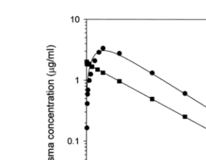
Parent FLU levels in plasma declined slowly after intravascular dosing and were
Ž . Ž .
Ž .
Fig. 1. Mean parent FLU concentrations in plasma after intravascular dosing at 1 mgrkg B or oral dosing at
Ž . Ž .
5 mgrkg Ø in channel catfish ns5 .
ŽTable 1 . Other pharmacokinetic parameters Table 1 indicate moderate distribution of. Ž .
Ž . Ž
FLU outside of the vascular system V , 527 mlss rkg , and slow clearance Cl , 14.9b .
mlrhrkg .
Ž .
After oral dosing, parent FLU was measurable in plasma within 15 min Fig. 1 . Peak plasma concentration was 3.1 mgrml at 14 h. Absorption and elimination half-lives
Ž .
were 4.9 and 22 h, respectively Table 1 . The bioavailability of FLU was 44%. FLU levels were not quantifiable beyond 168 h.
3.2. Plasma protein binding
Plasma protein binding of14C-FLU in spiked plasma was extensive, but saturable,
Ž .
declining from 88% at 0.125mgrml to 55% at 8mgrml Fig. 2 . Similar values were
Table 1
Pharmacokinetic values for parent FLU in channel catfish
Ž . Ž .
Intravascular 1 mgrkg Oral 5 mgrkg
Parameter Unit Value Parameter Unit Value
t1r2el h 24.6 t1r2abs h 4.94
AUC mgPhrml 67.3 t1r2el h 21.8
Clb mlrhrkg 14.9 AUC mgPhrml 149
Vss mlrkg 527 Cmax mgrml 3.06
MRT h 35.5 tma x h 13.7
F % 44.3
Pharmacokinetic parameter abbreviations: t1r2el, elimination half-life; AUC, area under the plasma concentra-tion-time curve; Cl , total body clearance; V , apparent volume of distribution at steady-state; MRT, meanb ss
Fig. 2. Plasma protein binding of FLU as determined in vitro.
found for residual14C in plasma of orally dosed animals, depending on concentration or
Ž .
time after dosing data not shown . For example, mean binding values were 67% at 24 h
Ž 14 .
peak C levels and 89% at 168 h.
3.3. Tissue distribution and elimination
After oral dosing with 14C-FLU, total residue concentrations in the tissues were
Ž . Ž .
highest at 12–24 h Table 2 . At 24 h, liver had the highest concentrations 6.2 mgrg
Ž .
and muscle had the lowest 1.8 mgrg of any tissues analyzed. Concentrations in plasma, trunk kidney, head kidney, and spleen were similar, ranging from 3.3 to 4.4 mgrg at 24 h. Rates of elimination of radioactive residues were also similar between individual tissues. In muscle,14C concentrations declined below the limit of
determina-Table 2
Ž . a
Concentration of radioactive residues mg equiv.rg or ml in the bile and tissues of catfish after oral
14 Ž .
administration of C-FLU 5 mgrkg
Ž .
Tissue Time after dosing h
3 6 12 24 72 168 336
Bile 0.48"0.24 6.30"7.52 12.2"6.18 33.9"15.3 119"71.1 91.0"34.9 82.4"27.7 Liver 2.15"2.12 4.62"2.91 5.46"2.01 6.15"1.24 1.77"0.74 0.34"0.20 0.08"0.04 Plasma 1.71"0.46 3.16"0.96 4.00"1.97 4.10"0.89 3.00"1.20 0.49"0.44 0.02"0.02 Trunk kidney 0.95"0.24 2.92"1.35 3.78"1.54 4.41"1.16 1.54"0.52 0.25"0.17 0.05"0.02 Head kidney 0.75"0.27 2.42"1.12 3.80"1.75 3.37"0.82 1.07"0.38 0.22"0.16 0.07"0.03 Spleen 0.70"0.27 2.42"1.16 3.35"1.26 3.34"0.78 1.07"0.46 0.24"0.18 0.11"0.06 Skin 0.42"0.12 1.32"0.74 1.84"0.73 2.18"0.72 0.84"0.20 0.26"0.16 0.09"0.05
b
Muscle 0.28"0.12 1.03"0.68 1.80"0.91 1.84"0.47 0.42"0.19 0.07"0.06 –
a
Values are the mean"s.d. of five animals.
b Ž .
Fig. 3. Parent FLU concentrations in muscle after oral dosing at 5 mgrkg in channel catfish. Values are the mean"s.d. of five animals.
Ž .
tion by LSC -0.005mgrg at 336 h. In skin, concentrations were initially similar to those in muscle, but were eliminated more slowly. Residue levels in bone, as estimated by solvent extraction, were 0.11mgrg at 3 h, 0.66mgrg at 24 h, and 0.03mgrg at 336
Ž .
h data not shown .
Ž .
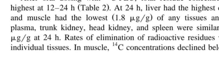
Parent FLU concentrations in muscle, as determined by HPLC Fig. 3 , were nearly
Ž .
identical to total residue concentrations, by LSC Table 2 . Parent FLU levels were 1.8
Ž . Ž
mgrg at 24 h and 0.07 at 168 h Fig. 3 . Parent FLU was not quantifiable -0.01 .
mgrg beyond 168 h. Estimated half-life for elimination of FLU in muscle was 26 h.
Ž . 14
Fig. 4. Renal excretion of FLU and its metabolites total residues after oral dosing with C-FLU at 5 mgrkg
Ž .
3.4. Excretion
Residues were highly concentrated in bile, relative to the tissues. Total residue concentrations increased rapidly from 0.48 mgrml at 3 h to 119 mgrml at 72 h, and
Ž . Ž .
then declined slowly to 82mgrml at 336 h Table 2 . At peak levels 72 h , the amounts of14C contained in the gall bladder represented 3–4% of the administered oral dose.
Ž .
In urine, total residue concentrations were highest 0.88 mgrml at the 12–24 h
Ž . 14
sampling interval Fig. 4 . By 168 h, C levels in urine were 0.04mgrml and at 336 h,
-0.01 mgrml. Cumulatively, 4.9% of the oral dose was excreted in urine over the 2-week sampling period.
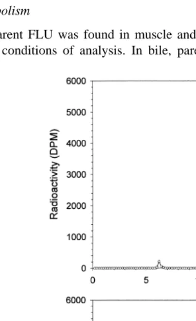
3.5. Metabolism
Only parent FLU was found in muscle and plasma after oral dosing with14C-FLU, under our conditions of analysis. In bile, parent FLU was a minor component at the
Ž .
earlier sampling times e.g., 12% of total residues at 24 h , but was not detectable beyond 72 h. Residues in bile were mostly composed of a single, polar metabolite with a
Ž .
retention time of ;13 min Fig. 5 . The molecular weight of this metabolite was 368, Ž
by LCrMS. This finding, along with a significant fragment ion at mrz 80 80% relative .
abundance in the negative ion MSrMS spectrum, is consistent with the metabolite being a taurine conjugate of FLU.
Ž .
In urine, four radioactive peaks were found Fig. 5 . Parent FLU comprised 72% of total radioactive residues at 3 h, 55% at 24 h, and 38% at 168 h. The taurine conjugate of FLU also was identified, comprising 11% of total residues at 3 h, 32% at 24 h, and
Ž
59% at 168 h. Two minor metabolites, which eluted as a doublet peak retention time, .
9.5–10 min by HPLC, comprised -10% of total radioactivity. These minor metabo-lites were both identified as hydroxyl-FLU, and were probably structural isomers. Liver
Ž .
and kidney contained mostly )80% parent FLU at 24 h.
4. Discussion
Parent FLU levels declined slowly after intravascular and oral dosing, with elimina-tion half-lives of 25 and 22 h, respectively, at 248C. Much longer half-lives were reported in other freshwater species. Elimination half-lives in European eel, common
Ž
carp, and African catfish, were 451, 104, and 59.5 h, respectively, at 248C Van der
. Ž .
Heijden et al., 1994 . Boon et al. 1991 reported a terminal elimination half-life of 256 h in European eel at 238C. In freshwater rainbow trout, FLU half-life in plasma was 569
Ž .
h at 38C and 137 h at 138C Sohlberg et al., 1994 . In seawater, elimination of FLU in fish is generally more rapid than in freshwater, even at lower temperatures. In Atlantic salmon held in saltwater, FLU half-lives were 21–23 h in studies conducted at 5–108C ŽRogstad et al., 1993; Elema et al., 1994; Martinsen and Horsberg, 1995 . Elema et al.. Ž1995 found half-lives of 30–40 h in Atlantic salmon at 6–8. 8C. In Atlantic salmon smolts, FLU half-life was 170 h in freshwater compared with 140 h in seawater, at 118C ŽHiney et al., 1995 . Sohlberg et al. 1996 reported elimination half-lives of 67 and 38 h. Ž . in cannulated and non-cannulated Atlantic salmon, respectively, after intravascular dosing at 118C. In Atlantic halibut, terminal half-life of FLU was 43 h, at 98C ŽSamuelsen and Ervik, 1997 ..
A bi-exponential equation provided the best fit of experimental data after
intravascu-Ž .
lar dosing, but with low confidence in the initial distribution phase. A mono-exponen-tial equation could also be applied with virtually no change in those pharmacokinetic values reported in Table 1. Plasma protein binding of FLU was extensive and saturable in the range of concentrations observed in vivo, which possibly influenced FLU
Ž .
distribution kinetics under our dosing conditions. Boon et al. 1991 found concentra-tion-dependent changes in plasma protein binding of FLU in eel, although binding
Ž . Ž
values 30–40% were much lower than in catfish. Other processes e.g., metabolism,
. Ž .
excretion may also be non-liner with respect to dose or concentration of FLU. Pharmacokinetic values for FLU in channel catfish indicate moderate distribution
Ž . Ž
outside of the vascular system V , 0.527 lrss kg and slow clearance Cl , 0.015b .
Ž
from 0.005 to 0.2 lrhrkg Rogstad et al., 1993; Sohlberg et al., 1994,1996; Elema et .
al., 1995; Martinsen and Horsberg, 1995 . In Atlantic halibut, Vss and Cl values wereb
Ž .
2.3 lrkg and 0.052 lrhrkg, respectively Samuelsen and Ervik, 1997 . Although not reported in the latter studies, plasma protein binding may be an important variable influencing distribution and clearance of FLU.
Ž .
After oral dosing in channel catfish, peak levels Cma x, 3.1mgrml of parent FLU in
Ž .
plasma occurred at 14 h tma x , with an absorption half-life of 4.9 h. Bioavailability of FLU was 44%. Oral absorption is also fairly rapid, but incomplete, in other fish species.
Ž
However, species differences occur even under identical dosing conditions Van der .
Heijden et al., 1994 . In some freshwater species, peak concentrations were found within
Ž .
2 h after dosing Van der Heijden et al., 1994 . In Atlantic salmon, plasma FLU levels Ž typically peak at 12–24 h after oral dosing, with a bioavailability of 35–55% Rogstad
. et al., 1993; Elema et al., 1995; Martinsen and Horsberg, 1995; Sohlberg et al., 1996 . In
Ž
Atlantic halibut, tmax was 20 h and bioavailability was 31% Samuelsen and Ervik, .
1997 . Absorption of FLU may be lower in seawater by its complex with divalent
Ž .
cations, as found with the fluoroquinolone drug difloxacin Elston et al., 1994 . FLU is very stable in marine sediments, and its application and incomplete absorption in
Ž
medicated feeds may have environmental consequences Ervik et al., 1994; Samuelsen .
et al., 1994 .
Total residue concentrations in the tissues of channel catfish were highest at 12–24 h after dosing and rates of elimination were generally similar. At 24 h, tissue:plasma concentration ratios ranged from 0.45:1 in muscle to 1.5:1 in liver, which were congruent with pharmacokinetic analyses suggesting moderate extravascular distribution of FLU. In Atlantic salmon, tissue penetration of FLU appears more extensive than that
Ž
in catfish, with higher tissue:plasma concentration ratios Rogstad et al., 1993; Elema et
. Ž
al., 1994 and larger volumes of distribution. In some fishes e.g., Atlantic salmon, sea .
bream , FLU is highly concentrated in skin and especially bone, relative to muscle ŽSteffenak et al., 1991,1994; Malvisi et al., 1997 . These tissues represent a deep storage. compartment prolonging overall elimination of FLU. From the food safety standpoint, FLU may be released from these sites during normal processing and cooking of the
Ž .
marketed product Steffenak et al., 1994 . In channel catfish, total residue concentrations in skin were initially comparable to those in muscle, but were eliminated more slowly. Residue levels in bone, as estimated by solvent extraction, were mostly lower than in
Ž muscle. Possibly, solvent extraction of bone underestimates total residue content i.e.,
. bound drug .
Parent FLU was the only residue detected in muscle of catfish and was eliminated with a half-life of 26 h at 228C. By comparison, half-life of FLU in muscle of rainbow
Ž .
trout was 11 h at 148C Malvisi et al., 1994 . In Atlantic salmon, half-life was 18 and 21
Ž .
h after 6- and 8-day medication periods, respectively, at 88C Elema et al., 1994 . In Atlantic salmon smolts, half-life in muscle was 81 and 41 h for fish held in freshwater
Ž .
and seawater, respectively Hiney et al., 1995 . In juvenile Atlantic halibut, FLU
Ž .
half-life in muscle was 10 h at 128C Samuelsen and Lunestad, 1996 . Muscle and Ž plasma data indicate significant species differences occur in elimination of FLU in
.
Renal excretion cumulatively accounted for 5% of the oral dose of radioactivity in
Ž .
catfish. In bile, 4% of the oral dose was found at 72 h tma x . Bile was not continuously collected and amounts of drug found in the gall bladder probably underestimates total drug excreted by this route, while enterohepatic circulation of FLU is also possible. FLU
Ž .
may be eliminated by other routes e.g., branchial . Evidence of branchial absorption of FLU was provided by bath exposure studies in freshwater fish, in which significant
Ž
levels of FLU were found in the tissues after treatment O’Grady et al., 1988; Hiney et
. Ž .
al., 1995 . Boon et al. 1991 suggested gill surface area as a possible limiting factor in the elimination of FLU in eel. However, the contribution of the gills in absorption and elimination of FLU remains undetermined.
We identified a taurine conjugate of FLU in bile and urine of catfish and hydroxy-FLU in urine. We did not find metabolites in plasma or muscle tissues. In mammalian
Ž species, FLU glucuronide and 7-hydroxy-FLU are principal urinary metabolites
Harri-.
son et al., 1986; Mevius et al., 1990; Vree et al., 1992 . Possibly, FLU glucuronide was unstable in urine and tissues during processing under our conditions; conversely, bile was directly analyzed with no evidence of the glucuronide. Previous studies in fish, while not examining excretory fluids, reported only trace levels of metabolites in tissues ŽHaagsma et al., 1993; Van der Heijden et al., 1994; Samuelsen and Ervik, 1997 ..
Ž .
In summary, FLU was moderately absorbed by the oral route bioavailability, 44% and residues were evenly distributed and eliminated in tissues of channel catfish. Parent FLU was the only residue found in edible flesh and was eliminated with a half-life of 26 h. Parent FLU is an appropriate target analyte for residue monitoring programs, for which rapid analytical methods are available. Current data suggest that estimation of withdrawal periods following FLU administration in fish requires individual study for each species under defined environmental and dosage conditions.
Acknowledgements
Ž . Ž .
We thank F.A. Bencsath FDA for technical advice and G. Stehly USGS for reviewing this manuscript.
References
Boon, J.H., Nouws, J.M.F., Van der Heijden, M.H.T., Booms, G.H.R., Degen, M., 1991. Disposition of
Ž .
flumequine in plasma of European eel Anguilla anguilla after a single intramuscular injection. Aquacul-ture 99, 213–223.
Elema, M.O., Hoff, K.A., Kristensen, H.G., 1994. Multiple-dose pharmacokinetic study of flumequine in
Ž .
Atlantic salmon Salmo salar L. . Aquaculture 128, 1–11.
Elema, M.O., Hoff, K.A., Kristensen, H.G., 1995. Bioavailability of flumequine after oral administration to
Ž .
Atlantic salmon Salmo salar L. . Aquaculture 136, 209–219.
Grave, K., Markestad, A., Bangen, M., 1996. Comparison in prescribing patterns of antibacterial drugs in salmonid farming in Norway during the periods 1980–1988 and 1989–1994. J. Vet. Pharmacol. Therap. 19, 184–191.
Haagsma, N., Van Roy, M., Gortemaker, B.G.M., 1993. High performance liquid chromatographic determina-tion of flumequine and its hydroxy metabolite in fish tissue. In: Haagsma, N., Ruiter, A.,
Czedik-Eysen-Ž .
berg, P.B. Eds. , Residues of Veterinary Drugs in Food. University of Utrecht, Utrecht, The Netherlands, pp. 337–341.
Harrison, L.I., Schuppan, D., Rohlfing, S.R., Hansen, A.R., Gerster, J.F., Hansen, C.S., Funk, M.L., Ober, R.E., 1986. Disposition of radiolabeled flumequine in rat and dog. Drug Metab. Disp. 14, 555–558. Hiney, M.P., Coyne, R., Kerry, J., Pursell, L., Samuelsen, O.B., Smith, P., 1995. Failure of flumisol bath
treatments during commercial transport of Atlantic salmon smolts to prevent the activation of stress inducible furunculosis. Aquaculture 136, 31–42.
Malvisi, J., Della Rocca, G., Anfossi, P., Giorgetti, G., 1997. Tissue distribution and depletion of flumequine
Ž .
after in-feed administration in sea bream Sparus aurata . Aquaculture 157, 197–204.
Malvisi, J., Giorgetti, G., Raspa, M., Guiliani, A., Tomasi, L., Roncada, P., 1994. Kinetics of flumequine in
Ž .
rainbow trout Oncorhynchus mykiss tissue. Riv. Ital. Acquacolt. 29, 121–128.
Martinsen, B., Horsberg, T.E., 1995. Comparative single-dose pharmacokinetics of four quinolones, oxolinic
Ž .
acid, flumequine, sarafloxacin, and enrofloxacin, in Atlantic salmon Salmo salar held in seawater at 108C. Antimicrob. Agents Chemother. 39, 1059–1064.
Mevius, D.J., Breukink, H.J., Guelen, P.J.M., Jansen, T., De Greve, B., 1990. Pharmacokinetics, metabolism and renal clearance of flumequine in veal calves. J. Vet. Pharmacol. Therap. 13, 159–169.
O’Grady, P., Moloney, M., Smith, P.R., 1988. Bath administration of the quinoline antibiotic flumequine to brown trout Salmo trutta and Atlantic salmon S. salar. Dis. Aquat. Org. 4, 27–33.
Plakas, S.M., El Said, K.R., Bencsath, F.A., Musser, S.M., Hayton, W.L., 1998. Pharmacokinetics, tissue
Ž .
distribution and metabolism of acriflavine and proflavine in the channel catfish Ictalurus punctatus .
Xenobiotica 28, 605–616.
Plakas, S.M., El Said, K.R., Bencsath, F.A., Musser, S.M., Walker, C.C., 1999. Determination of flumequine in channel catfish by liquid chromatography with fluorescence detection. J. AOAC Int. 82, 614–619. Plakas, S.M., El Said, K.R., Stehly, G.R., 1994. Furazolidone disposition after intravascular and oral dosing in
the channel catfish. Xenobiotica 24, 1095–1105.
Rogstad, A., Ellingsen, O.F., Syvertsen, C., 1993. Pharmacokinetics and bioavailability of flumequine and oxolinic acid after various routes of administration to Atlantic salmon in seawater. Aquaculture 110, 207–220.
Samuelsen, O.B., Ervik, A., 1997. Single dose pharmacokinetic study of flumequine after intravenous,
Ž .
intraperitoneal and oral administration to Atlantic halibut Hippoglossus hippoglossus held in seawater at 98C. Aquaculture 158, 215–227.
Samuelsen, O.B., Lunestad, B.T., 1996. Bath treatment, an alternative method for the administration of the quinolones flumequine and oxolinic acid to halibut Hippoglossus hippoglossus, and in vitro antibacterial activity of the drugs against some Vibrio sp. Dis. Aquat. Org. 27, 13–18.
Samuelsen, O.B., Lunestad, B.T., Ervik, A., Fjelde, S., 1994. Stability of antibacterial agents in an artificial marine aquaculture sediment studied under laboratory conditions. Aquaculture 126, 283–290.
Sohlberg, S., Aulie, A., Soli, N.E., 1994. Temperature-dependent absorption and elimination of flumequine in
Ž .
rainbow trout Oncorhynchus mykiss Walbaum in fresh water. Aquaculture 119, 1–10.
Sohlberg, S., Martinsen, B., Horsberg, T.E., Soli, N.E., 1996. Evaluation of the dorsal aorta cannulation
Ž .
technique for pharmacokinetic studies in Atlantic salmon Salmo salar in sea water. J. Vet. Pharmacol. Therap. 19, 460–465.
Steffenak, I., Hormazabal, V., Yndestad, M., 1991. Reservoir of quinolone residues in fish. Food Add. Contam. 8, 777–780.
Steffenak, I., Hormazabal, V., Yndestad, M., 1994. Effect of cooking on residues of the quinolones oxolinic acid and flumequine in fish. Acta Vet. Scand. 35, 299–301.
Stehly, G.R., Plakas, S.M., 1993. Pharmacokinetics, tissue distribution, and metabolism of nitrofurantoin in the
Ž .
channel catfish Ictalurus punctatus . Aquaculture 113, 1–10.
Ž .
flumequine in European eel. In: Haagsma, N., Ruiter, A., Czedik-Eysenberg, P.B. Eds. , Residues of Veterinary Drugs in Food. University of Utrecht, Utrecht, The Netherlands, pp. 357–361.
Van der Heijden, M.H.T., Keukens, H.J., Van den Nieuwboer, W.H.F.X., Mengelers, M.J.B., Boon, J.H.,
Ž .
1994. Plasma disposition of flumequine in common carp Cyprinus carpio L., 1758 , African catfish
ŽClarias gariepinus Burchell, 1822 and European eel. ŽAnguilla anguilla L., 1758 after a single peroral.
administration. Aquaculture 123, 21–30.