ISSN 0854-8587
Solubilization, Activation and Partial Purification of a Sialidase
from Horse Liver
KRISHNA PURNAWAN CANDRA1‡*, PETER ROGGENTIN1,2, ROLAND SCHAUER1
1Biochemistry Institute of Christian-Albrechts University, Olshausenstr. 40, D-24098 Kiel, Germany
2Hygiene Institut Hamburg, Marckmannstr. 129a, D-20539 Hamburg, Germany
Diterima 30 Juli 2003/Disetujui 21 Juni 2005
Using sialyl-methylumbelliferyl ααααα-glycoside as substrate, sialidase in horse liver was detected as a membrane-bound enzyme. A yield of about 50% of sialidase activity was found in supernatant when solubilized in 0.1 M sodium-phosphate buffer pH 5.5, containing 0.15 M NaCl, 0.25 M sucrose, and 0.5% Triton X-100. Sialidase in the solubilisate could be activated by incubating in acidic pH at 37 oC. Incubation of this solubilized enzyme at 37 oC for 1.5 h at pH 5.0 led to 10% increase of activity and to the precipitation of about 50% of contaminating protein. Using cation-exchange chromatogra-phy on S-Sepharose FF and affinity chromatograchromatogra-phy on p-aminophenyl oxamic acid-agarose following solubilization and activation, about 6% of total sialidase activity was recovered with the purification factor of about 500. The pH and temperature optimum were measured at pH 4.3 and between 37-45 oC, respectively. Neu5Ac2en was a strong inhibitor, while p-aminophenyl oxamic acid had only a weak inhibitory effect.
___________________________________________________________________________
_________________
‡Alamat kini: Laboratory of Chemistry and Biochemistry,
Agricultural Faculty of Mulawarman University, Jalan Tanah Grogot, Samarinda 75123
∗ ∗∗
∗∗Penulis untuk korespondensi, Tel. +62-541-749347,
Fax. +62-541-738341, E-mail: [email protected] INTRODUCTION
Sialidases (EC 3.2.1.18) play an important role in metabolism of sialic acid, an amino sugar with pyranose structure and 9-atom carbon and posseses a negative charge due to the carboxyl group at C-atom 1. It catalyzes the release of the α -glycosidically bound sialic acids from terminal sialo-oligosaccharides, gangliosides, or sialo-glycoproteins (Corfield & Schauer 1982). In eucaryotic cells, sialidase can be found in almost all of subcellular components, i.e. cytosol, intra-lysosomes and lysosomes (Miyagi et al. 1992), nuclear membrane (Saito et al. 1996), and Golgi membrane (Kishore et al. 1975).
Purification of sialidase, in particular lysosomal sialidase, was proven difficult because it usually was associated with
β-galactosidase (EC 3.2.1.23), carboxypeptidase A (EC 3.4.16.1), and N-galactosamin-6-sulfate sulfatase (EC 3.1.6.4) sulfatase, besides this enzyme was also membrane-bound characteristic and less stable if it was separated from the membrane (Pshezhetsky & Potier 1996). Some lysosomal sialidases can be activated in vitro by incubating glycoprotein fraction in acidic buffer at 37 oC
for 1-2 hours. The endogenous protease activities were believed to involve in the activation phenomenon of the sialidase (Hiraiwa et al. 1993).
In the case of sialidase, acetylation of the hydroxyl group at C-4 of the substrate molecule (sialic acid) was shown to block most of the enzyme activity except for some virus sialidases (Kleineidam et al. 1990). Therefore, it is interesting
to understand the sialic acid metabolism (catabolism) of outstanding sialic acids like O-acetylated sialic acid. The finding that horse liver sialidase was unable to hydrolyse Neu4,5Ac2, even though high levels of Neu4,5Ac2 are present, leads the first report for the existence of esterase in horse liver, which is involved in the 4-O-acetylated sialic acid catabolism (Schauer et al. 1988).
Studying sialidase from horse liver is still interesting because sialidase, in particular, lysosomal sialidase has not been characterized. The protein component of lysosomal sialidase following purification performed by some groups still cannot elucidate the catalytic subunit of this protein (Hiraiwa et al. 1996). Isolation, purification, and determination of factors affecting horse liver lysosomal sialidase activities need to be conducted in order to further investigate the properties of this enzyme. This report described localization, isolation and activation of lysosomal horse liver sialidase.
MATERIALS AND METHODS
Enzyme and Protein Assays. Sialidase and β-galactosidase were assayed as procedure described by Potier et al. (1979) using MU-Neu5Ac in acetat buffer pH 4.3 and MU-Gal in acetate buffer pH 4.0, respectively. N-acetylgalactosamine-6-sulfate sulfatase was assayed using MU-Gal-6-S as described by Pshezhetsky and Potier (1996). The protein content was determined using spectrophotometer at 280 nm or by the Bradford method (Bradford 1976).
Localization of Sialidase Activity. The homogenate of 65 g frozen horse liver was centrifuged at 100,000 g for 60 minutes following homogenization in 240 ml water with an ultra turrax (3 x 1 minute) and homogenization for 10 strokes in
Table 1. Distribution of the sialidase activity in horse liver
Total sialidase activity1,2
mU %Homogenization in water
Specific activity of the sialidase (µU/mg protein)
Homogenate
After centrifugation at 100,000 g for 60 minute Supernatant fraction
1Mean + SD from 5 separate experiments, 2Sialidase was assayed with MU-Neu5Ac at 37 oC for 1 h, *Activity was absent in supernatant after a
second ultra-centrifugation step
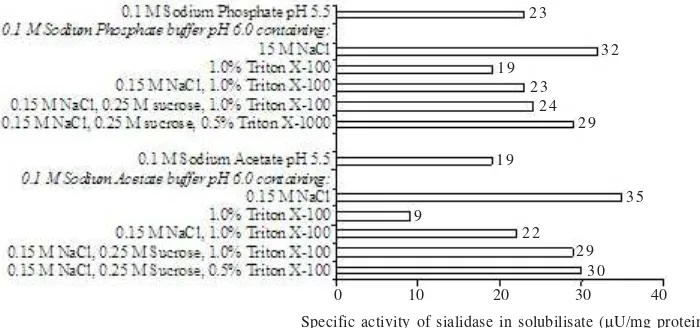
Solubilization of Sialidase. A 0.5 ml of membrane preparation of 20 mg/ml protein concentration was added to an Eppendorf cap (end protein concentration was 10 mg/ml), to it some other reagents such as water, buffers (acetate and phosphate), detergents (Triton X-100, cholic acid, and taurocholic acid) and sucrose were added. This mixture was first mixed well and after that salt (NaCl or KCl) was added. The end volume of this solution was 1 ml, where end concentration of each substance in the buffer showed in legend of Figure 1 and 2. The solution was then shaken gently at 4 oC, and after 1 h was ultra-centrifuged. Sodium phosphate
buffer of 0.1 M pH 5.5 containing 0.15 M NaCl, 0.25 M sucrose, and 0.5% Triton X-100 (buffer A) was applied for further experiment.
Activation. The 100,000 g supernatant (solubilisate) of membrane fraction following solubilization was titrated with 2 M HCl in order to obtain pHs between 4.8 and 5.6, the suspension was then incubated at 37 oC. After a given time of
incubation, the suspension was centrifuged at 12,000 g for 30 min. Three incubation times of 1, 1.5, and 2 hours were observed. As a control, suspension with pH of 5.6 was also incubated on ice.
Isolation and Purification of Sialidase. All isolation and purification procedures were carried out at 4 oC unless
otherwise specified.
Solubilization and activation of sialidase: Aproximately 65 grams of frozen horse liver was homogenized 3 times, each for 1 min in 240 ml cold water with an Ultra turrax and then centrifuged at 100,000 g for 1 hour to obtain a raw membrane preparation (pellet). The pellet was solubilized in buffer A using a Potter-Elvehjem apparatus (10 strokes) and then centrifuged at 100,000 g for 1 hour. The solubilisate was titrated with 2 M HCl to pH 5.0 and incubated at 37 oC for 1.5 hours
followed by centrifugation at 12,000 g for 30 minutes. The activated soluble sialidase was dialyzed over night against 10 mM sodium phosphate buffer pH 5.0 (buffer B).
Cation-exchange chromatography on S-Sepharose FF: The dialyzed sample described in step 1 was applied at a flow rate of 1 ml/min to a 50 ml S-Sepharose FF column (3.5 x 5.2 cm) that had been equilibrated with buffer B. The column was washed with 150 ml buffer B, followed by washing with 150 ml 0.3 M sodium phosphate buffer pH 5.0 and then again with 150 ml buffer B. The sialidase was then eluted using a concentration gradient of 300 ml sodium phosphate buffer pH 7.0 from 10 to 500 mM. The sialidase was eluted at the phosphate buffer concentration of about 200 mM, whereby 5 ml-fractions were collected. The sialidase positive fractions
were pooled and dialyzed over night against 10 mM sodium phosphate buffer pH 5.5 (buffer C).
Affinity chromatography on PAOX-agarose: The dialyzed sample described in step 2 was added with MnCl2 and CaCl2 to get a final concentration of the divalent cations of 1 mM, and was then applied at a flow rate of 0.5 ml/min to a 7 ml PAOX-agarose column (1.4 x 4.5 cm) that had been equilibrated with buffer C containing 1 mM of MnCl2 and CaCl2 (buffer D). The column was washed with 50 ml of buffer D. The sialidase was eluted by shifting the pH and concentration with bicarbonate buffer (NaHCO3) pH 9.1 using a concentration gradient of the buffer (80 ml) between 10 and 300 mM, and 4 ml-fractions were collected. Four ml of 300 mM sodium phosphate buffer pH 5.0 was added to each fraction prior the elution started to adjust the basic eluate to a pH-value of about 6.0. The fractions containing sialidase activity were pooled and dialyzed over night against buffer B, and used for characterization for temperature-, pH-optimum and inhibitory effects.
RESULTS
Localization of Sialidase. About 20-30% of total sialidase activity was found in supernatant while the main activity was detected in the pellet fraction (Table 1). Theoretically, the enzyme activity found in the supernatant after ultra centrifugation should be a soluble enzyme, however the sialidase activity from the supernatant could be totally pelleted after re-ultra-centrifugation for 60 minutes. These results strongly suggest that the sialidase in horse liver is a membrane-bound enzyme, or a large soluble complex.
and NaCl of the same concentration showed the same effect on solubilization of sialidase.
It is very important to see the specific activity of sialidase in solubilisate because the highest specific activity will lead the easier purification step, however the yield is also become consideration. The specific activity of sialidase solubilized was outlined in Figure 2.
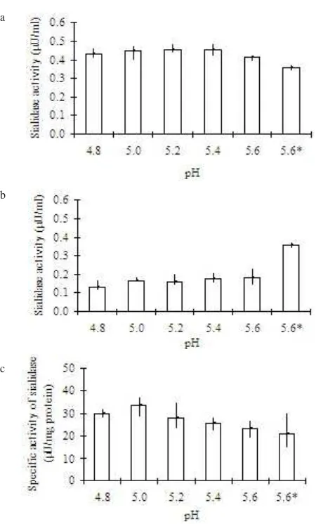
Activation of Sialidase. The sialidase activity detected prior to centrifugation at 12,000 g (solubilisate) increased by approximately 10% upon incubation at 37 oC, pH 5.0 (Figure
3a). Removal of precipitated proteins via centrifugation resulted in a slight loss of activity in the soluble fraction (Figure 3b), however an increase in specific activity of approximately 50% was obtained (Figure 3c). Incubation of the soluble sialidase suspension at pH 5.0, 37 oC for 1.5 hours
showed the best activation condition for this sialidase. In this step the sialidase activity was increased by about 50% over the solubilisate with a purification factor of 1.4 and a yield of about 40% against solubilisate.
Purification of Sialidase. In cation-exchange chromatography step, washing with 0.3 M phosphate buffer pH 5.0 increased the purification factor to about 2-fold in comparison to when the wash was omitted. After this step the sialidase was purified with purification factor of about 78 and yield of 9%. On the other hand, addition of concentrated
phosphate buffer pH 5.0 onto eluate of affinity chromatography was very effective and easier to handle to keep the eluate at pH around 6.0 than the method described before in Cuatrecasas and Illiano (1971). The chromatogram of sialidase and β-galactosidase activity following affinity chromatography on PAOX-agarose is shown in Figure 4. The
β-galactosidase detected in this purification step was eluted earlier than sialidase and the both enzymes were found to be separate. This finding suggests that the sialidase isolated did not occur as an enzyme complex with β-galactosidase. Following this purification with PAOX-agarose, a purification factor of 500 and yield of 6% for the sialidase were resulted (Table 2).
Temperature and pH Optimum. The temperature optimum of the sialidase activity was detected between 37-45 oC and
the pH assay optimum was detected at 4.3 (Figure 5). Inhibition on Sialidase Activity. The inhibitory effect on sialidase activity by p-aminophenyl oxamic acid (PAOX) and Neu5Ac2en can be seen in Figure 6. PAOX was found to be a moderate inhibitor, with about 20 and 40% inhibition occurring at PAOX concentration at 1 mM and 5 mM, respectively. Neu5Ac2en, on the other hand, is a strong inhibitor of sialidase, with about 70% of sialidase activity being inhibited by the addition of 0.1 mM Neu5Ac2en. At Neu5Ac2en concentration of 1 mM only 5% of sialidase activity remained. 0 10 20 30 40
2 3
9
3 5 1 9
2 9 2 4 2 3 1 9
3 2
2 2 2 9
3 0
Specific activity of sialidase in solubilisate (µU/mg protein)
Figure 2. Specific activity of sialidase found in solubilisate following 100,000 g centrifugation of enzyme solution in different solubilization buffers.
0 10 20 30 40 50 60 Percentage of sialidase activity in solubilisate
2 0
4 7 5 2 3 9
1 4 2 4 7
4 8 5 2 4 5 3 5 2 8
Figure 3. Activity and specific activity of sialidase of solubilisate following activation by incubating at 37 oC for 1.5 h.
Control of pH value was stated with asterisk, solubilisate incubated on ice for 1.5 h. a. Sialidase activity of activated solubilisate, b. Sialidase activity found in supernatant following 12,000 g c e n t r i f u g a t i o n f o r 3 0 m i n . o f a c t i v a t e d s o l u b i l i s a t e , c . Specific activity of sialidase found in supernatant following 12,000 g centrifugation for 30 min. of activated solubilisate.
a
b
c
sialidase β-galactosidase protein
Figure 4. Chromatogram of sialidase and β-galactosidase activity following affinity chromatography using PAOX-agarose. Percentage of enzyme activity in each fraction was calculated on the base of enzyme activity in the original sample of (enzyme pool following ion-exchange using S-Sepharose FF), 27.957 mU of sialidase activity and 3.368 mU of β-galactosidase activity.
a
b
Figure 5. Influence of temperature and pH to the sialidase activity. a. temperature experiment, b. pH experiment.
Table 2. Purification of sialidase
Specific activity Purification (µ U/mg) factor Fractions Protein
(mg) Yield (%)
Solubilisate “Activation” S-Sepharose FF PAOX-agarose
8401 2494 10 1
15 21 1,163 7,441
1 0 0 41 9 6
1.0 1.4 78.0 500.0
Figure 6. Influence of inhibitors to the sialidase activity.
DISCUSSION
considering approaches for the solubilization of membrane-bound proteins attention must be given not only to detergents but also to the use of salts and high ionic strength buffers such as phosphate buffer (Schägger 1994). In this study, for example, we observed that the solubilization of sialidase activity from membrane preparation of horse liver was, in many cases, solubilization effects of NaCl on sialidase was better than detergents. This was observed not only in the case of Triton X-100 but also for cholic acid or taurocholic acid. The concentration of NaCl usually used to solubilize membrane-bound proteins is around 150 mM, however before using this salt it is important to check its inhibitory effect on the enzyme in question, because Na+ may inhibit the activity (Tulsiani &
Carubelli 1970). KCl is another alternative, which can be used in solubilization experiments.
The addition of sucrose in solubilization buffers can also be used to reduce the detergent concentration required to solubilize membrane-bound enzymes. Sucrose can also stabilize membrane-bound enzymes in solutions as has been shown for glycerol (Schägger 1994). The action of sucrose in reducing the amount of detergent required was also observed for the solubilization of sialidase from horse liver in this study. A mild activation of sialidase activity in soluble fraction was observed when the solubilisate was incubated under acidic pH conditions at 37 oC for about 1.5 h. This activation
maybe caused by the endogenous protease activities in the soluble fraction as described before that leupeptin inhibited the activation of sialidase from the crude glycoprotein fraction of human placenta (D’Agarosa & Callahan 1988). This involve of protease was clearly understood when the purified sialidase-β-galactosidase-carboxypeptidase A could not be activated in acidic pH after incubated at physiological temperature for 90 minutes unless cathepsin C was added and this activation was also inhibited by leupeptin. This activation by protease seems to be involved in the physiological function in the lysosome. However, sialidases of some tissues could not be activated with this method such as bovine, chicken, rat, and rabbit liver sialidases (Hiraiwa et al. 1993).
Ion-exchange chromatography is frequently used as the first step in enzyme purification because it gives a high degree of purification, even from crude extracts, and can be used easily for membrane-bound enzymes. The “classical” first step in purification, ammonium sulfate precipitation, however, is difficult to be used for membrane proteins because in many cases the protein remains insoluble following this method, even when the ammonium sulfate is removed. However, ammonium sulfate together with detergents, such as cholic acids, can be used in the purification of membrane-bound proteins (Schägger 1994). In this study, ammonium sulfate with 1.6% cholic acid was trialed, but the protein containing sialidase activity was pelleted and remained insoluble after the removal of ammonium sulfate, even the washed pellet was resuspended in phosphate buffer containing 50 mM NaCl. It may be possible that an irreversible change on the conformation of the sialidase during ammonium sulfate precipitation caused the insolubility of the sialidase.
During the purification of sialidase using cation-exchange chromatography on S-Sepharose FF, we observed that sialidase activity bound very tightly to the beads and could not be eluted even with 2 M NaCl in 10 mM phosphate buffer, pH 5.5. However, sialidase activity could be successfully eluted only when a concentrated phosphate buffer at pH 7.0 was used. An elution method using phosphate buffer containing 50 mM citrate was also trialed but was found to be unsuccessful, even though this elution method was successful in the case of sialidase from Trypanosoma brucei (Engstler et al. 1992).
The affinity chromatography medium PAOX-agarose was successfully used in this study for the purification of horse liver sialidase. According to Cuatrecasas and Illiano (1971) Vibrio cholerae sialidase bound to this column could be eluted using NaHCO3 buffer, pH 9.1. We were also able to elute sialidase activity using this elution method, however, instead of using 0.1 M HCl to lower the pH of the eluent, we used 0.3 mM phosphate buffer, pH 5.0. This method allowed a better recovery of sialidase activity, as well as being easier and more reproducible.
There were two major β-galactosidase isozymes reported in human tissue, a β-galactosidase A, which is optimally active between pH 3.5 and 4.5 and localized in lysosomes and the second one, which is optimally active near pH 6.5 and referred to as “neutral” β-galactosidase (Shows et al. 1979). In horse liver homogenate, β-galactosidase activity was detected with pH optimum at 6.0. Amazingly, the β-galactosidase obtained after PAOX-agarose is an acid β-galactosidase, which has an optimum pH at 4.0. To elucidate the possibility whether a sulfatase also occurs as enzyme-complex with sialidase-β -galactosidase-carboxypeptidase A as shown in Pshezhetsky and Potier (1996), a N-acetylgalactosamine 6-sulfate sulfatase test was performed. The sulfatase activity was not detected in the sialidase positive fractions following affinity chromatography on PAOX-agarose, however it was found in the homogenate of horse liver (in water). The sialidase positive fraction was first dialyzed against acetate buffer prior sulfatase test because phosphate inhibited its activity.
On the other hand, the evidence that lysosomal sialidase did not occur as enzyme complex with β-galactosidase was also reported, even the sialidase was co-purified with
β-galactosidase, both enzymes can be separated and the sialidase remained to be active (Yamamoto & Nishimura 1987). This evidence is perhaps also facing for the lysosomal sialidase isolated from this horse liver, as was shown that β -galactosidase activity could be separated from sialidase.
The acidic optimum pH of the sialidase isolated suggests that this sialidase is a lysosomal enzyme, since it has a similar pH optimum (4.3) to lysosomal sialidases previously reported (Hiraiwa et al. 1997), as well as the fact that the pH of lysosomes themselves is between 4.5 and 5.0 (Koolman & Röhn 1994).
Neu5Ac2en possessed different inhibitory characteristics depending on the type of substrates being used. For example, it did not inhibit sialidase when fetuin was the substrate, but was a strong inhibitor when II6Neu5-AcLac was used as
substrate. On the other hand, p-aminophenyl oxamic acid (PAOX) is only moderate inhibitory for the sialidase isolated, this is unlike the sialidases from T. brucei (Engstler et al. 1992) and V. cholerae (Engstler et al. 1994) which were strongly inhibited by PAOX. However, PAOX is not an inhibitor for rat sialidases (Miyagi et al. 1993).
ACKNOWLEDGMENT
The authors would like to express their gratitude to Lohr from NFZ Slaughterhouse, Lübeck, for his help in obtaining the horse liver used in these investigations. The author is grateful to Joe Tiralongo for his kind discussion and suggestion in preparation of this manuscript. The financial assistance of the German Academic Exchange Service (DAAD), Bonn, to the first author is gratefully acknowledged.
REFERENCES
Bradford M. 1976. A rapid sensitive method for the quantitation of microgram quantities of protein utilizing the principle of protein-dye binding. Anal Biochem 72:248-254.
Corfield AP, Schauer R. 1982. Occurence of Sialic Acids. In: Schauer R (ed). Sialic Acids - Chemistry, Metabolism and Function. Cell Biology Monographs. Wien: Springer. p 5-50.
Cuatrecasas P, Illiano G. 1971. Purification of neuraminidases from
Vibrio cholerae, Clostridium perfringens and influenza virus by
affinity chromatography. Biochem Biophys Res Commun 44:178-184.
D’Agarosa RM, Callahan JW. 1988. In vitro activation of neuraminidase in the β-galactosidase-neuraminidase-protective protein complex by cathepsin C. Biochem Biophys Res Commun
157:770-775.
Engstler M et al. 1994. N-(4-Nitrophenyl)-oxamic acid and related N -acylanilines are non-competitive inhibitors of Vibrio cholera
sialidase but do not inhibit Trypanosoma cruzi or Trypanosoma
brucei trans-sialidase. Helvetica Chimica Acta 77:1166-1174.
Engstler M, Reuter G, Schauer R. 1992. Purification and characterization of a novel sialidase found in procyclic culture forms of Trypanosoma brucei. Mol Biochem Parasitol 54:21-30. Hiraiwa M et al. 1993. Activation of human lysosomal sialidase. J
Biochem (Tokyo) 114:901-905.
Hiraiwa M et al. 1996. A sialidase complex from chicken liver: Characterization of a multienzyme complex with β-galactosidase and carboxypeptidase A. Comp Biochem Physiol 115B:541-546. Hiraiwa M et al. 1997. Protective protein in the bovine lysosomal β
-galactosidase complex. Biochim Biophys Acta 1341:189-199. Kishore CS, Tulsiani DR, Bhavanandan VP, Carubelli R. 1975.
Membrane-bound neuraminidases of rat liver. Neuraminidase activity in Golgi apparatus. J Biol Chem 250:2655-2659. Kleineidam RG, Furuhata K, Ogura H, Schauer R. 1990.
4-Methylumbelliferyl-α-glycosides of partially O-acetylated N -acetylneuraminic acids as substrates of bacterial and viral sialidases.
Biol Chem Hoppe Seyler 371:715-719.
Koolman J, Röhn KH. 1994. Taschenatlas der Biochemie. Stuttgart: Thieme Publ.
Michalski JC, Corfield AP, Schauer R. 1986. Properties of human liver lysosomal sialidase. Biol Chem Hoppe Seyler 367:715-722. Miyagi T, Hata K, Hasegawa A, Aoyagi T. 1993. Differential effect of
various inhibitors on four types of rat sialidase. Glycoconj J 10:45-49.
Miyagi T, Hata K, Konno K, Tsuiki S. 1992. Multiple forms of mammalian sialidase: altered expression in carcinogenesis. Tohoku
J Exp Med 168:223-229.
Miyagi T, Tsuiki S. 1986. Evidence for sialidase hydrolyzing gangliosides GM2 and GM1 in rat liver plasma membrane. FEBS Lett 206:223-228.
Potier M, Mameli L, Belisle M, Dallaire L, Melancon SB. 1979. Fluorometric assay of neuraminidase with a sodium (4-methylumbelliferyl-α-D-N-acetylneuraminate) substrate. Anal
Biochem 94:287-296.
Pshezhetsky AV, Potier M. 1996. Association of N-acetylgalactosamine-6-sulfate sulfatase with the multienzyme lysosomal complex of β-galactosidase, cathepsin A, and neuraminidase. Possible implication for intralysosomal catabolism of keratan sulfate. J Biol Chem 271:28359-28365.
Saito M, Fronda CL, Yu RK. 1996. Sialidase activity in nuclear membranes of rat brain. J Neurochem 66:2205-2208.
Schauer R, Reuter G, Stoll S. 1988. Sialate O-acetylesterases: key enzymes in sialic acid catabolism. Biochimie 70:1511-1519. Schägger H. 1994. Chromatographic techniques and basic operations
in membrane protein purification. In: von Jagow G, Schägger H (ed). A Practical Guide to Membrane Protein Purification. San Diego: Academic Pr. p 23-57.
Shows TB, Scrafford-Wolff LR, Brown JA, Meisler MH. 1979. GM1-gangliosidosis: chromosome 3 assignment of the β -galactosidase-A gene (βGALA). Somatic Cell Genet 5:147-158.
Tulsiani DRP, Carubelli R. 1970. Studies on the soluble and lysosomal neuraminidase of rat liver. J Biol Chem 245:1821-1827. Yamamoto Y, Nishimura K. 1987. Copurification and separation of β
-galactosidase and sialidase from porcine testis. Int J Biochem