Non functional adrenocortical carcinoma
A case report
Adianto Nugroho, Memet Nataprawira
Abstrak
Karsinoma adrenokortikal merupakan kasus yang jarang dijumpai dengan insidens 1-2 kasus per 1 juta penduduk per tahun. Gejala utama adalah timbulnya massa pada abdomen dengan atau tanpa disfungsi endokrin. Namun demikain, tampilan klinisnya dapat tidak khas sehingga menyulitkan dalam diagnosa dan penentuan terapi bahkan adakalanya diperlukan metode pemeriksaan tambahan seperti imunohistokimia untuk menentukan diangosis pasti karsinoma adrenokortikal. Karena sifat keganasannya yang tinggi, prognosis adreno kortikal karsinoma tidak terlalu baik, apalagi jika pengangkatan tumor tidak dimungkinkan karena ukurannya yang besar. Dilaporkan satu kasus karsinoma adrenokortikal di RSCM yang ditegakkan dengan pemeriksaan imunohistokimia pasca operasi pada pasien dengan tampilan klinis awal berupa tumor intra-abdomen berukuran besar (kista mesenterial ). (Med J Indones 2008; 17: 203-9)
Abstract
Adrenocortical carcinoma is a rare disease with an incidence of approximately 1-2 per million populations per year. The main symptoms are abdominal mass with or without endocrine dysfunction. However, there are dificulties in the diagnosis and treatment of this case due to its unspeciic clinical presentation which sometimes require additional diagnostic methods such as immunohistochemistry. The prognosis is often poor, especially when it’s impossible to resect the entire tumor. We present one case of adrenocortical carcinoma in RSCM which was diagnosed through immunohistochemistry examination postoperatively in patient with giant intra-abdominal tumor (mesenteric cyst). (Med J Indones 2008; 17: 203-9)
Keywords: Adrenocortical carcinoma, giant intra-abdominal tumor, mesenteric cyst, immunohistochemistry, resection
Adrenocortical carcinoma (ACC) is a rare tumor with poor prognosis.1-3 Disease stage and completeness of resection are primary determinants of outcome for this disease.2
Adrenocortical tumors are classiied as either functional or non functional.1,3-5 Patients with non functional Adrenocortical carcinoma usually came with a large tumour mass accompanied by symptoms related to the size of the tumor and invasion to adjacent organs. 1, 3, 5
We report a case of Adrenocortical carcinoma which came with a large intra abdominal mass.
CASE ILLUSTRATION
Male, 30 year old with a mass in his abdomen, came with complaints of dyspepsia, fatigue and weight loss that has been going on for about 11 months. Previously, he has had medication from another hospital, but there was no improvement on the symptoms. He never had dificulties nor abnormalities of defecation and also never felt pain on his abdomen. On physical examination, the abdomen was tender, venectation (+), dullness on percussion, with normal bowel sound. The abdominal circumference was 106 cm. (Figure 1)
The ultrasound revealed a well deined cystic mass, 15 cm in diameter at the central region of the abdomen. And the CT scan showed a cystic mass with multiple septums, extending from the minor pelvis to the right renal sinus, that pushed the gastro-intestinal tract to the left with normal Department of Surgery, Faculty of Medicine University of
distribution of bowel gas. This igure was matched with mesenterial cyst. (Figure 2)
Explorative laparotomy was conducted, a large reddish-black cystic mass was found inside the abdominal cavity, measuring 34 x 30 x 11 cm. There were blood vessel dilatations at the surface of the mass, with the feeding artery from vasa lienalis and gastroepiploic. The tumor was excised carefully from the surrounding tissues. Its weight was 10 kg. (Figure 3).
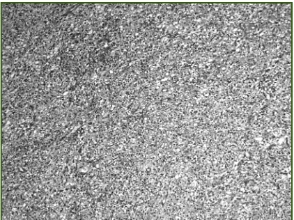
Histopathologic examination showed that the tumor mass was lobulated, separated with multiple thin septums of ibrous tissue, some parts were solid, and the others were trabecular. The nuclei was pleomorphic, hyperchromatic with eosinophilic cytoplasm. No mitosis was found. In
the central region, there was positive smear with PAS. (Figure 4) Conclusion: Grade II sarcoma with GIST, mesothelioma and poorly differentiated adenocarcinoma as the differential diagnosis.
To further prove the exact diagnosis, immuno histo-chemistry examination was done. The result was EMA (+) 90%, Chromoganin (+) 80%, Vimentin (+) positive 90%, Cd-117 (+) positive weak, and Cam 5.2 (+) positive 70%. Conclusion: Adrenocortical carcinoma.
The patient’s condition gradually improved and he went home 1 week postoperative. The clinical as well as CT scan assessment 4 months after the operation showed promising result with no recurrent mass and normal biochemistry parameter.
Figure 1. Male, 30 y.o. Pre- and Post- Operative
Figure 2. CT Scan showed a cystic mass with Figure 3. Intra operative tumor mass, 34 x 30 x 11 cm, 10,000 g multiple septums, from the minor pelvis to
Figure 4. Histopathologic Result Figure 5. PAS staining
DISCUSSION
Adrenocortical carcinoma (ACC) is a rare malignant neoplasm with poor prognosis.1,2,6,7 The annual incidence is 0.5 – 2 cases per 1 million and mortality is 0.04 – 0.2 % of all cancer deaths.1,3,4,7 This disease is more common in women, with female/male ratio of 1.75.1,7 The age
distribution is bimodal with a irst peak in childhood and a higher second peak in the 4th to 5th decade.3, 5 This tumor was classiied into two distinct types, functional and non functional.1,3-5 The clinical features of ACC include endocrine dysfunctions and symptoms due to the enlarging of tumor mass inside the abdominal cavity.1
Functioning ACC (approximately 60% of cases) often presents with signs and symptoms of adrenal steroid hormone excess, although hypersecretion of androgens in males or oestrogens in females may go unnoticed.3 Cushing’s syndrome (CS) with or without virilization is the most frequent presentation in functioning ACC.1, 3 Rapid development of CS with skin atrophy, muscle weakness, hyperglycaemia, hypertension and psychiatric symptoms is common, due to the excess of steroid, androgen, estrogen and mineralocorticoids.3 Adrenocortical carcinomas are ineficient in steroido genesis and may not show obvious clinical syndromes of excess even with large bulky disease.4
Patients with a nonfunctioning ACC usually present with symptoms related to the local effect like abdominal fullness, pain, indigestion, nausea and vomiting. In a minority of patients, weight loss, low-grade fever and
weakness may also occur. Due to the large tumour size at diagnosis, an abdominal mass may be palpable in a signiicant percentage of patients. The initial manifestation may also be related to metastatic disease (e.g. pathologic fracture, bone pain). A substantial and apparently increasing fraction of patients is diagnosed incidentally by abdominal imaging. Forty percent of adult patients with ACC present with a nonsecretory mass detected incidentally or during evaluation for abdominal or lank pain.3 Other study by Schulick and Brennan in 1999 found 45% non functional AC which came with pain, palpable abdominal mass and weight loss. The average tumor size was 14 cm in diameter weighing 750 g (range 4-2600 g).4 Non functional type was more prevalent in male then female.7
One type of non functional ACC is Oncocytic adreno-cortical carcinoma. A rare, non-functional tumors found in adults. Oncocytic neoplasms are comprised exclusively of oncocytic tumor cells, which are characterized by abundant granular eosinophilic cyto-plasm by light microscopy. Song et al in 2004 described four cases of oncocytic adrenocortical carcinomas with detailed clinicopathological, immuno histochemical and ultrastructural indings. They suggested that oncocytic adrenocortical carcinoma may be a low-grade malignancy and should be excised completely.8
radiological examination or autopsy. Symptoms such as abdominal pain and increasing girth occur only when the tumor grows large. Akamatsu et al report a case of a giant adrenal myelolipoma in a 51-year old man who presented with a huge abdominal mass and abdominal pain. The resected tumor weighed 6000 g and could represent the largest such tumor ever documented in the literature.9
Hormonal evaluation is mandatory in all patients with suspected ACC. Unfortunately, hormone concentrations are usually of limited help in predicting malignancy. However, in the presence of an adrenal lesion, elevated serum dehydroepiandrosterone sulphate (DHEAS) levels suggest an ACC, as benign adrenocortical tumours often exhibit low DHEAS concentrations. In addition, elevated serum 17β-oestradioal is a rare but rather typical marker of oestrogen-secreting ACC in men. Accordingly, in male patients with an adrenal tumour and elevated serum 17-oestradioal, an ACC should be assumed until proven otherwise.3
The size of the adrenal mass, as measured by computed tomography (CT) or magnetic resonance imaging (MRI) remains the single best indicator of malignancy. In a recent series from France (Icard et al, 2001), mean tumour size at diagnosis was 12·0 ± 6·0 cm (n= 223) and mean tumour weight was 689 ± 822 g (n = 202).10 Adrenal malignancies usually measured more than 6 cm.11 The likelihood of ACC increases to 35–98% in patients with an adrenal mass > 6 cm.3
Our patient was a 30 year old male, who came with enlarged abdominal mass along with fatigue and weight loss. There was no sign of Cushing’s syndrome in this patient. On admission, the abdominal mass has become so large that localisation of the origin was dificult, even with imaging technique.
Most of the patients came when the tumor has developed into a very large mass, because they didn’t recognized it, or because they have tried to ind other treatment, either surgical or non surgical. And that was also what happens to this patient, which came after 11 months of symptoms and has a history of previous medication. From international literature, there was no strict deinition of when a tumor is called “giant”.12
Kastomo report a study in Dharmais Hospital during 1993-2003, of which 24 giant intra abdominal tumor, intra peritoneal and retroperitoneal which do not originate from gastrointestinal tract, had been operated. Total extirpation was succesfull in 15 cases, 7 cases
near total, and 2 cases of malignant lymphomas were unresectable due to iniltration to the retroperitoneal lymph node. The largest resected tumor was Malignant
Fibro Histiocytoma measuring 70x50x40 cm, with 11
kg in weight.12
Almost all giant intra abdominal tumor can be seen macroscopically, so it can be diagnosed with a careful examination. Imaging techniques such as CT scan remains necessary to locate the origin of the tumor and its relations with adjacent organs. These information is important for the preparation of surgery.12
The CT scan result from our patient showed a mesenterial cyst, which is also a relatively rare case. The incidence of mesenterial cyst is only 1 case per 140,000 people.13 Omental cyst, an even rarer entity which is one of the differential diagnosis of mesenterial cyst, was irst described by Murata in 1906, only accounts for 130 cases in Japan since it was irst reported. Clinical indings include abdominal distension and pain and a palpable mass due to tumor growth. Disorders of the respiratory or urinary system, in addition to symptoms caused by compression of the portal vein, are often seen because of cyst enlarge ment. Between 11% - 19% of patients present with acute abdominal symptoms due to torsion, bleeding, or rupture of a cyst.
Surgical resection represents the deinitive diagnosis and treatment for intra abdominal tumor, including omental and mesenterial cyst.13,14 Percutaneous aspiration and biopsy are not warranted because they are not likely to provide a deinitive diagnosis and may lead to infection or rupture of the cyst.
The main dificulty in this operation is the relation between tumor mass and adjacent organs which might force us to excise the normal organs. The objective of the surgery should be to remove the tumor as cleas as possible. The excision of the whole mass (curative) or near total (debulking/palliative) could be very useful to reduce the pain and preparation for chemotherapy.12
Even when the tumor has been adequatelly excised, the diagnosis still remains a dificult problems. The tumor weight and size could be an important factor for the diagnosis, but in this patient, the tumor weight of 10,000 g, seems unreliable. Adenoma of adrenal cortex is usually weights 20-50 g, whereas ACs are 100 g or more.3
Like in other endocrine diseases, the diagnosis of AC is sometimes dificult, even when the resection has already been carried out. There were no single characteristic pathological feature to ensure its diagnosis.6 Combination of some pathological features as shown in Weiss score, could help in the diagnosis of malignancy2,6 (Table 1). Besides that, immuno histo chemistry could also play a role in the diagnosis with analysis of certain cells using antigen-antibody reaction.
Table 1. Histopathologic diagnosis criteria of adrenocortical carcinoma
Kriteria Derajat Weiss et al
(1989)
Atipia Nuclear Menengah sampai kuat 1
Mitosis > 5/50 LPB 1
Mitosis atipikal Ada 1
Clear Cells < 25% volume 1
Arsitektur Difus 1
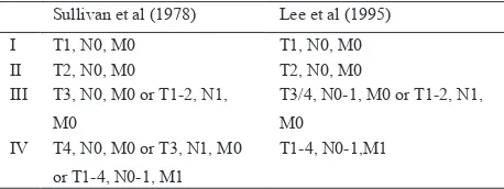
Table 2. TNM Classiication of Adrenocortical Carcinoma
Sullivan et al (1978) Lee et al (1995)
I T1, N0, M0 T1, N0, M0
T3: tumour iniltration locally reaching neighbouring organs
T4: tumour invasion of neighbouring organs N1: positive lymphnodes
M1: distant metastasis
Immunohistochemistry is a method of localizing speciic antigens in tissues or cells based on antigen
antibody recognition; it seeks to exploit the speciicity provided by the binding of an antibody with its antigen at a light microscopic levels.15
Various monoclonal antibodies have been suggested as useful in recognizing ACC, and are still under evaluation. Neuron speciic enolase (NSE), chromoganin A, S-100 protein, Leu-7, vimentin, epithelial membrane antigen (EMA), keratin, KL1, AE1, AE2, and adrenal 3 binding protein have been proposed as indicators of malignancy. However, although immnuno histochemical data are helpful in distinguishing ACC from other morphologically similar extra adrenal neoplasm, they are not able to differentiate malignant from benign adrenocortical tumors.16 Recently, it has been reported that immunoreactivity for D11 and melan-A antibody may help to differentiate ACC from adrenal metastases of medullary tumor. ACC also showed a neuroendocrine differentiation, which is shown by its immunoreactivity to sinapthophysin, neuroilamen and NSE. Unlike pheochromocytoma, ACC showed a negative result to chromoganin. The oncocytic carcinomas were frequently positive for synaptophysin (50%), although their intensity was weak and the proportion focal. Only 33% of conventional carcinomas showed focal and weak immunoreactivity only for synaptophysin. There was no immunoreactivity for chromogranin in both the oncocytic and conventional carcinomas.8
Markers of adrenocortical cells including steroidogenic enzymes, monoclonal antibody D11, adrenal 4 binding proteins (Ad4BP), A103 (melan-A), calretinin and inhibin.17
Busam reported a 100% immunoreactivity to A103 of ACC and not in kidney canacer, hepatocellular carcinoma, pheochromocytoma and other epithelial tumors. Other markers that can be used including immuno-reactivity to cytokeratine, which is relatively high in adrenal cortex neoplasm, especially CAM5.2. ACC also positive for vimentin. Adrenal metastases was shown by immunoreactivity to CEA, CD15 and EMA. Whereas ACC showed a negative result to those three markers. ACC is positive for vimentin, but inconsistent with keratine.18
present in all cases. At immunohistochemistry, all ACs were strongly positive for vimentin, faintly positive for keratin, and negative for EMA, CEA, and chromogranin, which confirmed the diagnosis of malignant adrenal tissue. Chromogranin negativity confirmed the cortical origin of the lesion.1
Immunohistochemistry result from our patient showed EMA (+) 90%, Chromoganin (+) 80%, Vimentin (+) positive 90%, Cd-117 (+) positive weak, and Cam 5.2 (+) positive 70%, which was inally concluded as Adrenocortical carcinoma.
Because the objective of ACC therapy is a complete resection of the tumor mass, the therapy for this case is suficient enough. A good inal result of ACC therapy can only be reached through a complete resection of the tumor.3,6Tumor resectability during the first surgery
was associated with a better overall survival.1 A margin-free resection (R0 resection) is a strong predictor of long-term survival. These result can be achieved by an experienced surgeon through transabdominal or even thoraco-abdominal approach.3
Meanwhile, spillage of tumor cells during surgery may be the cause of recurrence.1 To avoid tumour spillage, the tumour capsule must remain intact. Invasion by or adherence of the carcinoma into adjacent organs often requires en bloc excision of those organs. But in the case of metastatic disease, the role of tumour debulking is still a matter of debate. Incomplete resection of the primary tumour or metastatic disease not amenable to surgery is associated with a particular poor prognosis. However, tumour debulking may help to control hormone excess and may in individual cases facilitate other therapeutic options.3
Resection is the only treatment with a curative potential in ACC. According to Schulick and Brennan, patients undergoing a complete primary resection had a median survival of 74 months (5-year survival, 55%), whereas patients undergoing an incomplete primary resection had a median survival of 12 months (5-year survival, 5%; P.001).4
Due to the high rate of locoregional or metastatic recurrence after seemingly curative resection adjuvant treatment options are clearly needed.3 The role of
radiotherapy in ACC has not been well deined and is usually regarded as of limited beneit.3,6 Medical treatments are also developed, including mitotane and other systemic chemotherapy, such as etoposide,
cysplatin and doxorubicin.7 Mitotane or 1,1
dichloro-2(ochlorophenyl)2 (pchlorophenyl) ethane was irst introduced by Bergenstal et al. in 1959, are adrenolytic compound with speciic activity on the adrenal cortex. Its therapeutic effects depend on intradrenal metabolic transformation. However, its therapeutic effect on ACC remains disputed.3
Because in our case, the diagnosis of ACC was conirmed after immunohistochemistry analysis after the surgery, it is dificult to compare the management of ACC in this case to the conventional management of ACC, where ACC has been suspected from the beginning.
Staging of ACC according to MacFarlane classiication (1958) and modiication by Sullivan et al (1978) are shown in Table 2.
Retrospectively, according to MacFarlane, this case are classiied into stage II (T2, N0M0) with non functional status. Other study by Icard et al showed that stadium, curative resection, age and functionality have a positive relation to survival.10 On the other hand, tumor size according to Weiss et all is not a good prognostic factor for survival. However, if primary tumor size is classiied into two categories (≤ 12 cm and >12 cm), patients with completely resected large (> 12 cm) tumors have signiicantly reduced survival rate (P <
0.03). The presence of more than six abnormal mitotic igures was a negative prognostic feature when compared with the presence of 0 to 6 abnormal mitotic igures per highpowered ield (5year survival of 13% versus 51%). Hemorrhage into the tumor was also a negative prognostic factor when compared with lesions without intratumoral hemorrhage (5-year survival of 22% versus 53%).4 Regarding those factors, the prognosis of this patients is good.
But still, even after a complete resection, there is a possibility of recurrences or metastases of ACC.4 Therefore, a close follow up is mandatory.1,3 The objective is to detect recurrences at the point where resection is still possible. Unfortunately, this part of management is rarely being done.
If a recurrence or a metastatic lesion is found, The only effective treatment for recurrent adrenocortical carcinoma is reoperation.4 CT scan and MRI could give the necessary information about tumor respectability.3, 4 In the case of recurrences, resection provide a good
survival. The most frequent indication for reoperation is loco-regional recurrences (>65%). Surgery-related mortality has improved but remains substantial (5%; Icard et al., 2001).3
Our patient had a routine follow up every month and had undergone CT evaluation 4 months after surgery. From those examinations, we concluded that the patient’s condition was good, with no sign of recurrent mass or metastatic lesions. Patient were still subjected to a routine follow up. Mitotane supplementation after surgery was not given to our patient due to its low effectivity and high cost. A study by Alolio et al reveal that the use of mitotane after complete resection still give a bad result, due to its side effects to the gastrointestinal tract (diarrhoea, anorexia, nausea) and central nervous systems (lethargia, somnolence, ataxia). Due to its adrenolytic activity, long-term mitotane treatment induces adrenal insuficiency.3 Medical treatment with mitotane as an adjuvant showed unreliable result. Partial responses were only found in 35% of cases. While other study found partial responses in 13% and complete response in 15% of cases.4
CONCLUSION
One case of Adrenocortical Carcinoma has been reported with an initial clinical appearance of a giant intra abdominal tumor which was inally diagnosed as Adrenocortical Carcinoma through integration of multidisciplinary modality, including clinical assessment, radiology, histopathology and immuno-histochemistry. Immunohistochemistry plays an important role in the dispute cases. Alongside with the principle of intra abdominal tumor management, the management of Adrenocortical Carcinoma is mainly consist of complete resection of the tumor. The effectiveness of radiotherapy and chemotherapy is still very limited. Therefore, post operative follow up is mandatory to detect recurrent lesion as well as metastatic lesion as early as possible. Further study is necessary to understand the characteristic and epidemiology of Adrenocortical Carinoma in Indonesia.
REFERENCES
1. Tauchmanova L, et al. Adrenocortical Carcinomas. World J Surg, 2004. 28: p. 896-903.
2. Stojodinovic A, et al. Adreocortical Carcinoma: Clinical, Morphologic and Molecular Characterization. J Clin Oncol 2002. 20(4): p. 941-50.
3. Allolio B, et al. Management of adrenocortical carcinoma. Clin Endocrine, 2004. 40: p. 273-87.
4. Schulick R and M. Brennan. Long-term survival after complete resection and repeat resection in patients with adrenocortical carcinoma. Annals of Surgical Oncology, 1999. 6(8): p. 719-26.
5. Kirschner L. Review: Emerging treatment strategy for Adrenocortical Carcinoma: A New Hope. J Clin Endrocrin and Metab 2005. 91(1): p. 15-21.
6. Libe R, A. Fratticci, and J. Bertherat. Review: Adrenocortical cancer: pathophysiology and clinical management. Endocrine-Related Cancer, 2007. 14: p. 13-28.
7. Norton J, Adrenal Tumors. In: Cancer: Principles and Practice of Oncology, VT DeVita, S. Hellman, and SA Rosenberg, Editors. 2005, Lippincott Williams & Wilkins: Philladelphia. p. 1528-40.
8. Song S, et al. Oncocytic adrenocortical carcinomas: A pathological and immunohistochemical study of four cases in comparison with conventional adrenocortical carcinomas. Path Int, 2004. 54: p. 603-10.
9. Akamatsu H, et al. Giant Adrenal Myelolipoma: Report of A Case. Surg Today, 2004. 34: p. 283-5.
10. Icard P, et al. Adrenocortical Carcinomas: Surgical Trends and Results of a 253-Patient Series from the French Association of Endocrine Surgeons Study Group. World J. Surg, 2001. 25: p. 891-7.
11. Vaughan E, et al. The Adrenals, in Campbell’s Urology, P.C. Walsh, et al., Editors. 2002, Saunders.
12. Kastomo D and A. Soemardi. Tumor Besar Intra Abdomen, in Bedah Digestif Onkologi, D. Kastomo, Editor. 2006, Penerbit FKUI: Jakarta. p. 50-7.
13. Evers B. Disease of the peritoneum, retroperitoneum, mesentery and omentum, in Yamada’s Textbook of Gastroenterology, T. Yamada, et al., Editors. 2003, Lippincott Williams & Wilkins Publishers: Philladelphia. 14. Masashi U, et al. Omental Cyst: Report of a Case. Surg
Today, 2001. 31: p. 1104-6.
15. Taylor C, et al. Techniques of Immunohistochemistry: Principles, Pitfalls, and Standarization in Diagnostic Immunohistochemistry, D. Dabbs, Editor. 2006, Elsevier Inc: Philladelphia. p. 1-42.
16. Miccole P and G. Bernini. Adrenocortical Carcinoma, in Surgical Endocrinology G. Doherty and B. Skogseid, Editors. 2001, Lippincott Williams & Wilkins: Philladelphia. p. 263-72.
17. DeLellis R and S. Shin. Immunohistology of Endocrine Tumors, in Diagnostic Immunohistochemistry, D. Dabbs, Editor. 2006, Elsevier Inc: Philladelphia. p. 261-329. 18. Busam K, et al. Immunoreactivity for A103, an Antibody