Chromosomal
Abnormalities
in
Mentally
Retarded
Boys
Other
than Down's
Syndrome
in
Indonesian Special
Schools
M.H. Sultanu*, P.R.L. Lam-po-Tangï, Hariyono Suyitno+, Ag. Soemantri+
Abstrak
Kelait
ental- Kelainan genetik pada retardasi ntental terserittgadalah
triso
sindront "fragilix' neipakan penyakit retardasinteltnl
yang
dapat
nl?uan krontosont pada anak-anak-retardasi ntental belutn pertruhditeliti di Indonesia. Makalah ini nelaporkatr penelitian pada anak yang ntenderita retardasi nrental, nrurid dari 6 sekolah luar bi4stt uittuk retardasi nental ringan (sLB-c) di setnarang dan dua kota kabupaen di Jawa Tengah (cilacap dan purbalingga). Dari 6 sLB-c telah diamati secara klinik 46o nurid/sub1,ek Petneriksaan sitogenetika dikerjakan pada 164 siswi laki-laki subysk. Dari 340 subyek di Sernarang didapatkan secara klinik 42 anak (12,3%) dengan sindrotn Down, sedangknn di purbalingga dan Cilacap didapatkatr ak ( 12,6%) dari 63 nurid. peueriksaan sitogenetika pada subl.ek
(3,1%) dan 7 anak (5,46%) dengan bentuk aneuploidi lainnya. ',:,',:"::f:.*t DNA' Peneril<saan sitogenetika pada sanpel vtutg
Abstract
chronosonal abnontnlities is a leading cause of tnental retardatiott. The tnost conunon genetic cause of tttental retarclation is trisotttl' 21, followed by fragile-X s1'ndrone which is inherited i.e passed onfrottr generation to generatiott. Chrorrtosotnal
disorders itt nentalll' retarded children have not been stutlied in Indonesia. This paper ,"pnrt, a survey of chrontosotttal abtrorrnalities in tttetrtalll, retarded children attending special schoolsfor tnild tnental retardation (q'pe c) in settnrang and tv,o towns in cetrtral Java, Itttlrtresia (Cilacap and Purbalingga). A total of 460 pupils as subject lfrottr 6 schoiis) were clinically screened. A c),toge,1etic study wo.s ttn1e ott
(12.3%)
h
nend 8
(12.6
in) other
an
lç.NA study- cytogenetic study in the satnpres frotn ru.rar are' ottr,
Keywords : Mental retardatiott, central Java, setnarang, purbalingga, cilacap, Dowtt's Sl,trdrotne, Fragile_X
The cause
of childhood
mental retardation could be obstetric problemsat birth, prematurity,
perinatal trauma, infections/intoxications, metabolic disorders, chromosome abnormalities (genetic disorders) and psychiatric disorders. Trisomy-2 I (Down,s syndrome) as a chromosomal abnormality is the most common2031, Australi&
+
Departtnent of Pediatrics, Faculq, of Medicitte, Diltonegoro
U ni v e rs i t1', Se narang, I nd o nesi a
124
Sultana et al.Chromosomal disorders in mentally retarded children
have not been studied in Indonesia. This paper reports
a survey
of
chromosomal abnormalitiesin
mentally retarded boys other than Down syndrome attending special schools for mild mental retardation (type C) inSemarang (4 schools) and two towns (rural area) in
Central Java, Indonesia,
METIIODS
Selection of children
in
special schoolsfor
mental retardationDuring
March-November 1994,340pupils
(187 males, 153 females) from 4 special schools (type C)for the mentally retarded in Semarang were surveyed
(Table 1). In June 1995 there was a further survey in
two special schools in two rural areas: Purbalingga 57 pupils (41 males, 16 females) and Cilacap 63 pupils (32 males, 31 fernales) (Table 1). Their ages varied between 6 to 24 years (school age), mostly of them between 6 to 15 years. These pupils had been admitted
to the school after medico-psychological evaluation and IQ testing. Only those with an IQ score of above
50 (mild mental retardation) were admitted to C type
classes.
Clinical examination
Limited physical examination
of
all subjects (males and females) was undertaken by assessing their faciesand obvious deformities and were classified as Down's syndrome, suspected Down's syndrome and normal or
Med J Indones
with minimal dysmorphism, When an abnormal clini-cal finding was found, a further examination was un-derdaken including measurement
of
testicular size(comparative palpation) using teslicular models of
known volume (orchidometer).'
Blood collection
Ten ml of heparinized peripheral blood were drawn
from boys without classical Down's syndrome facies,
for
cytogenetic andDNA
analysis. Blood samples were also drawn from female siblings as well as fromthe parents of the boys, who has chromosomal
abnor-malities and familial mental retardation,
for
familystudies. Blood samples were obtained from 128 boys (Semarang), 19 boys (Purbalingga), l7 boys (Cilacap)
without the clinical features of Down's syndrome or
obvious congenital abnormalities. Boys
with
sus-pected Down's syndrome who have minimal stigmata were included in the study.Cytogenetic studies
Lymphocyte culture and metaphase preparations were performed according to standard methods using folate
free media in order to detect fragile-X. Fifty unbanded cells were microscopically scanned.
If
a fragile site in the C group were found, a further 50 cells were scanned and the slides were destained and banded to confirm the fragile-X. Six further fresh banded cells were analyzed and another 14 cells were counted to assessother chromosomal abnormalities.
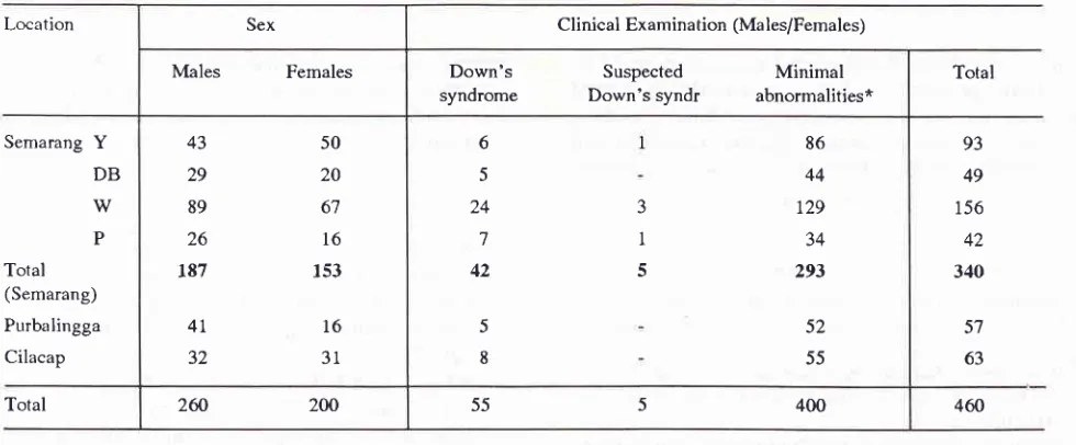
Table l. Clinical surrey in special schools for the mentally retarded in Semarang city, Purbalingga and Cilacap
Location Sex Clinical Examina tion (Ma les/Females)
Males Females
Down's
Suspected
Minimalsyndrome
Down'ssyndr
abnormalities*Total
Semarang Y
DB
w
P
Total
(Semarang)
Purbalingga Cilacap
43
29
89
26
187
50
20
6'l l6
153
4L
1632
316
5
24
7
42
5
8
J
I 5
86 44 r29 34
293
52
55
93
49
156
42
340
5'7
63
Total
2û
200 55 5 400 460 [image:2.595.54.544.516.719.2]Molecular studies
Southern blotting of Pstl digested genomic DNA
(restriction fragment length polymorphism/RFlP) was performed. Using the FRAXA probe (pfxa3) and an X-chromosome single locus control probe (pS8), (both
kindly supplied by Professor G. Sutherland, Adelaide), the cytogenetically positive fragile-X cases were con-firmed.
RESULTS
A
totalof
128 males from Semarang were studiedcytogenetically and the results are summarized in Table 2. There were 11 children with chromosomal abnormalities, four were positive fragile-X (with the
following number of positive cells,3% inDm,4% in
Pr,3% in Iw and 77o inZn) and seven with abnormal
karyotypes namely
two
47,XY+21, 46,XY,-7,+der(7 )t(7 ;?) (p22
;?),
46,XY,dup(9)
(p 1 3 - > p24),4S,XXXY, 46,XYl46XY,r(22) and 46,XY,+mar 15
(confirmed by fluorescence
in
situ
hybridizationstudies). The two 47 ,XY+2I were found from the five
cases with suspected Down's syndrome. Both cases
had minimal stigmata of Down's syndrome such as a
big tongue and upslanting eyes in one case and
hyper-telorism and stubby hand in the other case, The four fragile-X positive cases were found in two families.
DNA analysis
by
Southern blotting showed amosaicism pattern 1.6 kb and2.6 kb band inDm,2.2
kb band in Pr from family 1, and2.L kb band in Iw from
family 2,
which confirmed the affected fragile-Xchildren to have
full
mutation and 1.3 kb band in Znfrom family 2, which confirmed the affected fragile-X
child to
have premutation. There wasonly
one chromosomal abnormality in the samples from rural area (Cilacap), namely 46,XYl47,XYY (see Table 3).Only half of the boys from the two rural schools were included in the study since they were absent during the post exam and four boys refused to have blood
collec-tion
(two from each school).In
Semarang specialschools there were difficulties in seeing those children
as they were scared and rejected. There were 22 boys rejected i.e. eight from Y school, five from DB school,
five from W school and four from P school.
Table 2. Cytogenetic study in boys without classical Down's syndrome in Semarang city
Special Schools in Semarang
Cytogenetics findings
Fragile X positive Others 46,Xv Total
Y
DB
w
P31
l8
55 13
34 20 59
l5
2 2
4
2
r28 Lt7
Total
Table 3. Cytogenetic study in boys without classical Down's syndrome in Purbalingga and Cilacap
Cytogenetic findings Location
Fragile X positive Others 46,XY Total
Purbalingga
Cilacap
l9
t7 t9
l6
126
Sultana et al.DISCUSSIONS
In
our survey we found 30% excessof
males over females (2601200)in
mentally retarded institutions (Table 1). This male preponderance is similar to thatfound
in
other surveys of mentally retarded persons where there was an excess of 20% which was ascribedto
X-linked mental retardation (XLMR).2 Thein-cidence of Down's syndrome with typical clinical fea-tures was 12.3% of the population in Semarang schools and
ll%
of the population in rural area schools, whichwas lower when
comp
diesi.e.l8%o in Taiwan
(19
sianstudies.a There
were
and5.46% other chromosomal abnormalities in Semarang
mentally retarded school boys, compared to the 3.8%
both fragile-X and other chromosomal abnormalities
seen in Taiwan study.3 Comparison with other Asian
fragile-X
screeningstudy
(Japan) showed 10%Down's syndrome and 5.3% Fragile-X syndrome in institutionalized mentally retarded males.s About 40%
of
XLMR
and 47o of all mental retardation has beenattributed to fragile-X syndrome.6'7 Institutionalized populations studies in different countries such as
Ger-many (1983), Finland (1983), UK (1986), USA (1984) showed that the incidence
of
fragile-X syndromeamoong males were 6.27o, 4%,7 .9%, 4.1%
respective-ly." A
study of mentally retarded population in Australia (Sutherland 1985) found -3.35% fragile-X syndrome in mentally retarded boysg which was verysimilar with our
studyin
Semarang. Cytogeneticscreening studies for the male individuals attending adult and child facilities for intellectually handicapped
in New South Wales found positive fragile-X 6.5%, 70.6% and 18.6% in the programme 1984-1985, 1986-1988 and 1989-1990 respectively.l0 Overall srudies in New South Wales found an additional 2% with other
chromosome anomalies of those tested for fragile-X and lO% chromosome anomalies out
of
suspectedchromosome disorders. The Autralian, European and
American figures showed
a
higher prevalenceof
fragile-X than Asian figure found in the literature and
in our study, but this trend cannot be analyzed statisti-cally.
Our study
in
therural
areas identifiedonly
onechromosomal abnormality without any fragile-X case.
The sampling was incomplete because the number
of
children studied in rural areas were too small to pro-vide useful analysis. Most of the children in rural areaspecial schools have been stayed in school dormitory, their parents tend to pick them up and allow them to
stay home in post exam (end of academic year). The lower incidence in Indonesia could be due to cultural
Med J Indones
factors that allow children to stay at home and caused
an incomplete sampling. The boys from six mentally
retarded school surveyed were not all included in this
study since some of them refused to have blood collec-tion. Some mentally retarded children and adult was
not covered
in
this study since they did not attendspecial school
or
sheltered workshopfor
economicreasons.
The frequency
of
positive fragile-X cells (1-4%) in fragile-X patients -appears to be lower thanin
other studies (2-507o).6'6It
is
possible that although low folate medium was used, there was enough folate in foetal bovine serum (FBS) of the medium to reduce theincidence
of
fragile-X expression. The sizeof
thetrinucleotide repeat expansion (CGG repeat) is a causal factor
in
the expressionof
fragile site.ll
Th"
foo,fragile-X positive cases were found in two families.
Southern blotting showed a mosaicism pattern i.e. 1.6
kb and 2.6 kb band
in Dm,2.2
kb band in Pr from familyI
and 2.1 kb band in Iw from family 2, which confirmed the affected fragile-X children to have full mutation. Thosefull
mutation cases showed typical fragile-X syndrome such as high forehead, prominentjaw,
long ears and shyness (reportedin
previouspaper).'' One positive fragile-X case (Zn) found as premutation
wjth
1.3kb
band(-90
CGG repeats) showed only l% fragile-Xpositive cells cytogenetical-ly. In general, males with premutation i.e. the size of trinucleotide repeat 60-200 repeat(.
t.Okb
band)almost do not show phenotypic abnormalities and
called as normal transmiting males (NTM;.13'la Ou,
case with the 1.3 kb band showed normal looking but
he was mentally retarded. It was presumably caused by
methylation
in
CpG islandof
FMR-1g"ne."
DNAanalysis by using Southern blotting of Pst 1 product of fragile-X cases showed bands of more than 1.6 kb in affected individual
(full
mutation). This pattern wasthe same as in Caucasian cases. l6
Acknowledgment
This work was supported
by
theSix
Universities Development and Rehabilitation Project of the Depart-ment of Education and Culture, ADB loan no.l0l3-INO. We wish to thank the parents, the heads and staff ofthe special schools for providing records and clinical
data, allowing us to examine the children and collect the blood samples. Particular thanks are extended to
the laboratory staff of Telogorejo Hospital Semarang for their assistance in the collection of samples and
preliminary
preparationof
samples, andto
thelaboratory staff of the Prince of Wales Hospital,
R.EFERENCES
1. Prader A. Testicular size: Assesrnent and clinical funpor-tance. Triangle L966;7 :240-3.
2. Turner G, Turner B. Xlinked mental retardation. Am I Med Gen 1974;11:109-13.
3. Li SY, Tsai CC, Chou MY, Lin JK. A cytogenetic study of
mentally retarded school children in Taiwan with special teference to the fragile
X
chromosome. ÉIum GenetL988:79:292-6.
4. Gorlin RI, Cohen MM, I-evin LS. Syndrome of the head and
neck. 3rd ed. Oxford: Oxford University press, 1990.
5. Arinami T, Kondo I, Nakajima S. Frequency of the fragile
X syndrome in Japanese mentally retarded males. Hum Genet 1986;73:3O9-12.
6. Connor IM, Ferguson-Smith MA. Essential Medical
Genetics. 4th ed. Oxford: Blackwell Scientific publications,
1993.
7. Mikkelsen M. Sexlinked mental retardation. trn: Vogel F,
Sperling K, editots. Human Genetics. Berlin: Springer-Ver-lag,1987;441-9.
8. Davies KE, editor. The Fragile Xsyndrome. Oxford: Oxford University Press, 1989.
9. Sutherland GR. Heritable fragile sites on human chromosomes XIL Population Cytogenetics. Annals of Hum Gen 1985;49:153-61.
10. Tumer G, Robinson H, I:ing S, Van den Berk M, Colley A, Goddard A et al. Population Screening for fragile X. The Laneet 1992;339 : 12 I G3.
11. De Vries BBA, Wiegers Alvt De Graaff
I
et al. Menral status and fragile X expression in relation to FMR-I genernutation. Eur J Hum Genet 19931,l:72-9.
12. Sultana, Soemantri Ag, I-am-Po-Tang pRL, Wright F,
Lin-deman R, Purvis-Smith S. Fragile-X mental retardation in an
Indonesian family. Med J Indones 1995;4:17-23.
13. Rousseau F, Heitz D, Tarleton J, Biancalana V, Blumenfeld S, Kretz C et al. Direct diagnosis by DNA analysis on the fragile X syndrome of mental retardation. N Engl J Med l99t;325,1673-81.
14. Oberle I, Rousseau F, Heitz D, Kretz C, Devys D, Hanauer
A, et al. Instability of a 550-base pair DNA segment and
abnormal methylation in fragile
X
syndrome. Science l99l;252:l@7 -1O2.15. Smeets HIM, Smits API Verheij CE, Theelan IpG, Willem_
sen R, Van den Burgt I, et al. Normal phenotype in two brothers with a full FMR-I mutation. Hum Mol Genet
1995;4:2103-8.
16. Sutherland GR, Gedeon A, Komman L, Donnelly A, Byard RW, Mulley IC, et al. Prenatal diagnosis of fragile X syndrome by direct detection ofthe unstable DNA sequence.