Applicability of an oligonucleotide probe in radioisotope
32P-based dot blot
hybridization for detection of hepatitis C virus in large sample numbers:
a preliminary study
Maria L. Rosilawati,1 Andi Yasmon2
1 Center for Application of Isotopes and Radiation Technology, National Nuclear Energy Agency, Jakarta, Indonesia 2 Department of Microbiology, Faculty of Medicine, Universitas Indonesia, Jakarta, Indonesia
Abstrak
Latar belakang: Penelitian ini bertujuan untuk merancang dan menganalisis pelacak oligonukleotida apakah dapat diterapkan dalam hibridisasi dot blot menggunakan radioisotop 32P untuk mendeteksi virus hepatitis C.
Metode: Sampel yang digunakan adalah 46 plasma darah. Plasma diekstraksi untuk mendapatkan RNA genom virus sebagai cetakan reaksi RT-PCR dan amplikon digunakan untuk nested PCR. Genom HCV berjumlah 24 diunduh dari GeneBank dan penderetan sekuen DNA dilakukan dengan Software Bio Edit versi 7.0.9.0. Pelacak oligonuklueotida dirancang berdasarkan daerah lestari genom HCV yang terletak pada sekuen internal di antara 2 primer yang digunakan pada nested PCR. Homologi oligonukleotida HCV dianalisis menggunakan teknik Blast di GeneBank. Radioisotop 32P digunakan untuk melabel oligonukleotida. Oligonukleotida berlabel diaplikasikan untuk produk nested PCR menggunakan metode hibridisasi dot blot. Konirmasi hasil ampliikasi dan hibridisasi dot blot dilakukan menggunakan metode sekuensing DNA.
Hasil: Hasil analisis Blast menunjukkan homologi yang tinggi untuk HCV (100%). Hasil nested PCR menunjukkan tiga pola fragmen DNA. Tiga pola tersebut masing-masing adalah genotip HCV 1, 2, dan 3. Primer yang digunakan dalam nested PCR tidak spesiik dinyatakan dengan adanya tiga fragmen DNA sehingga sulit diinterpretasikan. Hasil hibridisasi dot blot menggunakan oligonukleotida yang didesain dalam penelitian ini menunjukkan intensitas dot yang tebal. Semua pola fragmen hasil nested PCR menunjukkan hasil positif dot blot. Hasil hibridisasi dot blot sesuai dengan hasil sekuensing DNA.
Kesimpulan: Pelacak oligonukleotida menunjukkan kriteria yang sangat memuaskan secara bioinformatika. Hasil hibridisasi dot blot menggunakan 32P menunjukkan intensitas dot yang tebal dan lebih mudah diinterpretasi dibandingkan dengan hasil nested PCR. (Med J Indones. 2012;21:71-6)
Abstract
Background: This study aimed to design and analyze the applicability of an oligonucleotide probe in radioisotope 32P-based dot blot hybridization for detection of hepatitis C virus.
Methods: Forty-six of plasma samples were used. The plasma was extracted to obtain viral RNA genome as template for RT-PCR and the amplicon was used for nested PCR. Twenty-four HCV genomes were retrieved from GeneBank DNA sequence and alignment was performed by Bio Edit Software version 7.0.9.0. An oligonucleotide probe was designed based on a highly conserved region that is located on internal sequence between two primers used for nested PCR. Blast analysis on GeneBank was performed to obtain homology of the oligonucleotide for HCV. The oligonucleotide was then labeled with 32P and dot blot hybridization was applied for nested PCR products. DNA Sequencing was performed to conirm the amplicon and dot blot hybridization results.
Results: Blast analysis showed high homology (100%) for HCV. Nested PCR resulted in three patterns of DNA fragments
representing HCV genotypes 1, 2, and 3, respectively. The primers used in nested PCR were not speciic and resulted in DNA fragments dificult to be interpreted. Dot blot hybridization using the designed oligonucleotide showed high intensity
dots. All nested PCR fragments showed the dot blot positive. The dot blot results were in accordance with DNA sequencing
that conirmed three patterns of DNA fragments as different HCV genotypes.
Conclusion: The oligonucleotide showed excellent bioinformatically criteria. 32P-based dot blot hybridization yielded high intensity dots and was easier to be interpreted than nested PCR assay. (Med J Indones. 2012;21:71-6)
Keywords: Dot blot hybridization, hepatitis C virus, oligonucleotide, radioisotope
Correspondence email to: [email protected]
contamination from re-used needles and syringes, multiple-use medication vials, and infusion bags has been reported.4 Moreover, transmission of HCV is less eficient through accidental needlesticks,4 but the transmission is most eficient through injecting drugs, transfusion or transplantation from infectious donors.5-7 In developing countries, unsafe medical injections and transfusions are predominant routes of HCV transmissions.6
cross-With the advancement of technologies, diverse methods are available to detect HCV. Serological assays, detecting anti-HCV antibodies and/or HCV antigens, can be performed on any kind of blood samples. In the case of detecting anti-HCV antibodies, the assay can yield false negative results for people with early phase of infection (window period) and immunological dysfunction. Prevention of HCV transmission through blood products will not be achieved if the blood screening only counts on serological assays since the assays can yield false negative results; therefore, complementary testing, such as molecular techniques, is most needed to overcome the limitation of serological assay.
The molecular techniques for HCV detection have been developed including conventional and real time RT-PCR. The conventional RT-PCR is not sensitive.8 The real time RT-PCR is very sensitive but it is very expensive because of special reagents and thermal cycle machine. In addition, health care services in developing countries including Indonesia rarely have the real time PCR machine. Therefore, in this preliminary study we designed an oligonucleotide probe by a radioisotope 32P-based dot blot hybridization method that can be applied to nested PCR products. The method is highly sensitive,9 can detect DNA targets of 96 samples in one test and is suitable for screening large sample numbers and for surveillance purposes.
METHODS
The research was carried out at the molecular biology laboratory of Radiation Processing Department, Center for Application of Isotopes and Radiation Technology, National Nuclear Energy Agency, Jakarta.
Clinical specimen and transport
Forty-six plasma samples from suspected patients with HCV infection were obtained from January to December 2009 in the government hospital in Jakarta. To avoid the possibility of RNA or viral destructions caused by transportation, the samples were transported by using a special cool box containing ice packs. After arrival in the laboratory, the samples were immediately stored in -20oC without thawing until used.
Viral RNA extraction
Two hundred microliter of plasma were extracted to obtain the HCV genome RNA by QIAamp Viral RNA Mini Kit (Qiagen) according to manufacturer’s instruction, with 50 μL of inal elution and immediately used as template for RT-PCR.
RT-PCR and nested PCR
Primers used for RT-PCR and nested PCR from non coding region (NCR) were previously reported by Garson et al.10 The outer sense and anti-sense primers for RT-PCR were 5’ – CCA CCA TAG ATC TCT CCC TGT and 5’ – ATA CTC GAG GTG CAC GGT CTA CGA GAC CT , respectively. The RT-PCR reaction mixture was prepared by using one-step RT-PCR kit (Qiagen). The reaction mixture contained 10 μL viral RNA, 1x one-step RT-PCR buffer, 2.5 mM MgCl2, 1x Q solution, 400 μM each of dNTP mix, 0.6 μM each of primers, 2 μL of one step RT-PCR enzyme mix, 10 U RNAse inhibitor and sterile distilled water to a total volume of 50 µL. The RT-PCR was performed in a Master Gradient Thermal Cycler (Eppendorf) with the irst step at 50oC for 30 minutes; 35 cycles of 96oC for 30 seconds, 48oC for 45seconds and 72oC for 1 minute; and extended extension at 72oC for 7 minutes. The nested primers used for the second PCR were 5’- AGA TCT TCA CGC AGA AAG CGT and 5’- CAC TCT CGA GCA CCC TAT CAG GCA GT as inner sense and anti-sense primers, respectively. The reaction mixture of nested PCR consisted of 1x hot star Taq DNA polymerase buffer, 2.5 mM MgCl2, 1x Q solution, 200 μM each of dNTP mix, 0.4 μM each of primer, 1.5 U hot star Taq DNA polymerase (Qiagen), 2 μL of RT-PCR product and sterile distilled water to a total volume of 50 µL. The reactions were run on thermal cycler using the following conditions: 15 minutes at 95oC; 30 cycles of ampliication 96oC for 30 seconds, 48oC for 45 seconds, and 72oC for 1 minute; and extended extension at 72oC for 7 minutes. The nested PCR products were analyzed on 1.5% agarose and visualized on ultraviolet transilluminator.
Design of oligonucleotide probe
Twenty-four HCV genomic sequences were retrieved from GeneBank (Figure 1). The sequences were aligned by using Bio Edit version 7.0.9.0.11 A highly conserved sequence located on internal sequence between two primers used for nested PCR was chosen for oligonucleotide sequence. The reliability and homology (speciicity) of the oligonucleotide was analyzed by primer designer-V2.0 serial number: 50053 (scientiic & educational software 1991) and GeneBank Blast, respectively.
Oligonucleotide labeling and dot blot hybridization
at 37oC for 30 minutes and terminated by incubating at 72oC for 10 minutes. The dot blot hybridization was performed: 10 µL of nested PCR product was added with 190 µL of the dot buffer (0.4 N NaOH and 25 mM EDTA). The mixture was heated at 100oC and immediately placed on ice, then blotted on Hybond N+ membrane by means of dot blotter (Bio-Rad). The DNA on membrane was ixed by heating at 80oC for 2 hours. The membrane was immersed in hybridization solution (5x SSPE, 5x Denhardt and 0.5% SDS) and shaken at 64oC overnight (16-18 hours). Forty milliliter of the hybridization solution containing 32P-labeled oligonucleotide was reacted with the membrane at 64oC for 1-2 hour. The membrane was washed twice by washing buffer (2x SSPE and 0.1% SDS) at room temperature for 30 minutes, each time followed by a inal washing (1x SSPE and 0.1% SDS) at 62oC for 15 minutes. The formed dots on the membrane were detected with autoradiography.
DNA sequencing
To conirm the exactness of speciic DNA ampliication and dot blot hybridization, three products of nested PCR
AN ....|....| ....|....| ....|....| ....|....| ....|....| ....|....| ....|....| 150 160 170 180 190 200 210 D63821 ATAGTGGTCT GCGGAACCGG TGAGTACACC GGAATCGCCG GGTTGACCGG GTCCTTTC-- TTGGAACTA- D63822 ... ... ... ...T...A ..AC... ...-- ...T.A.- NC_0098 ... ... ... ...T. ..G... ...-- ...G.A.- NC_0098 ...A.... ... ... ...T...A ..AC... ...CA ...T.A.A EU15532 ... ... ... ...T...A ..AC... ...-- ...TTA.- EU15521 ... ... ... ...T...A ..AC... ...-- ...TAA.- EF42462 ... ... ... ...T...A ..AC... ...-- ...T.A.- EF42462 ... ... ... ...T...A ..AC... ...-- ...T.A.- AY46020 ... ... ... ...T.... ..AA...T.. ...-- ...T.A.- AF13959 ... ... ... ...T...A ..AC... ...-- ...T.A.- U89019 ... ... ... .-...T...A ..AC... ...-- ...T.A.- U45476 ... ... ... ...T...A ..AC... ...-- ...TTA.- U16362 ... ... ... ...T...A ..AC... ...-- ...T.A.- U01214 ... ... ... .A...T...A ..AC... ...-- ...T.A.- M58335 ... ... ... ...T...A ..AC... ...-- ...T.A.- M84754 ... ... ... ...T...A ..AC... ...-- ...T.A.- L02836 ... ... ... ...T...A ..AC... ...-- ...T.A.- AM42293 --- --- --- --- --- --- --- DQ48052 ... ... ... ...T...A ..A... ...CA ...T.A.A D49374 ... ... ... ... ..A... ...-- ...A.- EU23406 ... ... ... ...T...A ..AC... ...-- ...TAA.- EU23406 ... ... ... ...T...A ..AC... ...-- ...T.A.- AY05129 ... ... ... ...T...A ..AC... ...-- ...T.A.- AF48326 ... ... ... ...T...A ..AC... ...-- ...T.A.-
Figure 1. The DNA sequence (GTCTGCGGAACCGGTGAGTACA) from nucleotides 147-168 was used as oligonucleotide probe for dot blot hybridization. The sequence was located between two primers used for nested PCR. AN= accession numbers of HCV genomic sequences that were retrieved from GeneBank
assays were sequenced. The DNA sequencing results were blasted on GeneBank to obtain the taxonomical reports of the sequences.
RESULTS
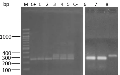
Positive nested PCR was deined by a 259 bp DNA band visualized at the right position on 1.5% agarose gel. Of 46 plasma samples, thirty-nine showed 3 different patterns of DNA fragments and 7 showed negative results. Pattern 1 was one speciic DNA band (Lane 1, 2, 6, 7, Figure 2), pattern 2 was one DNA band higher in size than the positive control (Lane 8, Figure 2,), and pattern 3 were multi-DNA bands (Lane 3-5, Figure 2).Of 39 samples, thirty-three, ive, and 1 showed pattern 3, 1, and 2, respectively. To conirm the ampliication products, one of each pattern was sequenced and blasted on GeneBank (Figure 3). The results showed that pattern 1, 2, and 3 were HCV genotypes 2, 1, and 3, respectively. Results as mentioned above indicated that nested PCR primers used in this study are not speciic for HCV genotype 1 and 3 because of resulting unspeciic DNA band
400 1000
100
M C+ 1 2 3 4 5 C- 6 7 8
300 200 bp
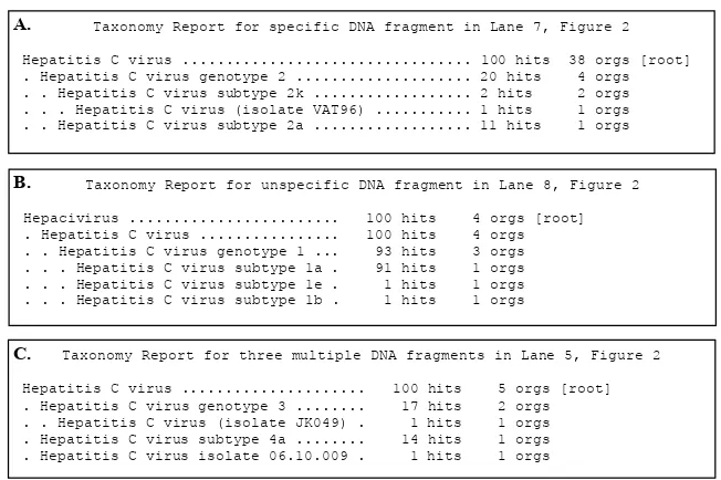
C. Taxonomy Report for three multiple DNA fragments in Lane 5, Figure 2 Hepatitis C virus ... 100 hits 5 orgs [root] . Hepatitis C virus genotype 3 ... 17 hits 2 orgs . . Hepatitis C virus (isolate JK049) . 1 hits 1 orgs . Hepatitis C virus subtype 4a ... 14 hits 1 orgs . Hepatitis C virus isolate 06.10.009 . 1 hits 1 orgs
A. Taxonomy Report for specific DNA fragment in Lane 7, Figure 2
Hepatitis C virus ... 100 hits 38 orgs [root] . Hepatitis C virus genotype 2 ... 20 hits 4 orgs . . Hepatitis C virus subtype 2k ... 2 hits 2 orgs . . . Hepatitis C virus (isolate VAT96) ... 1 hits 1 orgs . . Hepatitis C virus subtype 2a ... 11 hits 1 orgs
B. Taxonomy Report for unspecific DNA fragment in Lane 8, Figure 2
Hepacivirus ... 100 hits 4 orgs [root] . Hepatitis C virus ... 100 hits 4 orgs . . Hepatitis C virus genotype 1 ... 93 hits 3 orgs . . . Hepatitis C virus subtype 1a . 91 hits 1 orgs . . . Hepatitis C virus subtype 1e . 1 hits 1 orgs . . . Hepatitis C virus subtype 1b . 1 hits 1 orgs
Figure 3.Blast analysis on GeneBank for three ampliication products, speciic (A), unspeciic (B), and three multiple DNA fragments
(C). A, B, and C: samples resulting speciic (Lane 7, Figure 2), unspeciic (Lane 8, Figure 2), and three multiple DNA fragments
(Lane 5, Figure 2), respectively.
and multi-DNA bands, respectively, leading to the confusing interpretation of nested PCR. Other HCV genotypes 4-6 are not known in this study, they may include one of those patterns or different patterns; therefore, the applicability of nested PCR using primers in this study is questionable to detect all HCV genotypes, speciically.
To overcome the limitation of nested PCR assay, an oligonucleotide probe was designed and evaluated for all nested PCR results. Based on the analysis,
Table 1. Analysis result of oligonucleotide reliability using primer designer-V2.0 serial number: 50053
Criteria Criteria Setting Actual Meets Criteria
% GC Min 50, Max 60 59 Yes Tm (oC) Min 55, Max 80 77 Yes No Hairpins Energy cutoff 0.0 kcal -- Yes No 3’ Dimers Reject >= 3 matches at 3’ end 2 Yes No Dimers Reject >= 7 adj homol bases 6 Yes No Runs Reject >= 3 base runs 2 Yes No 3’GC runs Reject >= 3 G or C at 3’ end 0 Yes
GC= guanine and cytosine, Tm= melting temperature, Hairpins= loop formation on single oligonucleotide
the oligonucleotide probe showed excellent criteria (Tabel 1). Technically, hairpins and 3’GC runs on oligonucleotide will inluence the speciicity, while dimers will inluence the sensitivity. The percentage of GC and melting temperature of the oligonucleotide were within normal ranges. Based on Blast analysis on GeneBank, the homology of the oligonucleotide was very high (100%) for HCV (data not shown).
All ampliication products of nested PCR assays were probed by radioisotope 32P-labeled oligonucleotide using
A
B
C
D
1 2 3 4 5 6 7 8 9
the dot blot hybridization method. Of 46 tested samples, thirty-nine samples that showed all three patterns of DNA bands by nested PCR were also dot blot positive. Seven samples with nested PCR negative were also dot blot negative (Figure 4). The results showed that dot-blot hybridization using the oligonucleotide designed in this study could be used for detection of HCV genotypes 1, 2, and 3 speciically, compared with nested PCR showing multi-DNA fragments dificult to be interpreted.
DISCUSSION
RT-PCR followed by nested PCR for detection of HCV was applied for 46 plasma samples. Of these samples, thirty-nine showed the three patterns of DNA bands, namely one speciic and one unspeciic DNA band, and three multi-DNA bands (Figure 2). Such three patterns of DNA bands were conirmed by DNA sequencing and showed HCV with different genotype (Figure 3). Accordingly, the nested PCR used in this study resulted in unspeciic or multi-DNA fragments confusedly to be interpreted. This indicates that primers used in nested PCR are not speciic for HCV genotypes 1 and 3. The problem may be caused by different DNA sequences among HCV genotypes/subtypes or HCV quasispecies in one infected patient. HCV isolates can be highly divergent and have been classiied into 6 major genotypes and more than 30 subtypes.11 Like other RNA viruses, HCV circulates in vivo as a highly polymorphic population of genetically closely related variants.12 In consequence, it is dificult to detect or identify all HCV genotypes in a single detection system like using a primer pair of PCR.
As known, PCR relies on the eficient attachment of primers to the DNA target. Mismatches between the primer and the DNA target can inluence duplex stability and limit the assay to amplify target sequences. Several factors causing the mismatches are primer length, nature and position of mismatches, hybridization temperature, presence of co-solvents (such as DMSO), and concentrations of primers and monovalent and divalent cations.12-14 For different HCV genotypes with different genomic RNA sequences, the partial mismatches between primers and DNA target and the presence of insertion/deletion mutation or recombination can also result in unspeciic DNA fragments.
To overcome the limitation of nested PCR, an oligonucleotide of 22 nucleotides in length was designed and its applicability was evaluated. Hybridization probes can be applied with lengths from around 20 up to several hundreds of nucleotides. However, longer oligonucleotides lead to more mismatches. To minimize the mismatches, the length
of our oligonucleotide was determined in a conserved region (Figure 1). The homology of oligonucleotide by Blast analysis on GeneBank showed 100% homology for HCV (data not shown) indicating technically high speciicity of the oligonucleotide for HCV. The oligonucleotide should be unique for the target sequence and fulill certain criteria such as the length, GC%, annealing and melting temperature (Tm), 5’ end stability, 3’ end speciicity.14 Annealing the 3’ end and/or the 5’ end among oligonucleotide segments will interfere the optimal attachment of oligonucleotide to DNA target. An important factor to consider in designing an oligonucleotide probe is the presence of hairpin loops, which reduces the hybridization eficiency by limiting the ability to bind to the DNA target.15 The oligonucleotide that was designed in this study showed all criteria and it could be applied bioinformatically (Tabel 1).
The oligonucleotide was labeled by 32P isotope and tested for all nested PCR products by dot blot hybridization method. Of 46 samples, thirty-nine were dot blot positive (Figure 4). All nested PCR fragments (speciic, unspeciic, multi-DNA fragments) were dot blot positive. The results are in accordance with DNA sequencing in that all three patterns of DNA fragments were different HCV genotypes (Figure 3). This indicates that the oligonucleotide designed in this study can detect all nested PCR fragments of HCV genotype 1, 2, and 3. The important differences between nested PCR and dot blot hybridization are that the nested PCR resulted in multi-DNA fragments that are dificult to be interpreted, while the dot blot hybridization showed high intensity dots that are easy to be interpreted. The high intensity results from an autoradiography signal of the radiation emitted by the radioisotope. The 32P has been used widely in dot blot hybridization, Southern blot hybridization, colony and plaque hybridization because it emits high energy β-particles which afford a high degree of sensitivity of detection.16 For these reasons, numerous researchers have used this approach to detect and identify pathogenic microorganisms.17-19 Therefore, the dot blot hybridization using oligonucleotide designed in this study is suitable for detection of HCV, particularly for screening of large sample numbers and for surveillance purposes. Other researchers have used the method for large sample numbers for detection and identiication of Dendrobium species,20 multi-drug resistant Mycobacterium tuberculosis,9 and Echoviruses.21 However, the oligonucleotide designed in this study should be evaluated in the future for larger sample numbers to obtain the validity of the speciicity and sensitivity.
for RT-PCR instead of nested PCR products) is cost effective compared with nested PCR and real time PCR, giving signiicant health care-associated cost savings. The conventional PCR is much cheaper than dot blot hybridization, nested PCR and real time PCR; however, it is less sensitive. A limitation of using radioisotope is that a special radioisotope laboratory is needed and the laboratory technicians must have additional knowledge and skills to handle the radioisotope.
In conclusion, an oligonucleotide probe speciic for HCV was successfully designed and evaluated by using a 32P radioisotope-based dot blot hybridization method. This method is suitable for detection of HCV for large sample numbers and surveillance purposes. The oligonucleotide showed excellent criteria bioinformatically to be applied for probing HCV from plasma samples. The method yielded high intensity dots that are easier to be interpreted than nested PCR assay with multi-DNA fragments. In future research, the dot blot hybridization will be optimized for RT-PCR instead of nested RT-PCR products and evaluated for large sample numbers.
Acknowledgments
This research was supported by Center for the Application of Isotopes and Radiation Technology, National Nuclear Energy Agency, Indonesia. We thank our highly dedicated technicians, Rika Heryani and Almaida for assistance in performing the research activities.
REFERENCES
1. World Health Organization. Hepatitis C [Internet]. 2011 [cited Dec 11]. Available from: http://www.who.int/ mediacentre/factsheets/fs164/en/
2. Shepard CW, Finelli L, Alter MJ. Global epidemiology of hepatitis C virus infection. Lancet Infect Dis. 2005;5:558-67. 3. Alzahrani AJ, Obeid OE, Al-Ali A, Imamwardi B. Detection
of hepatitis C virus and human immunodeiciency virus in
expatriates in Saudi Arabia by antigen-antibody combination assays. J Infect Dev Ctries. 2009;3:235-8.
4. Williams IT, Perz JF, Bell BP. Viral hepatitis transmission in ambulatory health care settings. Clin Infect Dis. 2004;38:1592-8.
5. Tugwell BD, Patel PR, Williams IT, Hedberg K, Chai
F, Nainan OV, et al. Transmission of hepatitis C virus to
several organ and tissue recipients from an antibody-negative donor. Ann Intern Med. 2005;143:648-54.
6. Nelson PK, Mathers BM, Cowie B, Hagan H, Des Jarlais D, Horyniak D, et al. Global epidemiology of hepatitis B and
hepatitis C in people who inject drugs: results of systematic reviews. Lancet. 2011;378:571-83.
7. Teo M, Hayes P. Management of hepatitis C. Br Med Bull. 2004;70:51-69.
8. Pillay A, Radebe F, Fehler G, Htun Y, Ballard RC. Comparison of a TaqMan-based real-time polymerase chain reaction with conventional tests for the detection of Trichomonas vaginalis. Sex Transm Infect. 2007;83:126-9.
9. Rosilawati ML, Yasmon A. Detection of multidrug-resistant
mycobacterium tuberculosis directly from sputum samples of patients from Jakarta, Indonesia by radioisotope-based PCR dot blot hybridization. Southest Asian J Trop Med Public Health. 2012;43:89-95.
10. Garson JA, Ring C, Tuke P, Tedder RS. Enhanced
detection by PCR of hepatitis C virus RNA. Lancet.
1990;336:878-9.
11. Ayesh BM, Zourob SS, Abu-Jadallah SY, Shemer-Avni Y. Most common genotypes and risk factors for HCV in Gaza strip: a cross sectional study. Virol J. 2009;6:105.
12. Bracho MA, Garcia-Robles I, Jimenez N, Torres-Puente M, Moya A, Gonzalez-Candelas F. Effect of oligonucleotide primers in determining viral variability within hosts. Virol J. 2004;1:13.
13. Sipos R, Szekely AJ, Palatinszky M, Revesz S, Marialigeti K, Nikolausz M. Effect of primer mismatch, annealing temperature and PCR cycle number on 16S rRNA gene-targetting bacterial community analysis. FEMS Microbiol Ecol. 2007;60:341-50.
14. Abd-Elsalam KA. Bioinformatic tools and guideline for PCR primer design. Afr J Biotechnol. 2003;2:91-5.
15. Singh VK, Govindarajan R, Naik S, Kumar A. The effect of hairpin structure on PCR ampliication eficiency. Mol Biol
Today. 2000;1:657-9.
16. Strachan T, Read AP. Human molecular genetics. 2nd ed:
New York: Wiley-Liss; 1999.
17. Yusuf RU, Omar SA, Ngure RM. The effect of point mutations in dihydrofolate reductase genes and multidrug resistance gene 1-86 on treatment of falciparum malaria in Sudan. J Infect Dev Ctries. 2010;4:61-9.
18. McMahon JM, Simm M, Milano D, Clatts M. Detection of hepatitis C virus in the nasal secretions of an intranasal drug-user. Ann Clin Microbiol Antimicrob. 2004;3:6.
19. Okuse C, Rinaudo JA, Farrar K, Wells F, Korba BE.
Enhancement of antiviral activity against hepatitis C virus in vitro by interferon combination therapy. Antiviral Res. 2005;65:23-34.
20. Xu H, Ying Y, Wang ZT, Cheng KT. Identiication of
Dendrobium species by dot blot hybridization assay. Biol Pharm Bull. 2010;33:665-8.