Morphological and genetic variability within
Aedes aegypti
in Niakhar, Senegal
Christophe Paupy
a,b,*
, Ce´cile Brengues
b, Ousmane Ndiath
c, Ce´line Toty
b, Jean-Pierre Herve´
b,
Fre´de´ric Simard
b,daCentre International de Recherches Me´dicales de Franceville (CIRMF), P.O. Box 769, Franceville, Gabon
bInstitut de Recherche pour le De´veloppement (IRD), RU016, P.O. Box 64501, 34394 Montpellier, France
cInstitut de Recherche pour le De´veloppement (IRD), RU077, Centre de Hann, P.O. Box 1386, 18524 Dakar, Senegal
dInstitut de Recherche en Sciences de la Sante´ (IRSS), P.O. Box 545, Bobo Dioulasso, Burkina Faso
1. Introduction
The mosquitoAedes aegypti(Linne´, 1762) is a major vector of Yellow Fever (YF), Dengue (Gubler, 2002) and Chikungunya viruses (Charrel et al., 2007; Yergolkar et al., 2006) throughout most tropical areas in the world. It is arguably one of most domestic mosquito vectors, feeding predominantly on man, mating and resting indoors and breeding in man-made containers in and around human habitations, especially in urban environments (Morisson et al., 2008).
In Africa however, which is considered the native range of the species (Chistophers, 1960), the biology of Ae. aegyptiis some-times less dependent on the presence of man than in other tropical regions. Indeed sylvan populations ofAe. aegyptibreeding in natural water collections such as rock pools, tree holes and leaf axils have been reported (Mattingly, 1957; Paupy et al., 2008). Morphological and bionomical differences were extensively described in Kenya (East Africa), between a dark, sylvan form
and a light, domestic form which appeared genetically differ-entiated using isoenzymatic markers (Petersen, 1977; Tabachnick and Powell, 1979; Wallis et al., 1983). In East Africa, the dark form, also known asAe. aegypti formosus(Aaf) is considered indigenous and ancestral, whereas the presence of the derived light form, also calledAe. aegypti aegypti(Aaa) is thought to result from secondary introduction.Tabachnick (1991)presented a verbal argument for the role of demographic history and vicariance in generating two subspecies ofAe. aegyptiin Africa. The author proposed that the transition from a ‘‘green Sahara’’ to the present hyperarid desert, which started in mid-Holocene (between 6000 and 4000 years ago,Kro¨pelin et al., 2008), promoted the isolation of two genetic pools from the original sylvan form (Aaf) on both sides of the desert. This model argues thatAe. aegyptideveloped its domestic habits north of the Sahara desert, once separated from the core of the species range in sub-Saharan Africa, where the species persisted as ancestral. The domesticAaa further spread out of Africa, and was later re-introduced in some costal regions of sub-Saharan Africa (e.g., Kenya). In East Africa, gene flow between
Aaf and Aaa was shown to be restricted to the peridomestic habitat where both species co-occur and hybrids with inter-mediate phenotypes (e.g., morphology and behavior) can be found (Trpis and Hausermann, 1978).
A R T I C L E I N F O
Article history:
Received 12 October 2009
Received in revised form 26 February 2010 Accepted 2 March 2010
Available online 16 March 2010
Keywords: Aedes aegypti Morphological variation Microsatellite Population genetics Senegal
A B S T R A C T
Aedes aegypti (Linne´, 1762) is a major vector of arboviruses such as Yellow Fever, Dengue and
Chikungunya. In Africa, where the species exhibits major variations in morphology, ecology, behavior and vector competence, two subspecies have been described: a light form, namedAe. aegypti aegypti (Aaa) with highly domestic and anthropophilic habits and a cosmotropical distribution; and a dark form, referred to asAe. aegypti formosus(Aaf), which is endemic to Africa and thrives in sylvan environments. In East Africa, both forms were described to occur in sympatry whereas onlyAafwas reported from Central/ West Africa. However, recent findings suggestAaawas also common in Senegal. Here, we report on a longitudinal survey of morphological and genetic variability of Ae. aegypti sampled in the rural environment of Niakhar, Senegal. In agreement with recent findings, most of specimens we analyzed were classified asAaasuggesting typicalAafwas scarce in the studied area. AmongAaa, significant temporal variations in abdominal pale scales pattern were detected. Depending on the season and the nature of larval breeding places, the specimens (particularly females) tend to segregate in two main morphological groups. Microsatellite-based estimates of genetic differentiation did not provide any clear evidence that the two groups were genetically distinct. Overall, these results improve our understanding of the diversity ofAe. aegyptiin West Africa, where data are crucially lacking.
ß2010 Elsevier B.V. All rights reserved.
* Corresponding author at: RU016 Biology and Control of Vectors, IRD, CIRMF, P.O. Box 769, Franceville, Gabon. Tel.: +241 67 70 92; fax: +241 67 72 95.
E-mail addresses:[email protected],[email protected](C. Paupy).
Contents lists available atScienceDirect
Infection, Genetics and Evolution
j o u r n a l h o m e p a g e : w w w . e l s e v i e r . c o m / l o c a t e / m e e g i d
The situation in West/Central Africa remains unclear, as very few investigations were conducted in this area. Until recently, the idea of Mattingly (1957), which considered Aaf as the only subspecies occurring in West and Central Africa, was largely admitted. However,Ae. aegyptiwas recently reported from both sylvan and anthropogenic environments, including large urban centers in Senegal, Cameroon and Gabon (Huber et al., 2008; Paupy et al., 2008, 2009), which represents much broader ecological preferences than typically admitted forAaf. In addition,McClelland (1974) described significant morphological variability within West/Central African populations ofAe. aegypti, with specimens showing a mixture of morphological characters that would not allow unambiguous classification asAaforAaa. Additional studies further supported the presence of populations ecologically (Diarrassouba and Dossou-Yovo, 1997) and morphologically (Hervy, 1977; Huber et al., 2008) similar toAaa. In a recent study carried out in Senegal, Sylla et al. (2009) unambiguously demonstrated that there was a clear northwest–southeast cline in the abundance ofAaa vs Aaf. Based on analyses of SNPs showing very little or no genetic differentiation between both subspecies, the authors also suggestedAafandAaawere monophyletic and thatAafis the ancestor ofAaain West Africa. In addition, indirect evidences for chromosomal polymorphism (inversions) in Aaf
strain originated in Senegal were recently discovered, which might impact on the distribution of molecular and phenotypic (morphol-ogy and behavior) polymorphisms within and among natural populations (Bernhardt et al., 2009).
The morphological characters used for the distinction ofAaa
andAaf– the pattern of pale scales on abdominal tergites and the color of integument of the body – were defined byMattingly (1957). This author stated the dark scaled parts of the body were generally blacker in Aaf, in which pale scales on the first abdominal tergite are never observed. The type formAaa, was defined as ‘‘either distinctly paler and browner (at least in females) than Aaf or with pale scaling on the first abdominal tergite or both’’. Among mosquitoes morphologically identified as Aaf, significant variation in the pattern of extension of abdominal pale scales was reported in Africa and particularly in areas were both subspecies were sympatric (McClelland, 1974; Hervy, 1977). This aspect of morphological variations in Ae. aegyptiand its biological significance remains understudied and poorly understood. Because vector competence and biological traits potentially involved in virus transmission to man vary according toAe. aegypti morphological form and geographical origin (Tabachnick et al., 1985; Failloux et al., 2002; Sylla et al., 2009), unambiguous characterization of the distinct genetic entities that make up Ae. aegypti in Africa would provide precious elements in risk assessment for viral diseases such as Dengue and Chikungunya which recently emerged in Africa (Peyrefitte et al., 2007; De Lamballerie et al., 2008; Paupy et al., 2009). As a step forward to this endeavor, we assessed morphological and genetic diversity in samples ofAe. aegypti
collected in Niakhar district in Senegal where several epidemics of YF and Chikungunya have occurred withAe. aegyptias a main vector (Thonnon et al., 1999). First, we estimated the level of local morphological variation based on patterns of abdominal scaling. Second, we estimated the level of genetic differentiation and gene flow between morphological and/or ecological groups, using microsatellite genotyping.
2. Materials and methods
2.1. Mosquito sampling
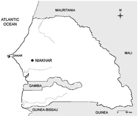
Mosquitoes were sampled as larvae in Niakhar district, a rural area of central Senegal (Fig. 1), typical of the Sahelian and
sub-Sahelian regions of Africa where the rainy season extends from July to October and mean annual rainfall is around 500 mm. Sampling was conducted in 2005 during the dry (May) and rainy (September) seasons in two neighboring villages 1 km apart, Kothiokh (14828.5020N; 16832.4240W) and Godel
(14829.5640N; 16833.3960W). The rainy season corresponds to
the highest season of exposure to the bites of Ae. aegypti
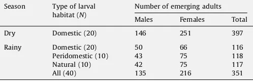
(Remoue´ et al., 2007). Aedes aegypti larvae were collected in three types of breeding sites: domestic, peridomestic and natural. Domestic breeding sites refer to water storage contain-ers (earthenware jars and metallic drums) regularly and consistently filled with water by inhabitants. Peridomestic (e.g., abandoned drums, tires, tin cans in the backyard of human dwellings) and natural (e.g., tree holes, leaf axils) larval development sites were flooded with rainwater during the rainy season only. All larvae from 20 indoor/outdoor domestic containers were sampled during the dry season. During the rainy season all larvae from 20 indoor/outdoor domestic, 10 peridomestic and 10 natural breeding sites were collected. Larvae were pooled according to the type of development sites they were collected from and reared to adults in the insectaries. Emerging mosquitoes were readily identified to species, sexed and stored individually at 208C, pending further morpholo-gical and molecular analyses.
2.2. Morphological analysis
Morphological analyses were performed only on well preserved specimens from both sexes. Specimens were observed and scored according to the method ofMcClelland (1974). Briefly, the method attributes an individual abdominal pattern value (PV) to each specimen, which calculation considers the number of abdominal tergites exhibiting pale scales spots (excluding apical bands and lateral spots of pale scales). The method differentiates tergites with speckled scales and those that harbor a complete medially pale band of scales. PV ranges from 1 (absence of pale scales on all tergites) to 15 (presence of pale scales on every tergite). Integument coloration, which we considered as subjective and easily biased depending on the investigator, was not considered in our analysis. Influence of season, sex and nature of breeding site on morphology variation was investigated. Comparisons of the distributions of PV across groups were performed using the Wilcoxon test implemented in R software (R Development Core Team, 2005).
2.3. Microsatellite analysis
DNA was extracted from adult mosquitoes of both sexes using DNAZOL (Invitrogen, USA) as described inHuber et al. (2002). DNA pellets were resuspended in 20
m
l of sterile water and stored at 208C until analysis. Individual genotypes were scored at eight microsatellite markers (Table 1): loci 34/72 and 38/38 (Huber et al., 2001), AEDGA, AED19, AEDC (Ravel et al., 2002) and A10, M313 and H08 (Chambers et al., 2007).The design, fluorescent labeling and Tm of primers used for microsatellite PCR amplification are givenTable 1. PCR amplifica-tions were performed using a 9600 thermocycler (PerkinElmer, USA) in 25-
m
l reactions containing 4m
l (2m
l for A10, M313 and H08) of a 1/5 DNA dilution, 2.5m
l of 10reaction buffer (Qiagen, USA), 1.2 mM of MgCl2(only for 34/72, 38/38, AEDGA, AED19 andAEDC), 125
m
M of each dNTP (Eurogentec, Belgium), 10 pmol of each primer and 0.5 U of Taq polymerase (Qiagen, USA). The 50endof the forward primer was labeled with a fluorescent dye (Table 1). Cycling temperatures were as follows for loci 34/72, 38/38, AEDGA, AED19, AEDC: 5 cycles of 2 min at 968C, 30 s at the annealing temperature (Ta in Table 1), 1.15 min at 728C, followed by 35
cycles of 30 s at 958C, 30 s atTa, 1.15 min at 728C, and a final
extension step at 728C for 30 min. For loci A10, M313 and H08, after 5 min at 948C, 35 cycles of 30 s at 948C, 30 s atTaand 30 s at
728C were performed, followed by 30 min at 728C.
PCR products were diluted 1/15 in water and pooled with other compatible products according to allele size range and fluorescent dye. Pools were prepared by adding to 1
m
l of each diluted amplification product, 0.4m
l of GS 500 Liz Internal Size StandardTM(Applied Biosystems, USA) and HD formamide for a total volume of 20
m
l. The mixture was heated at 948C for 3 min before migration in an automatic sequencer ABI PrismTM3100 (Applied Biosystems, USA). Microsatellite alleles were scored using GeneMapper soft-ware package (Applied Biosystems, USA).Genetic diversity by locus and sample and overall was characterized by estimating unbiased expected heterozygosity (He,Nei, 1987) and allelic richness (El Mousadik and Petit, 1996)
using the software FSTAT2.9.3.2 (Goudet, 1995). Linkage disequi-librium, deviations from Hardy–Weinberg equilibrium (HWE) and genetic differentiation indices were assessed using GENEPOP3.3 software (Raymond and Rousset, 1995).FISandFSTestimates were
calculated according toWeir and Cockerham (1984)and tested for
statistical significance with exact tests available in GENEPOP3.3. The overall significance of multiple tests was estimated by Fisher’s combined probability tests. Nominal significance levels for multi-ple testing were corrected using the sequential Bonferroni procedure (Holm, 1979).
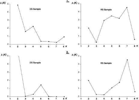
A Bayesian approach was used to infer the number of clusters (K) in the dataset without prior on the number of distinct populations in the dataset, as implemented in STRUCTURE 2.2 (Pritchard et al., 2000). A model where the allele frequencies were correlated within populations was assumed (
l
was set at 1, the default value). The software was run with the option of admixture, allowing for some mixed ancestry within individuals, anda
wasallowed to vary. We did 20 independent runs for each value ofK
(K= 1–9), with a burn-in period of 30,000 iterations and 100,000 replications. The method of Evanno et al. (2005) was used to determine the most likely number of clusters. This approach uses anad hocquantity,
D
K, based on the second order rate of change of the likelihood function between successive values ofK.3. Results
Rearing in the insectaries yielded 397 and 351Ae. aegyptiadults from larvae collected in the Niakhar area during the dry and rainy seasons, respectively (Table 2).
3.1. Morphological analysis
A total of 247 (156 females and 91 males) and 351 (216 females and 135 males) specimens collected during the dry and rainy season, respectively, were successfully scored. Among them, only
Table 1
Characteristics of the microsatellite markers used in the study.
Locus GeneBank accession no. Core repeat Primer designationa Primer sequence (50!30) T a(8C)
34/72 AF338656 GAAAA(GA)6CAGACAGGAAA 34/72-FOR-FAM CGT AGT GAT TCT GTG ATA 50 34/72-REV TGG CAT CAG ATT CAG TAA
38/38 AF338655 GCT(GTT)2GCTGTT(GCT)3(GTT)3GCT 38/38-FOR-VIC CGG TGG ACG AAT CAT 56 38/38-REV GAT GCC GCC TAG TCC AAT
AEDGA U28803 (GAA)3(GAC)4(GAA)3 AEDGA-FOR-VIC CCG AAG AAA TTG GGG TGA CC 55 AEDGA REV CCT CTC GGT GTT CGC TAA CC
AED19 U91680 GGAC(GGA)5 AED19-FOR-FAM GTA TGA CAA CTC TGG AAT GG 56
AED19-REV TTA TGG AAC TGG TAA GCC C
AEDC T58313 (GTA)6(ACG)(GTA)3 AEDC-FOR-FAM TGC AGG CCC AGA TGC ACA GCC 58 AEDC-REV TCC GCT GCC GTT GGC GTG AAC
A10 DU169901 (CT)3CGAT(CT)10TT(CT) A10-FOR-PET ATC CCG AAA ACA AAT CGT GA 58 A10-REV ATC GAA CAT CGC TTC CAA CT 30
M313 DU169909 ATG5(ATA)ATG M313-FOR-PET CAC CTC GTG ACA TAC AAA CAC C 60
M313-REV ACG TAC CCA AGC CAC GTA CA
H08 DU169903 TCG7 H08-FOR-NED AAA AAC CAC GAT CAC CGA AG 60
H08-REV ACG CGA TCA CAC ACT GAA AAT G
Ta: annealing temperature.
aFAM, VIC, NED and PET refer to fluorescent phosphoramidite dyes used to end label one of the two microsatellite markers.
Table 2
Description and yield ofAe. aegyptisamples collected in Niakhar, Senegal (2005).
Season Type of larval habitat (N)
Number of emerging adults
Males Females Total
Dry Domestic (20) 146 251 397
Rainy Domestic (20) 50 66 116
Peridomestic (10) 43 75 118
Natural (10) 42 75 117
All (40) 135 216 351
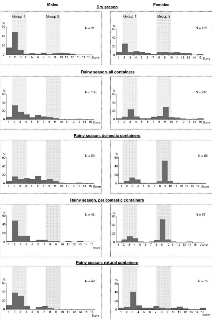
24 (dry season) and 13 (rainy season) corresponded toAaf(PV = 1), all other specimens correspondingAaa(PV>1). Mean PVs were
5.023.86 and 6.213.60 for mosquitoes from dry season (DS) and rainy season (RS), respectively.
3.1.1. Morphological variation between sexes
Frequency distributions of PV were plotted for both sexes according to the season of collection (Fig. 2). Important differences in the distribution of PV were observed between males and females collected in both seasons. These differences were highly significant (Table 3). Males and females were therefore considered indepen-dently in the following analyses.
3.1.2. Morphological variation between seasons
In the dry season collection, 70% of males exhibited a PV between 1 and 3, with a modal value located at PV = 2 (48% of the specimens). All other PV classes grouped together less than 10% of the specimens investigated (Fig. 2). While the modal value is also located at PV = 2 (33%) for males sampled during the rainy season, higher heterogeneity was observed than in the dry season. PV ranging from 1 to 3 encompassed only 57% of the specimens, whereas 11% of the specimens had PV = 4 and 10% had PV = 6. These distributions were significantly different (Table 3).
Females collected during the dry season harbored a modal value at PV = 2 (23% of the females), and all other PV classes grouped together less than 10% of the remaining females. In the rainy season, the distribution of PV in females was bimodal, with two
peaks at PV = 3 (23%) and PV = 9 (29%), respectively (Fig. 2). As observed in males, the distributions of PVs were also significantly different between females collected during the dry and the rainy season (Table 3).
3.1.3. Morphological variation according to larval ecology
Fig. 2 shows the distribution of PV in mosquitoes collected during the rainy season, broken down by sex and type of larval breeding site. In both sexes, significant differences were detected in all pairwise comparisons (Table 3), except between females sampled in domestic and peridomestic containers.
Males from peridomestic and natural containers showed a clear unimodal distribution with prevalence of specimens with low PV (PV = 2–4) whereas those from domestic containers showed a relatively flat distribution across the range of PVs. In females from domestic and peridomestic containers, two groups could be distinguished: a main one centered on PV = 9 (group 2) and a smaller one centered on PV = 3 (group 1). Interestingly, females from natural containers were mainly grouped in a single group centered on PV = 3.
3.2. Microsatellite analysis
3.2.1. Genetic variability and Hardy–Weinberg expectations
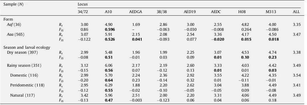
Genotypes at eight microsatellite loci were determined for 733
Ae. aegyptispecimens (males and females) collected in Niakhar in 2005 during the dry and rainy seasons (Table 4). All loci were polymorphic showing a number of distinct alleles ranging from 3 (AEDGA) to 9 (A10 and H08). The average allelic richness was assessed after grouping mosquitoes according to the form (Aaf vs Aaa), the season and type of larval development site. This grouping was based on studies from Kenya which evidenced that the level of genetic differentiation was linked to differences in larval ecology (Tabachnick and Powell, 1978). Across all loci, the average allelic richness ranged from 3.35 (groupAaf) to 3.49 (natural containers in the rainy season) and no statistical difference was detected across samples (Kruskall–Wallis’ test,P>0.05). Departure from Hardy–
Weinberg proportions associated to significant heterozygote deficits was detected in 16 out of 56 possible tests. One significant deviation was detected in theAafsamples (A10) and five inAaa
(A10, AEDGA, AEDC, H08 and M313). Four significant deviations were detected in the dry season (DS) sample (A10, AEDC, H08 and M313), and three in rainy season (RS) sample (A10, AEDC and M313). When RS was subdivided according to larval habitat,
Table 3
Pairwise comparisons of the abdominal pattern values (PVs) distributions inAe. aegyptiaccording to sex, sampling period and nature of the larval habitat.
Comparison P
<Dry season (91) vs ,Dry season (156) <10 6
<Rainy season (135) vs ,Rainy season (216) <10 6
<Dry season (91) vs <Rainy season (135) <0.01 <Nat (42) vs <Dom (50) <0.05 <Nat (42) vs <Peridom (43) <0.05
<Dom (50) vs <Peridom (43) <0.05
,Dry season (156) vs ,Rainy season (216) <0.01
,Nat (75) vs ,Dom (66) <0.05
,Nat (75) vs ,Peridom (75) <0.05
,Dom (66) vs ,Peridom (75) >0.05
<: male;,: female; in parentheses: sample size;P: probability of homogeneity (the Wilcoxon test); Nat, Dom and Peridom refer to the type of larval habitat, respectively, natural, domestic and peridomestic.
Table 4
Genetic variability and goodness of fit to Hardy–Weinberg expectations inAe. aegyptipopulations surveyed in Niakhar.
Sample (N) Locus
34/72 A10 AEDGA 38/38 AED19 AEDC H08 M313 ALL
Form
Aaf(36) Rs 3.00 4.90 1.69 2.86 3.00 2.55 4.82 4.00 3.35
FIS 0.86 0.596 – 0.063 0.030 0.008 0.264 0.086
Aaa(565) Rs 3.07 5.91 2.15 2.08 2.54 3.36 4.17 4.50 3.47
FIS 0.122 0.526 0.041 0.093 0.077 0.020 0.015 0.018
Season and larval ecology
Dry season (397) Rs 2.99 5.48 1.96 1.99 2.25 3.07 4.53 4.74 3.38
FIS 0.08 0.51 0.01 0.03 0.09 0.01 0.10 0.23
Rainy season (351) Rs 3.12 6.06 2.17 2.19 2.60 3.33 4.03 4.42 3.49
FIS 0.15 0.56 0.07 0.12 0.13 0.01 0.01 0.03
Domestic (116) Rs 2.99 5.70 2.24 2.36 2.92 3.55 4.22 4.35 3.54
FIS 0.20 0.64 0.23 0.14 0.32 0.01 0.11 0.01
Peridomestic (118) Rs 2.95 6.29 1.88 2.20 2.62 3.04 3.88 4.49 3.41
FIS 0.12 0.55 0.02 0.10 0.05 0.05 0.09 0.08
Natural (117) Rs 3.42 5.96 2.51 2.00 2.20 3.31 4.06 4.49 3.49
FIS 0.13 0.47 0.003 0.123 0.06 0.04 0.06 0.18
heterozygote deficits remained significant only at locus A10 in all samples. Significant linkage disequilibrium was revealed between locus A10 and loci 34/72 and M313 in the DS sample. A single significant association between locus 38/38 and AED19 was detected in the RS pooled samples. Due to the number of significant heterozygote deficits and linkage disequilibrium involving A10, analyses of genetic differentiation and population structure were performed excluding this locus from the dataset.
3.2.2. Genetic differentiation and population structure
Genetic differentiation was first assessed comparing mosqui-toes grouped according to the form (Aaf vs Aaa) and results suggested that the two forms were not differentiated when the two
sampling dates were considered together (FST= 0.0036,P>0.05)
or separately (dry season:FST= 0.0015,P>0.05; rainy season:
FST= 0.0091,P>0.05). When analyses were ran without
mor-phologic consideration, a very low level of genetic differentiation was detected between mosquitoes collected in the dry and rainy seasons (FST= 0.0003,P<10 4,Table 5). There was no evidence for
genetic structuring within the RS sample when mosquitoes were grouped according to the type of container they were colleted from (FST<0.0024, P>0.05,Table 5). However, significantFSTestimates
were detected between the dry season sample (DS) and mosquitoes collected during the rainy season in domestic (RS-DOM) and peridomestic (RS-PERI(RS-DOM) containers (FST>0.001,
P<10 4). No differentiation was detected between DS and RS-NAT
(Table 5).
Bayesian cluster analysis using STRUCTURE 2.2. was performed to estimate the most likely number of genetic clusters (K) in the dataset following the algorithm described byEvanno et al. (2005). Our results revealed the most likely number of cluster (K) was equal to 2 and 7 when we successively analyzing the global sample (i.e., males and females from both collection dates pooled), the DS and the RS sample (seeFig. 3) suggesting the number of genetic cluster was higher during the rainy season than during the dry season. Similarly, the number of genetic clusters (K) was also found to be higher during the rainy season when analyses were performed considering only those females for which the abdomi-nal morphology was checked. However, no correspondence between these genetic clusters and any grouping of mosquitoes based on either morphology or larval ecology (container type) was
Table 5
FSTestimates computed from seven microsatellite polymorphism for all pairs of samples subdivided according sampling date and ecological characteristics of larval habitat.
Sample DS RS RS-DOM RS-PERIDOM
DS –
RS 0.0007 –
RS-DOM 0.0010 NC –
RS-PERIDOM 0.0025 NC 0.0029 –
RS-NAT 0.0004 NC 0.0008 0.0013
DS: dry season; RS: rainy season; DOM, PERIDOM and NAT refer to domestic, peridomestic and natural larval development sites, respectively; statistical significance ofFSTestimates was assessed using theG-test of homogeneity of genotypic frequencies (Goudet et al., 1996). Underlined:P<0.0001. NC: not
computed.
detected, all groups appearing as a mixture of the genetic clusters identified (data not shown).
4. Discussion
We estimated the amount of morphological variation amongAe. aegyptispecimens from the area of Niakhar, Senegal. Relationships between morphological variation and factors such as sex, seasonality and larval ecology were explored. Population structure and the level of genetic differentiation between samples with contrasting larval ecology and/or adult morphology were assessed using a set of eight microsatellite markers.
Morphological analyses based on the pattern of abdominal scaling ofAe. aegyptirevealed significant variation according to sex. Here, the mean and range of pattern values (PVs) were significantly lower in males than in females. These results are consistent with previous observations (Mattingly, 1957; McClel-land, 1974) and suggest that the pattern of abdominal pale scaling is, at least in part, linked to sex. In Ae. aegypti, morphological mutant marker loci were reported across all three chromosomes (Munstermann and Craig, 1979; Severson et al., 2002). Some of them, involved in the coloration of the abdomen (e.g., ‘‘pale abdomen’’) were located on the first chromosome, within a linkage group containing one locus involved in sex determination (Anderson et al., 2001).
Mosquitoes from Niakhar mainly exhibited morphological characteristics which are typical of Ae. aegypti aegypti (i.e., PV>1). The specimens that would correspond to Ae. aegypti
formosus(i.e., blackish appearance of the thoracic and abdominal integument, and absence of pale scales on the first abdominal tergite, PV = 1) occurred at very low frequencies. In addition, our analyses indicated these ‘‘Aaf-like’’ mosquitoes were not ecologi-cally or genetiecologi-cally distinct from other morphotypes in our samples. Overall, these results suggest that this level of morpho-logical variation probably reflected intra-population variability rather than phenotypic markers of speciation. Hence, the typical
Aaf, such as observed in the rural gallery forests around Kedougou in Southern Senegal (Huber et al., 2008), tended to be rare in the anthropogenic area we investigated. Most of the specimens we analyzed would fell under the morphological description ofAaa, in agreement with the recent survey by Sylla et al. (2009). It is therefore clear from these results, as well as other reports (e.g.,
Hervy, 1977) thatAaais common in West Africa. Then, the question of its origins remains to be debated. According to Tabachnick (1991), these West African populations ofAaamight have been introduced back to sub-Saharan Africa after lineage splitting through vicariance following the apparition of the Sahara desert. However, the lack or very low levels of genetic differentiation observed using a wide range of molecular markers including isozymes (Huber et al., 2008), SNPs (Sylla et al., 2009) and microsatellites markers (this study) amongAe. aegyptipopulations from Senegal is at odds with this hypothesis. Rather, these results strongly suggest thatAaaandAafare monophyletic. In this context, it is tempting to establish a parallel between the evolution of anthropophily and more general domestic habits such as adapta-tion to anthropogenic environments (e.g., man-made, permanent breeding sites) in Ae. aegypti and in the more widely studied
Anopheles gambiae, the major malaria vector in Africa (Ayala and Coluzzi, 2005). Indeed, the history of human population settlement and climate changes in Africa may have provided ample opportunities and strong selection pressure for the evolution of anthropophily and domesticity in mosquitoes, especially through the ability of man to provide suitable development sites for the aquatic stages during the phases of ‘‘savannization’’ that occurred across most of the central Africa rain forest, ca. 2500 years ago (Willis et al., 2004). As such, ecologically driven selection might
well be the major evolutionary force driving radiation and lineage splitting within Ae. aegypti in Africa, rather than allopatric speciation followed by secondary contact. However, it is clear that a much broader assessment of morphological, ecological, behavioral and genetic variations that exist within African Ae. aegyptipopulations is needed for an in-depth understanding of the evolutionary history of the species both within and outside Africa. In Niakhar, significant morphological variations were evi-denced between the dry and the rainy season, particularly in females. Among mosquitoes collected during the dry season, a broad range of PVs was observed, but the females were mainly clustered into a single group centered on PV = 2. Interestingly, females collected during the rainy season segregated into two distinct morphological groups, one centered on PV = 3 (group 1) and one centered on PV = 9 (group 2). Such an observation could indicate the pattern of abdominal pale scales is a polymorphic trait for which the distribution of the different states (i.e., morphotypes) is variable rather than fixed across time and possibly randomly driven, due to stochastic processes in relation to demographic fluctuations. In South Vietnam indeed, it was suggested that seasonal fluctuations in rainfalls (which condition the availability of oviposition sites) affected the population size ofAe. aegyptiand induced great temporal variation in the allelic distribution of neutral genes (Huber et al., 2002). In Niakhar, the scarcity of suitable larval developmental places during the dry season possibly induced significant demographic crashes enhancing genetic drift that modulated the distribution of characters such as the abdominal morphology we surveyed. The temporal variation in distribution of morphotypes could alternatively be interpreted as the result of the co-occurrence of two morphological forms (or subpopulations) ofAaa. Additionally our results suggest the nature of larval breeding site greatly influences the pattern of abdominal scales. The relation between morphology and larval ecology indicated a possible segregation of forms according to the nature of breeding containers. However, microsatellite-based analyses failed to detect any evidence that the two morphological forms are genetically differentiated. Nevertheless, it was possible the set of microsatellite markers we employed was not sufficient to detected genetic differentiation. While such markers are not abundant in Ae. aegypti genome, a recent work increased the number of microsatellite suitable for population genetic studies up to 20 (Lovin et al., 2009). The recent study ofBernhardt et al. (2009), suggesting the occurrence of eight large paracentric inversions on three chromosomes of a Aafstrain from Senegal, introduced the idea that a chromosomal polymorphism possibly exists inAe. aegypti. These authors also discussed the possibility that genes controlling traits such as morphology, biology and vector competence would be located in inversions and then the traits that they condition would be maintained as correlated characters. To better tackle the heterogeneity of Ae. aegypti in Senegal, it would be crucial to study the distribution of such chromosomal inversions within natural populations.
Acknowledgements
We thank Franck Remoue´ and Bassirou Fall for significant contribution to the fieldwork. We are grateful to Franc¸ois Lacoste and ‘‘La Fondation des Treilles’’ for their financial support to CP trough postdoctoral fellowships. This study was funded by the French Institut de Recherche pour le De´veloppement (IRD).
References
Anderson, J.R., Grimstad, P.R., Severson, D.W., 2001. Chromosomal evolution among six mosquito species (Diptera: Culicidae) based on shared restriction fragment length polymorphisms. Mol. Phylogenet. Evol. 20, 316–321.
Ayala, F.J., Coluzzi, M., 2005. Chromosome speciation: humans, Drosophila, and mosquitoes. Proc. Natl. Acad. Sci. U.S.A. 102 (Suppl. 1), 6535–6542. Bernhardt, S.A., Blair, C., Sylla, M., Bosio, C., Black IV, W.C., 2009. Evidence of
multiple chromosomal inversions inAedes aegypti formosus from Senegal. Insect. Mol. Biol. 18 (5), 557–569.
Chambers, E.W., Meece, J.K., Mcgowan, J.A., Lovin, D.D., Hemme, R.R., Chadee, D.D., McAbee, K., Brown, S.E., Knudson, D.L., Severson, D.W., 2007. Microsatellite isolation and linkage group identification in the yellow fever mosquitoAedes aegypti. J. Hered. 98, 202–210.
Charrel, R.N., De Lamballerie, X., Raoult, D., 2007. Chikungunya outbreaks—the globalization of vectorborne diseases. N. Engl. J. Med. 356, 769–771. Chistophers, S.R., 1960.Aedes aegypti(L.): The Yellow Fever Mosquito. Cambridge
University Press, Cambridge, United Kingdom, pp. 1–739.
De Lamballerie, X., Leroy, E., Charrel, R.N., Ttsetsarkin, K., Higgs, S., Gould, E.A., 2008. Chikungunya virus adapts to tiger mosquito via evolutionary convergence: a sign of things to come? Virol. J. 5, 33.
Diarrassouba, S., Dossou-Yovo, J., 1997. Rythme d’activite´ atypique chezAedes aegyptien zone de savane sub-soudanienne de Coˆte d’Ivoire. Bull. Soc. Pathol. Exot. 90, 361–363.
El Mousadik, A., Petit, R.J., 1996. High level of genetic differentiation for allelic richness among populations of the argan tree [Argania spinosa(L.) Skeels] endemic to Morocco. Theor. Appl. Genet. 92, 832–839.
Evanno, G., Regnaut, S., Goudet, J., 2005. Detecting the number of clusters of individuals using the software STRUCTURE: a simulation study. Mol. Ecol. 14, 2611–2620.
Failloux, A.B., Vazeille, M., Rodhain, F., 2002. Geographic genetic variation in populations of dengue virus vectorAedes aegypti. J. Mol. Evol. 55, 653–663. Goudet, J., 1995. FSTAT (version 1.2): a computer software to calculateF-statistics. J.
Hered. 86, 485–486.
Goudet, J., Raymond, M., De Meeu¨s, T., Rousset, F., 1996. Testing differentiation in diploid populations. Genetics 144, 1933–1940.
Gubler, D.J., 2002. Epidemic dengue/dengue hemorrhagic fever as a public health, social and economic problem in the 21st century. Trends Microbiol. 10, 100–103. Hervy, J.P., 1977. Expe´rience de marquage-laˆcher-recapture, portant surAedes aegyptiLinne´, en zone de savane soudanienne ouest africaine II. Relations entre habitat, morphologie et comportement. Cah. ORSTOM Entomol. Me´d. Parasitol. 15, 391–400.
Holm, S., 1979. A simple sequentially rejective multiple test procedure. Scand. J. Stat. 6, 65–70.
Huber, K., Ba, Y., Dia, I., Mathiot, C., Sall, A.A., Diallo, M., 2008.Aedes aegyptiin Senegal: genetic diversity and genetic structure of domestic and sylvatic populations. Am. J. Trop. Med. Hyg. 79, 218–229.
Huber, K., Luu Le, L., Tran Huu, H., Ravel, S., Rodhain, F., Failloux, A.B., 2002. Temporal genetic variation inAedes aegypti(Ho Chi Minh City, Viet Nam) using microsatellite markers. Heredity 89, 7–14.
Huber, K., Mousson, L., Rodhain, F., Failloux, A.B., 2001. Isolation and variability of polymorphic microsatellite loci inAedes aegyptithe vector of dengue viruses. Mol. Ecol. Notes 1, 219–222.
Kro¨pelin, S., Vershuren, D., Lezine, A.M., Eggermont, H., Cocquyt, C., Francus, P., Cazet, J.P., Fagot, M., Rumes, B., Russell, J.M., Darius, F., Conley, D.J., Schuster, M., von Suchodoletz, H., Engstrom, D.R., 2008. Climate-driven ecosystem succes-sion in the Sahara: the past 6000 years. Science 320, 765–768.
Lovin, D.D., Washington, K.O., deBruyn, B., Hemme, R.R., Mori, A., Epstein, S.R., Harker, B.W., Streit, T.G., Severson, D.W., 2009. Genome-based polymorphic microsatellite development and validation in the mosquitoAedes aegyptiand application to population genetics in Haiti. BMC Genomics 10, 590.
Mattingly, P.F., 1957. Genetical aspects of theAedes aegyptiproblem. I. Taxonomy and bionomics. Ann. Trop. Med. Parasitol. 51, 392–408.
McClelland, G.A.H., 1974. A worldwide survey of variation in scale pattern of the abdominal tergum ofAedes aegypti(L.) (Diptera: Culicidae). Trans. R. Entomol. Soc. Lond. 126, 229–259.
Morisson, A.C., Zielinsky-Guitierrez, E., Scott, T.W., Rosenberg, R., 2008. Defining challenges and proposing solution for control of the virus vectorAedes aegypti. Plos Med. 5, 362–366.
Munstermann, L.E., Craig Jr., G.B., 1979. Genetics ofAedes aegypti: updating the linkage map. J. Hered. 70, 291–296.
Nei, M., 1987. Molecular Evolutionary Genetics. Columbia University Press, New York, pp. 1–512.
Paupy, C., Ollomo, B., Moutailler, S., Rousset, D., Kamgang, B., Demanou, M., Herve´, J.P., Leroy, E., Simard, F., 2009. Comparative role ofAedes albopictusandAedes aegyptiin the emergence of dengue and chikungunya in Central Africa. Vector Borne Zoonotic Dis., doi:10.1089/vbz.2009.0005.
Paupy, C., Brengues, C., Kamgang, B., Herve´, J.P., Fontenille, D., Simard, F., 2008. Gene flow between domestic and sylvan populations ofAedes aegypti(Diptera: Culicidae) in North Cameroon. J. Med. Entomol. 45, 391–400.
Petersen, J.L., 1977. Behavior differences in two subspecies ofAedes aegypti(L.) (Diptera: Culicidae) in East Africa. PhD thesis, University of Notre-Dame, IN. Peyrefitte, C., Rousset, D., Pastorino, B., Pouillot, R., Bessaud, M., Tock, F., Mansaray,
H., Merle, O.L., Pascual, A.M., Paupy, C., Vessiere, A., Imbert, P., Tchendjou, P., Durand, J.P., Tolou, H.J., Grandadam, M., 2007. Chikungunya virus, Cameroon. Emerg. Infect. Dis. 13, 768–771.
Pritchard, J.K., Stephens, M., Donnelly, P., 2000. Inference of population structure using multilocus genotype data. Genetics 155, 945–959.
R Development Core Team, 2005. R: A Language and Environment for Statistical Computing. R Foundation for Statistical Computing, Vienna, Austria. ISBN 3-900051-07-0 (http://www.R-project.org).
Ravel, S., Herve, J.P., Diarrassouba, S., Kone, A., Cuny, G., 2002. Microsatellite markers for population genetic studies inAedes aegypti(Diptera: Culicidae) from Coˆte d’Ivoire: evidence for a microgeographic genetic differentiation of mosquitoes from Bouake´. Acta Trop. 82, 39–49.
Raymond, M., Rousset, F., 1995. GENEPOP (version 1.2): population genetics soft-ware for exact tests and ecumenicism. J. Hered. 86, 248–249.
Remoue´, F., Alix, E., Cornelie, S., Sokhna, C., Cisse, B., Doucoure, S., Mouchet, F., Boulanger, D., Simondon, F., 2007. IgE and IgG4 antibody responses toAedes saliva in African children. Acta Trop. 104, 108–115.
Severson, D.W., Meece, J.K., Lovin, D.D., Saha, G., Morlais, I., 2002. Linkage map organization of expressed sequence tags and sequence tagged sites in the mosquito,Aedes aegypti. Insect Mol. Biol. 11, 371–378.
Sylla, M., Bosio, C., Urdaneta-Marquez, L., Ndiaye, M., Black, W.C., 2009. Gene flow, subspecies composition, and dengue virus-2 susceptibility amongAedes aegypti collections in Senegal. PLoS Negl. Trop. Dis. 3, e408.
Tabachnick, W.J., 1991. Evolutionary genetics and arthropod born disease. The yellow fever mosquito. Am. Entomol. 37, 14–24.
Tabachnick, W.J., Wallis, G.P., Aitken, T.H.G., Miller, B.R., Amato, G.D., Lorenz, L., Powell, J.R., Beaty, B.J., 1985. Oral infection ofAedes aegyptiwith yellow fever virus: geographic variation and genetic considerations. Am. J. Trop. Med. Hyg. 34, 1219–1224.
Tabachnick, W.J., Powell, J.R., 1979. A world-wide survey of genetic variation in the yellow fever mosquito,Aedes aegypti. Genet. Res. 34, 215–229.
Tabachnick, W.J., Powell, J.R., 1978. Genetic structure of East-African domestic populations ofAedes aegypti. Nature 272, 535–537.
Thonnon, J., Spiegel, A., Diallo, M., Diallo, A., Fontenille, D., 1999. Chikungunya virus outbreak in Senegal in 1996 and 1997. Bull. Soc. Pathol. Exot. 92, 79–82.
Trpis, M., Hausermann, W., 1978. Genetics of houses entering behaviour in east African populations ofAedes aegypti(L.) (Diptera: Culicidae) and its relevance to speciation. Bull. Entomol. Res. 68, 521–532.
Wallis, G.P., Tabachnick, W.J., Powell, J.R., 1983. Macrogeographic genetic variation in a human commensal:Aedes aegypti, the yellow fever mosquito. Genet. Res. 41, 241–258.
Weir, B.S., Cockerham, C.C., 1984. EstimatingF-statistics for the analysis of popula-tion structure. Evolupopula-tion 38, 1358–1370.
Willis, K.J., Gillson, L., Brncic, T.M., 2004. How ‘‘virgin’’ is virgin rainforest? Science 304, 402–403.