www.elsevier.com/locate/ibmb
Quantification of juvenile hormone III, vitellogenin, and
vitellogenin-mRNA during the oviposition cycle of the lubber
grasshopper
D.W. Borst
*, M.R. Eskew, S.J. Wagner, K. Shores, J. Hunter, L. Luker, J.D. Hatle,
L.B. Hecht
Department of Biological Sciences, Illinois State University, Normal, IL 61790-4120, USA
Received 31 October 1999; received in revised form 31 December 1999; accepted 25 January 2000
Abstract
The vitellogenic cycle of the lubber grasshopper (Romalea microptera) was studied by measuring levels of juvenile hormone (JH III), vitellogenin, and vitellogenin-mRNA through the first oviposition cycle. JH III and vitellogenin were measured by radioim-munoassay (RIA) and enzyme-linked immunosorbent assay (ELISA), respectively. To measure vitellogenin-mRNA, a partial (753 bp) cDNA fragment of vitellogenin was isolated from the fat body of vitellogenic animals. The sequence of this cDNA was related to vitellogenin sequences in other insect species. Using these sequence data, an RT–PCR (reverse transcriptase polymerase chain reaction) assay was developed to quantify vitellogenin-mRNA levels during the oviposition cycle. Vitellogenin-mRNA levels in the fat body tissue from virgin females were measured on specific days after eclosion and compared to hemolymph levels of JH III and vitellogenin from the same individuals. The levels of all three compounds (JH III, vitellogenin, and vitellogenin-mRNA) showed similar changes throughout the oviposition cycle, being undetectable or nearly undetectable initially (day 3), rising to maximum levels on days 23 and 28, and then dropped to lower or undetectable levels on the day of oviposition. The ability to measure these characteristics will be useful for studying the effects of hormonal and nutritional manipulations on reproduction.
2000 Elsevier Science Ltd. All rights reserved.
Keywords: Romalea microptera; Vitellogenin; Juvenile hormone; JH; Oviposition cycle; RT–PCR; Vitellogenin-mRNA
1. Introduction
Numerous studies have shown that hemolymph levels of vitellogenin are hormonally regulated, and there is a rich literature documenting how this regulation varies among insect species (Engelmann, 1983; Nijhout, 1994; Wyatt and Davey, 1996; Belles, 1998). In Orthoptera and related orders, the hemolymph concentration of juv-enile hormone (JH) appears to be the critical factor that determines the onset and continuation of vitellogenesis (Chinzei et al., 1982; Wyatt and Davey, 1996). JH directly induces vitellogenin synthesis in several species (Engelmann, 1983; Wyatt and Davey, 1996). InLocusta migratoria, JH has been shown to have both a priming
* Corresponding author. Fax:+1-309-438-7694.
E-mail address:[email protected] (D.W. Borst).
0965-1748/00/$ - see front matter2000 Elsevier Science Ltd. All rights reserved. PII: S 0 9 6 5 - 1 7 4 8 ( 0 0 ) 0 0 0 5 3 - 9
effect (Wyatt et al., 1996) and a direct stimulatory effect on the transcription of vitellogenin genes in the fat body tissue (Zhang et al., 1993; Glinka and Wyatt, 1996). In addition, JH has been shown to regulate the uptake of vitellogenin by the ovary by interacting with membrane receptors of the follicle cells (Abu-Hakima and Davey, 1977; Davey et al., 1993).
can be analyzed for a variety of proteins and hormones, allowing the effects of environmental changes on indi-viduals to be studied while controlling for individual variation. Indeed, a recent paper has shown that the level of nutrition and the timing of nutritional changes have marked effects on the reproductive output of this species (Moehrlin and Juliano, 1998). Studying the effects of nutritional and hormonal manipulations on reproductive responses should provide considerable insight into both the regulation of reproduction and the response of these organisms to variable environments.
As the groundwork for such studies, we have analyzed the levels of JH (by radioimmunoassay, RIA), vitellog-enin (by enzyme-linked immunosorbent assay, ELISA), and vitellogenin-mRNA (by quantitative reverse tran-scriptase polymerase chain reaction, RT–PCR). The sen-sitivity of these methods allowed all three factors to be measured in each animal in this study. Our data indicate that these factors begin to rise toward the end of the second week of the cycle, reach a peak during the fourth week of the cycle, and drop to low levels at oviposition. These results are consistent with previous results for other Orthoptera and provide us with a baseline by which to evaluate the effects of other hormones as well as environmental factors on the vitellogenesis of this spec-ies.
2. Materials and methods
2.1. Experimental design
The strategy used in this study was to measure the changes in JH, vitellogenin, and vitellogenin-mRNA lev-els in adult virgin females of the lubber grasshopper dur-ing reproduction. All three compounds were measured in each individual. Three to five grasshoppers were sacri-ficed at 3, 8, 13, 18, 23, 28, and 33 days after adult eclosion. Six additional individuals were sacrificed on the day of oviposition, which occurred around day 37.
2.2. Animals
A colony ofR. microptera was established at Illinois State University using animals obtained in Copeland, FL, USA, in 1996 and 1997. On the day of ecdysis, vir-gin adult females were marked and placed in a commu-nal cage without males. The cage was placed in an environmental chamber (photoperiod=14L:10D; 32:24°C) and the animals were fed Romaine lettuce and oatmeal ad libitum (Whitman, 1986). After 30 days, a sand container was provided for oviposition.
2.3. Hemolymph samples
Hemolymph was collected at the time of dissection. Samples for JH analysis were placed in 0.5 ml
acetonitr-ile and 1 ml 0.9% NaCl and extracted (2×1 ml) with hexane. The hexane phases were combined and held at
220°C until analysis. Hemolymph collected for vitellog-enin analysis was diluted (1:50) in hemolymph buffer (100 mM NaCl; 50 mM Tris; pH 7.5; 1 mM EDTA; 0.1 mM DTT; and 0.1% Tween-20) and stored at 220°C until analysis.
2.4. Quantification of JH III
The hexane extract of each hemolymph sample was dried, resuspended in 30µl methanol, and analyzed with a chiral-selective radioimmunoassay (RIA) for 10R-JH III as described previously (Hunnicutt et al., 1989; Huang et al., 1994). The amount of JH III present in each sample was calculated from the standard curve and expressed as ng 10R-JH III/ml hemolymph.
2.5. Quantification of vitellogenin
Lubber vitellin and vitellogenin were characterized and this work will be reported elsewhere. As part of this characterization, vitellin was isolated from freshly laid eggs or mature oocytes, purified by anion exchange chromatography, and injected (i.m.) into New Zealand white rabbits (Myrtle’s Rabbitry, TN, USA) with Freund’s complete adjuvant. Booster injections (every 2 weeks) used Freund’s incomplete adjuvant. As expected, the resulting anti-lubber vitellin antiserum cross-reacted strongly to its precursor, vitellogenin, allowing it to be used in an enzyme-linked immunosorbent assay (ELISA) for this protein. To this end, the IgG fraction of the antiserum was purified using protein A and a por-tion was derivatized with biotin using the procedure of Chang et al. (1998). Before use, both the underivatized and biotinylated IgG fractions were treated with an equal concentration of male lubber hemolymph and centri-fuged to remove precipitated protein.
determined by adding a chromogen solution (0.1 M cit-rate buffer; pH 4; containing 0.04% ABTS [Sigma Chemical Co.; 2,29 -azino-bis(3-ethylbenzthiazoline-6-sulfonic acid] and 0.006% hydrogen peroxide). The plate was covered with foil and incubated at room temperature until the color developed (approximately 15 min). The absorbance of the solution in each well of the microtiter plate was measured at 410 nm. The antiserum detected no immunoactivity in serum samples from adult males and juvenile males and females.
2.6. Cloning a cDNA for vitellogenin
Total RNA was isolated from fat body and ovarian tissue of adult females with Tri-Reagent (Molecular Research Center, Cincinnati, OH, USA) and used to pro-duce a cDNA library using the λTriplEx vector (Clontech, Palo Alto, CA, USA). Bacteria were infected with theλTriplEx library and the resulting plaques trans-ferred to pure nitrocellulose filters (Osmonics, Inc., Westboro, MA, USA) previously soaked in IPTG (Sigma Chemical Co, St Louis, MO, USA). Filters were blocked with 5% dry milk and treated with diluted (1:400) preabsorbed rabbit anti-vitellin antiserum. The filters were then treated with goat rabbit IgG anti-serum conjugated to horseradish peroxidase (Sigma Chemical Co.), washed, and then treated with a chromo-gen solution (PBS containing 2.8 mM 3.39 -diaminoben-zidine; 0.1% CoCl2; 0.03% hydrogen peroxide).
Plas-mids were isolated from the positive clones and cycle sequenced using the ABI 310 genetic analyzer (Perkin Elmer Biosystems, Norwalk, CT, USA). The sequence was compared with a BLAST search to other vitellog-enin sequences deposited in Genbank.
2.7. Measurement of vitellogenin-mRNA by RT–PCR
Total RNA was isolated from fat body tissue dissected from female and male animals using Tri-Reagent. The total RNA concentration was quantified at 260/280 nm and diluted to 0.5 mg/ml. The RNA (1 µg) was reverse transcribed (total volume of 20 µl) using 80 units MMLV reverse transcriptase (Promega, Madison, WI, USA), random hexamer primers (100 ng), dNTPs (25 µM), DTT (10 mM) and the buffer supplied by the manufacturer. After incubation at 37°C overnight, ali-quots of the RT reaction were amplified using primers (Operon Technologies, Alameda, CA, USA) that ampli-fied a 211 bp sequence of the cloned vitellogenin-cDNA fragment. The 59 primer (59 -TTCTGCGAATCTTGAAGACC) was unlabeled while the 39 primer (59-TTGGAGGAATCATGGACGAA) was labeled with 6-FAM at its 59end. An aliquot (2µl) of each RT reaction was added to 1 unit of Taq DNA polymerase (Perkin Elmer Biosystems), 0.2µM of each primer, and 20 µM dNTP. This mixture was amplified
by the polymerase chain reaction (PCR; 15 s at 94°C, 30 s at 50°C, 1 min at 72°C) for the indicated number of cycles. The fluorescently labeled products were diluted with dimethylformamide (DMF) and analyzed with an ABI 310 Genetic Analyzer using the GeneScan protocol. A calibration standard containing fluorescently labeled DNA fragments of known size (GS-500-TAMRA; Perkin Elmer Biosystems) was added to deter-mine the size of product produced by the sample.
The efficiency and linearity of the procedure was tested in three ways. First, cDNA samples with different amounts of vitellogenin-cDNA were amplified three times under identical conditions and the amount of pro-duct measured. For other studies, the cDNA fragment was amplified with the above primers and the product separated on a 2% agarose gel. The amplified band was removed, cleaned (GeneClean, Bio101, Vista, CA, USA), and quantified with a spectrophotometer. To determine the efficiency of the PCR amplification, a known amount of this purified cDNA (100–200 pg) was amplified in a double volume (40 µl) of PCR reactants for 15 cycles. After this and each of the next seven cycles, a small amount (2 µl) of the PCR reaction was removed and analyzed on the ABI310. In addition, known amounts of the purified cDNA (0.08 to 2.1 fmol) were amplified for 19 cycles using standard PCR con-ditions (see above) and the amount of fluorescent pro-duct determined. The slope of the resulting curve was used to determine the amount of vitellogenin-cDNA present in samples after reverse transcription.
2.8. Statistics
The natural or log transformed results were analyzed by one-way analysis of variance (ANOVA) followed by the Ryan–Einot–Gabriel–Welsch multiple range test (SAS Institute Inc., 1989).
3. Results
3.1. Cloning of a partial cDNA for vitellogenin
230) of theR. microptera sequence had a higher degree of similarity. For example, the lubber amino acid sequence was 49% and 47% identical to similar regions in the vitellogenins of Bombyx mori andAthalia rosae (Genbank accession # Q27309 and AB007850, respectively). Likewise, the lubber amino acid sequence was 46% identical to similar regions in the vitellogenins of Aedes aegypti and Anthonomus grandis (Genbank accession # Q16927 and M72980, respectively).
3.2. Measurement of vitellogenin-mRNA using RT– PCR
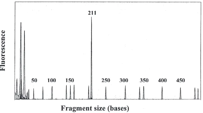
A 211 bp region of the lubber vitellogenin cDNA fragment was amplified by PCR using two 20-mer pri-mers, one of which was fluorescently labeled. When these primers were used in the RT–PCR analysis of total RNA from fat body tissue, preparations from vitellog-enic females yielded a fluorescent product with the anticipated size (Fig. 1). Total RNA from male fat body tissue (not shown) and newly eclosed (nonvitellogenic) females (see below) yielded no product.
For accurate quantification by RT–PCR it is necessary to measure product formation during the exponential phase of amplification, since amplification in the plateau phase is not quantitative (Freeman et al., 1999). Because of the relative abundance of vitellogenin-mRNA in vitel-logenic animals, it was anticipated that most samples would reach the plateau phase after relatively few cycles. Therefore, the optimum cycle number was determined by amplifying the purified cDNA fragment in amounts similar to fat body levels of vitellogenin-mRNA observed in preliminary experiments. This test showed
Fig. 1. RT–PCR analysis of vitellogenin-mRNA. Total RNA from the fat body of a vitellogenic female was reverse transcribed, amplified by PCR with a fluorescently labeled primer (6-FAM) and quantified with an ABI 310. The amplification yielded a single fluorescent product (211 bp). The small peaks of equal height were produced by the fluorescent (TAMRA) calibration standard, which has 16 DNA fragments from 35 to 500 bp.
that the PCR amplification was in the exponential phase from cycle 15 to 22 (Fig. 2; R2=0.9924). The slope of
this line indicated that the amplification factor for each cycle was 1.57. In addition, the linearity of the PCR reaction after 19 cycles was investigated with known amounts of the purified cDNA fragment. The level of fluorescence was linearly related to the amount of the cDNA being amplified (Fig. 3;R2=0.9701). The
quanti-fication of mRNA levels by this method was highly repeatable. Several samples from vitellogenic females
Fig. 3. The linearity of the PCR amplification. Known amounts of the vitellogenin-cDNA fragment were amplified for 19 cycles and ana-lyzed as in Fig. 1. PCR amplification was linear over the range of vitellogenin-cDNA amounts used.
were amplified three times under similar conditions. The average coefficient of variation in the results from these samples was 10% (Table 1).
3.3. Changes in animal weight and ovarian index (OI) during the oviposition cycle:
Animal weight increased from 3.3 (±0.5, SE) g on day 3 to 6.5 (±0.6) g on day 33, and then dropped to 5.1 (±0.3) shortly after oviposition (37.2±0.7 days). The OI (ovary wt/animal wt×100) was very low on day 3 (1.1±0.1) and did not significantly change through day 13 (2.0±0.1; P.0.05). Thereafter, the OI rose rapidly by day 18 (10.4±2), reached its maximum on day 33 (28.2±2.1) before eclosion, and then fell to a low level (6.1±0.6) shortly after oviposition. These results are similar to observations that we have made previously for this grasshopper (unpublished data) and for a related lubber grasshopper,Taeniopoda eques(Whitman, 1986).
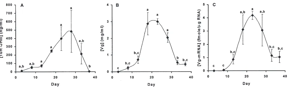
3.4. JH, vitellogenin, and vitellogenin-mRNA levels during the oviposition cycle
The levels of all three compounds (JH III, vitellog-enin, and vitellogenin-mRNA) changed in a parallel
Table 1
Precision of vitellogenin-mRNA analysis
Sample N Meana SE
1 3 2224 108
2 3 9599 325
3 3 3224 298
aArea of peak at 211 bp.
fashion throughout the oviposition cycle (Fig. 4). JH lev-els (Fig. 4a) were initially low, then rose modestly on days 8 and 13 to an intermediate plateau (approximately 60 ng/ml) that was about 5-fold higher than the initial level of JH. On day 18, JH levels began to rise again and reached maximum levels (approximately 450 ng/ml) on days 23 and 28. These values were 7-fold higher than the intermediate level observed on days 8 and 13 and over 30-fold higher than the initial level. Thereafter JH levels fell and were nearly undetectable at oviposition. Vitellogenin was first detectable (0.2 mg/ml) on day 8, and increased to a maximum (approximately 3 mg/ml) on days 18 and 23 (Fig. 4b). Thereafter, vitellogenin dropped to low levels (0.4 mg/ml) at oviposition. Finally, levels of vitellogenin-mRNA (Fig. 4c) were first detectable on day 8, and then rose to a maximum (about 4 fmol/µg total RNA) on day 23. Thereafter, vitellog-enin-mRNA levels fell to an intermediate level (1 fmol/µg total RNA) at oviposition.
4. Discussion
In this study we report the partial cDNA sequence for a third orthopteran vitellogenin. In previous studies, par-tial sequences for vitellogenin in L. migratoria (Locke et al., 1987) and Blattella germanica (Martin et al., 1998) have been reported. In addition, partial or com-plete cDNA sequences are now available for vitellogen-ins in a number of other vitellogen-insect species. Analysis of their deduced amino acid sequences indicates that insect yolk proteins fall into two major families (Hagedorn et al., 1998). Members of the first family are similar in sequence to vertebrate serum binding proteins such as apolipoprotein B and are usually referred to as vitellog-enins (Vgs). Members of the second family show some similarity to mammalian triacylglycerol lipase and are usually referred to as yolk proteins (Yps). Yolk proteins are typically synthesized in the ovary in contrast to Vgs that are usually synthesized and processed in the fat body and exported via the hemolymph to the ovary (Wyatt and Davey, 1996). Based on the sequence of the cDNA fragment and the expression of this mRNA in the fat body, the precursor protein for R. microptera vitellin appears to be a member of the insect vitellogenin family. The analysis of JH, vitellogenin, and vitellogenin-mRNA in the lubber grasshopper showed that these com-pounds all had similar profiles through the first ovi-position cycle. All three were low after eclosion, rose during the second week of the cycle, peaked during the fourth week of the cycle, and fell before oviposition. In view of the metabolic requirements for vitellogenesis, it is not surprising that these factors are tightly controlled during the cycle in order to maximize reproductive out-put.
vitellogenin-Fig. 4. JH III, vitellogenin, and vitellogenin-mRNA levels during the oviposition cycle. Virgin female lubber grasshoppers were analyzed on the indicated day after eclosion. Oviposition occurred on day 37. There were three to six individuals on each day. The results were analyzed by ANOVA; means with the same letter are not significantly different (P.0.05). (A) Hemolymph levels of 10R-JH III were measured by RIA (F7,22=6.53,P=0.0003). (B) Hemolymph levels of vitellogenin (Vg) were measured by ELISA (F7,22=16.37, P=0.0001). (C) Vitellogenin-mRNA (Vg-mRNA) levels in fat body tissue were measured by RT–PCR (F7,22=6.14,P=0.0005).
mRNA observed in lubber grasshoppers is similar to the regulation of these factors in L. migratoria (Chinzei et al., 1982; Chinzei and Wyatt, 1985; Glinka et al., 1995). Changes in the levels of vitellogenin-mRNA in the lub-ber grasshopper were generally consistent with changes reported forL. migratoria(Chinzei et al., 1982). A minor difference was observed at the end of the vitellogenic cycle. In the lubber grasshopper, vitellogenin-mRNA levels dropped to about 25% of the maximum at ovi-position, while vitellogenin-mRNA levels in the locust decreased to about 70% of the maximum. The reason for this difference is not clear. Vitellogenin-mRNA may be less stable in the lubber grasshopper. Alternately, this difference may reflect a difference in the length of the oviposition cycle (37 and 17 days for the lubber and locust, respectively), which would lengthen the period at the end of the cycle during which the vitellogenin-mRNA could be broken down. In any case, the relatively low level of vitellogenin in the hemolymph at ovi-position indicates that the remaining fraction of vitellog-enin-mRNA is not being efficiently translated at this per-iod. This suggests that the production of vitellogenin in lubber grasshoppers may be both transcriptionally and translationally controlled.
Our study does not address the relationship of JH lev-els to vitellogenin-mRNA and hemolymph levlev-els of vit-ellogenin in the lubber grasshopper. Nevertheless, in other Orthoptera (e.g.L. migratoria), JH both primes the fat body and stimulates production of vitellogenin from the hemolymph (Wyatt and Davey, 1996). In view of these results and the close correlation observed between the levels of JH, vitellogenin, and vitellogenin-mRNA, it seems likely that the intermediate levels of JH observed at days 8 and 13 stimulate the synthesis of vit-ellogenin-mRNA in the lubber. This in turn would lead to an increase in its hemolymph levels of vitellogenin. Likewise, we suspect that the higher levels of JH
observed later in the cycle (day 18) might stimulate vit-ellogenin uptake by the oocyte. This would be in agree-ment with the rapid rise in the OI that occurred between days 13 and 18. A similar requirement for different JH levels to stimulate vitellogenin synthesis and vitellog-enin uptake has been demonstrated inRhodnius prolixus (Davey et al., 1993). We are currently testing these hypotheses by treating animals with defined amounts of JH III and the JH analog methoprene.
Knowledge about the interactions among JH, vitellog-enin, vitellogenin-mRNA, and other factors that regulate reproduction is critical for understanding the molecular and physiological mechanisms that control this process. In addition, these interactions may also help explain how reproductive output is varied in response to variable and unpredictable environments. These issues are central to understanding the mechanisms of phenotypic flexibility (plasticity) and inflexibility (canalization) that occur dur-ing reproductive development (Scheiner, 1993; Schlicht-ing and Pigliucci, 1998). An analysis of some of these mechanisms and their relationship to the onset of repro-ductive canalization in the lubber grasshopper is given in a companion paper presented at this conference (Hatle et al., 2000). Ultimately, these mechanisms must have a major role in regulating reproductive output that occur in response to environmental variation.
Acknowledgements
References
Abu-Hakima, R., Davey, K.G., 1977. The action of juvenile hormone on the follicle cells ofRhodnius prolixus: the importance of volume changes. J. Exp. Biol. 69, 33–44.
Belles, W., 1998. Endocrine effectors in insect vitellogenesis. In: Coast, G.M., Webster, S.G. (Eds.), Recent Advances in Arthropod Endocrinology. Cambridge University Press, Cambridge, UK, pp. 71–90.
Chang, E.S., Keller, R., Chang, S.A., 1998. Quantification of crus-tacean hyperglycemic hormone by ELISA in hemolymph of the lobster, Homarus americanus, following various stresses. Gen. Comp. Endocrin. 111, 359–366.
Chinzei, Y., Wyatt, G.R., 1985. Vitellogenin titre in haemolymph of
Locusta migratoria in normal adults, after ovariectomy, and in response to methoprene. J. Insect Physiol. 31, 441–445.
Chinzei, Y., White, B.N., Wyatt, G.R., 1982. Vitellogenin mRNA in locust fat body: identification, isolation, and quantitative changes induced by juvenile hormone. Can. J. Biochem. 60, 243–251. Davey, K.G., Sevala, V.L., Gordon, D.R.B., 1993. The action of
juven-ile hormone and antigonadotropin on the follicle cells ofLocusta migratoria. Invert. Reprod. Dev. 23, 189–193.
Engelmann, F., 1983. Vitellogenesis controlled by juvenile hormone. In: Downer, R.G.H., Laufer, H. (Eds.), Endocrinology of Insects. Alan R. Liss, New York, pp. 259–270.
Freeman, W.M., Walker, S.J., Vrana, K.E., 1999. Quantitative RT– PCR: pitfalls and potential. BioTechniques 26, 112–125. Glinka, A.V., Wyatt, G.R., 1996. Juvenile hormone activation of gene
transcription in locust fat body. Insect Biochem. Mol. Biol. 26, 13–18.
Glinka, A.V., Kleiman, A.M., Wyatt, G.R., 1995. Roles of juvenile hormone, a brain factor and adipokinetic hormone in regulation of vitellogenin biosynthesis in Locusta migratoria. Biochem. Mol. Biol. Int. 35, 323–328.
Hagedorn, H.H., Maddison, D.R., Tu, Z., 1998. The evolution of vitel-logenins, cyclorrhaphan yolk proteins, and related molecules. Adv. Insect Physiol. 27, 335–384.
Hatle, J.D., Juliano, S.A., Borst, D.W., 2000. Juvenile hormone is a marker of the onset of reproductive canalization in lubber grass-hoppers. Insect Biochem. Mol. Biol. 30, 821–827.
Huang, Z.-H., Robinson, G.E., Borst, D.W., 1994. Physiological corre-lates of division of labor among similarly aged honey bees. J. Comp. Physiol. A 174, 731–739.
Hunnicutt, D., Toong, Y.C., Borst, D.W., 1989. A chiral specific anti-serum for juvenile hormone. Am. Zool. 29, 48a.
Locke, J., White, B.N., Wyatt, G.R., 1987. Cloning and 59end nucleo-tide sequences of two juvenile hormone-inducible vitellogenin genes of the African migratory locust. DNA 6, 331–342. Martin, D., Piulachs, M.D., Comas, D., Belles, X., 1998. Isolation and
sequence of a partial vitellogenin cDNA from the cockroach, Blat-tella germanica (L.) (Dictyoptera, Blattellidae), and characteriz-ation of the vitellogenin gene expression. Arch. Insect Biochem. Physiol. 38, 137–146.
Moehrlin, G.S., Juliano, S.A., 1998. Plasticity of insect reproduction: testing models of flexible and fixed development in response to different growth rates. Oecologia 115, 492–500.
Nijhout, F., 1994. Insect Hormones. Princeton University Press, Prin-ceton.
SAS Institute Inc., 1989. SAS/STAT User’s Guide, vol. 2. SAS Insti-tute Inc., Cary, NC, USA.
Scheiner, S.M., 1993. Genetics and evolution of phenotypic plasticity. Annu. Rev. Ecol. System. 24, 35–68.
Schlichting, C.D., Pigliucci, M., 1998. Phenotypic Evolution: A Reac-tion Norm Perspective. Sinauer, Sunderland, MA.
Whitman, D.W., 1986. Laboratory biology of Taeniopoda eques
(Orthoptera: Acrididae). J. Entmol. Sci. 21, 87–93.
Wyatt, G.R., Davey, K.G., 1996. Cellular and molecular actions of juvenile hormone. II. Roles of juvenile hormone in adult insects. Adv. Insect Physiol. 26, 1–155.
Wyatt, G.R., Braun, R.P., Zhang, J., 1996. Priming effect in gene acti-vation by juvenile hormone in locust fat body. Arch. Insect Biochem. Physiol. 32, 633.