Corresponding author: Haikal Halil, Department of Restorative Dentistry Kulliyyah of Dentistry Inter Islamic Univ Malaysia Kuantan Campus, Kuantan 25200, Pahang, Email: [email protected]
P-ISSN 1979-0201, e-ISSN 2549-6212 Available from:http://jurnal.unpad.ac.id/pjd/index DOI:
Submission: Oct 2017 Publishing: Nov 2017
Accuracy of CBCT as modality to identify the presence of
secondary mesiobuccal root canal in the maxillary irst molar
Haikal Halil*, Mazlifa Mahidin*, Nik Nur Farahiyah*, Khairul Bariah Chi Adam**
*Department of Restorative Dentistry Kulliyyah of Dentistry International Islamic University Malaysia, Malaysia
**Department of Oral Maxillofacial Surgery and Oral Diagnosis Kulliyyah of Dentistry International Islamic University Malaysia, Malaysia
ABSTRACT
Introduction: A successful endodontic treatment requires the clinician to be able to locate, disinfect, and obturate all canals presence in the root canal system to remove the infection and prevent re-infection. However, some canals often missed upon examination and the treatment, for example, the secondary mesiobuccal (MB) root canal. The success of locating these canals is determined by the methods used, for example, a periapical radiograph and Cone Beam Computed Tomography (CBCT). The purpose of this study was to examine the sensitivity and accuracy of the periapical radiograph (PA) and CBCT on determining the presence of the secondary MB root canal. Methods: As much as 40 intact crown and intact radicular
of the maxillary irst molars, without root caries, root restoration, and endodontic treated, were taken
as the samples. The presence of a secondary MB root canal was evaluated by a PA radiograph, CBCT, and clinical sectioning. All of the samples were undergone each test and sectioned after being completed the radiographic evaluation steps. The results from each test were then documented and analyzed by using SPSS® version 16. Results: CBCT radiograph was successfully identifying 62.5% secondary MB root canal presence, whilst the PA radiograph has detected only 20% of the samples. The sensitivity of CBCT and PA radiograph was compared with the gold standard method, resulting 86.2% and 27.6% respectively.
The statistical analysis showed that there was no signiicant diference between CBCT test and the gold
standard (p=0.00). Conclusion: CBCT was proven to be a reliable method to detect the presence of secondary MB root canals due to its sensitivity and accuracy as high as the clinical sectioning compared to the PA radiograph.
Keywords: Cone Beam Computed Tomography (CBCT), periapical (PA) radiograph, secondary mesiobuccal
INTRODUCTION
Endodontic treatment requires the clinicians
on having a thorough knowledge of the root
canal system. Hence, the success in endodontic treatment relies on the ability to locate, disinfect, and obturate all the root canal available in the root canal system. The endodontic procedure which needs the ability to locate all canals has been facilitated by the use of radiographic imaging. Hence it has been used as the major component in the management of the endodontic problems.1 Besides that, the periapical (PA)
radiograph only reveals a two-dimensional image that results in superimposition of the teeth and surrounding tissues. Fortunately, this problem can be overcome by using the cone-beam computed tomography (CBCT) radiograph that produces images in three-dimensional view.
CBCT is a cone-shaped source of ionizing radiation that creates images in sections and reproduced it into a three-dimensional image. Research done by Scarfe in 2007 stated that CBCT
works by a single rotation of 1800 that during each
exposure, hundreds of planar projections are
incorporated to the required ield of view (FOV).2
The advantage of the FOV is the improvement
of the diagnostic accuracy especially in the
evaluation of the accessory canals, calciied canal or missing canals. Besides that, the most signiicant
advantage of CBCT is that it produces the image in 3-D view. Apart from radiographic evaluation, the tooth morphology itself also plays a role in determining the success of endodontic treatment. The variations of the amount of roots canal and the morphology are among the important factors
will also be looked up so that the clinicians will
not miss any root canals during treatment. Due to the complexity of the pulp spaces in root canal system, many studies have been conducted to help in identifying the morphology of complex root canal, includesing the complexity of secondary
MB root canal system in maxillary irst molars.
Hence, it is appropriate to use the CBCT as the subject to study the complexity of the second MB
root canal in maxillary irst molars. These can be
correlated to the research conducted by Somma in 2008 that stated that the incidence of people having secondary MB root canal is as much as 80% with a variation of fused (58%) or independents
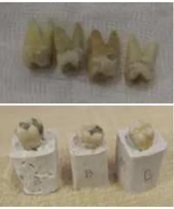
Figure 1: Right; Samples that had been cleaned, disinfected and autoclaved. GIC restoration was placed on the tooth with gross caries on enamel, Left; The teeth that had been mounted into a block made of Plaster of Paris with
sawdust, then labelled to ease the identiication process.
(42%) second canal corresponds to the irst
canal.3 Another study done by Blattner in 2010
has found that as much as 57.9% of secondary MB root canal was presented when using CBCT, while the gold standard was 68.4%.4 This result proved
that the CBCT is nearly as sensitive as the gold standard in detecting the presence or absence of secondary MB root canal. The purpose of this study was to examine the sensitivity and accuracy of the periapical radiograph (PA) and CBCT on determining the presence of the secondary MB root canal.
METHODS
For as much as 40 samples of extracted upper
maxillary irst molars were collected from several
dental clinics around the state of Kelantan. Besides those forty samples, another three samples were also collected and were used as pilot samples. These collected teeth associated with the purpose of the treatment and prevention of disease and not for this research study purpose. The teeth that included were all intact crown with root
structure, either right or left maxillary irst molars were taken as a sample. Any samples with root
caries or root canal treated teeth were excluded
as they might afect the outcome of the results.
in the Dettol® antiseptic solution. The teeth were then cleaned by removing the debris using a supersonic scaler. After that, all teeth that had gross enamel and dentine caries were restored using Glass Ionomer Cement (GIC) to restore the
tooth morphology for easier identiication process
(Figure 1) during radiographic tests. The tooth
blocks were made of the composition of Plaster of
Paris and sawdust to increase the contrast during the radiographic evaluation process.
Digital Periapical radiograph at imaging room Kulliyyah of Dentistry is used in this
research. The irst setup consisted of digital
sensor that was placed directly under the x-ray tube. The x-ray sensor was held by plasticine to avoid the sensor slipped. The x-ray sensor and x-ray tube were placed in the same position for
every PA radiograph evaluation for each tooth.
The blocks were placed directly onto the sensor in
a Buccal-Lingual orientation, and the buccal side of the tooth was facing the tube. Two views were
taken for every PA evaluation, which was buccal view and mesial SLOB (Side Lingual Opposite
Buccal Technique). For every mesial view, the tube was tilted mesially of the mesial side of each tooth (Figure 2a). The images from both buccal
and mesial SLOB technique were then evaluated
and documented (Figure 2b). The presence or absence of secondary MB root canal was noted as to compare in the CBCT radiograph and clinical sectioning.
Each tooth block then was incorporated into a piece of polystyrene block that a hole was made on top of the polystyrene to hold the block. The
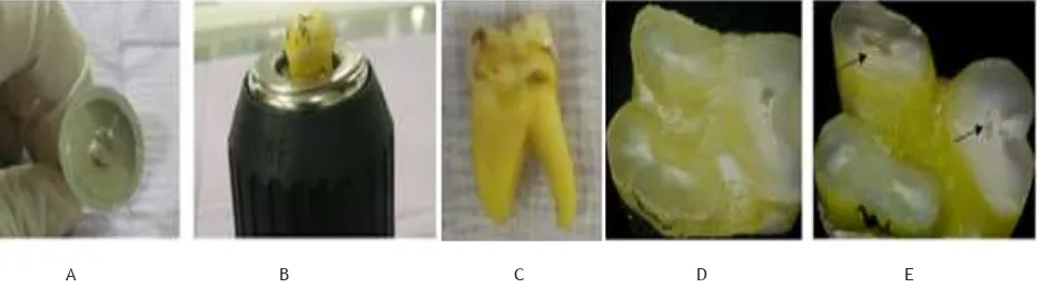
A B C D
Figure 2: (a) Mesial SLOB PA x-ray technique was done on each tooth by tilting the tube mesially, (b) showed the presence of secondary MB root canal detected (red arrows). The secondary MB root canal could be seen well in the Mesial SLOB
technique as compared to Buccal view, (c) The CBCT images that were produced into three diferent planes which were
sagittal, axial, and coronal views.
Figure 3. (a) Diamond disk bur was used for sectioning the samples, (b). The tooth was held during the sectioning process, (c) Tooth that has been immersed in an iodine solution for one minute and then dried before sectioning process, (d) Tooth
sample that showed the presence of Secondary MB Root Canal at 3mm after CEJ level. The arrows showed the irst and
second MB canal. (e) Sectioning was done at 3mm before the apex of the tooth. The arrows indicated the presence of the
irst and second MB canals.
polystyrene also functioned to elevate the tooth
blocks. The polystyrene was placed on top of the
chin rest of the CBCT machine. The machine was
then adjusted to make sure that the tooth was in
the middle position of the mesial crown. A preview
picture was taken to conirm the position of the
sample. The exposure time was minimal, with minimal exposure radiation which was 9 mA. The CBCT evaluations were operated by the students with the supervision from a radiographer in duty
during the evaluation process. The inal image was manipulated using every diferent cross section
planes to evaluate the root and the presence of the secondary MB root canal (Figure 2c). The results were evaluated by the students that later were
checked by the endodontist. The inter-reliability
tests were done between the endodontist and the students for every radiographic evaluation of PA and CBCT of every sample, including the pilot study test.
The samples were then removed from the
blocks and cleaned. The blocks were cut by using
stone trimmer in the laboratory. Every tooth was then cleaned again, and remnants of the Plaster of Paris were removed. Each sample was then placed into the iodine solution for one minute at room temperature, and dried before the clinical sectioning was done (Figure 3c).
Each tooth was then sectioned by using
diamond disk bur and the holder that functioned to
hold the tooth (Figure 3a & 3b). Every tooth was sectioned at the cemento-enamel junction (CEJ), and then 3mm after the CEJ, and 3mm coronal to the apex of the roots. All of the roots that were sectioned were then placed in their consecutive sample container that was initially labelled. The roots were then analyzed under the dental
surgical microscope (Omni Pico®). The number
of the roots present were then determined from the evaluation through a microscope (Figure 3d & 3e). The processes were then repeated for all forty samples.
RESULTS
All 40 samples were evaluated using PA, CBCT, and Clinical Sectioning methods. Periapical Xray (PA) 40 samples, Cone Beam Computed Tomography (CBCT) 40 samples, Clinical Sectioning (gold std) 40 samples.
Data analysis in Table 1, PA radiograph
successfully identiied eight samples presented with
MB2, while the CBCT radiograph evaluation found 24 samples were presented with MB2 canal, and their percentage was 20% and 62.5% respectively. The sensitivity of the tests, when compared to the Gold Standard, was 27.6% and 86.2% for both PA and CBCT respectively. The p-value for the CBCT test compared to CS was 0.00; meanwhile, the p-value for the PA test was 0.08. This result proved that the test from CBCT as compared to the gold standard method was statistically
more signiicant than PA radiograph (p < 0.05).
Speciicity and sensitivity of the test
between PA radiograph and CS were further evaluated using Kappa statistical analysis and the yields from the result were assessed by the
positive predictive value (PPV) and negative predictive value (NPV) from the Chi-Square test.
From Table 2, Kappa value correlation was only 0.173 between these tests. Besides that, it also
showed the reduced speciicity of PA test since the NPV of the PA radiograph was only 34.4%.
Table 1. Sensitivity diferences between PA and CBCT
radiograph in evaluating the presence of MB2 and their respective p-value.
PA CBCT CS - GOLD STANDARD
Has MB2 8 25 29 No MB2 32 15 11 p value 0.08 0.00
Table 2.: Correlation between the PA test and gold standard with their respective Kappa value
PA (in %) CS-GOLD STANDARD (in %) Kappa
Has MB2 8 (27.6%) 29 (72.5%) 0.173
PPV: 100% PPV: 100%
No MB2 32 (72.4%) 11 (27.5%)
NPV: 34.4% NPV: 100%
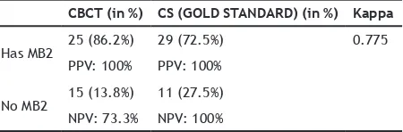
Table 3. Correlation between the CBCT radiograph and gold standard with their respective Kappa value
CBCT (in %) CS (GOLD STANDARD) (in %) Kappa
Has MB2 25 (86.2%) 29 (72.5%) 0.775
PPV: 100% PPV: 100%
No MB2 15 (13.8%) 11 (27.5%)
Data analysis comparing the sensitivity and
speciicity between the CBCT and CS was done by using Kappa and the yields of the result (PPV and NPV) from the Chi-Square test were tabulated in
Table 3. Kappa value between the CBCT and CS
was 0.775, and their NPV was 73.3%.
DISCUSSION
Radiographic imaging can be considered as an
essential investigation, speciically in endodontic
cases as it is an important method in diagnosis, treatment planning and also in for the evaluation after the endodontic treatment. According to the research conducted by Patel in 2007 that was agreed by American Association of Endodontist in 2011, the most outstanding advantage of CBCT is that it produces images in 3D views, the images will be presented into three planes it’s sagittal, axial and coronal, the images also can be zoomed without any distortion.1 Causative of these
factors, this study was aimed to prove that CBCT is more accurate as compared to PA radiograph in identifying the presence of Secondary MB root
canal in maxillary irst molars.
From the research, found that the percentage of secondary MB root canal present by the clinical sectioning evaluation it’s 72.5%, PA 20%, and CBCT 62.5%. Base on the data from the research found that the percentage of the secondary MB root canal evaluation by the CBCT was slightly lower than the literature, which has suggested that the prevalence of Secondary MB root canal in Malay population was 68%.5 However,
the smaller sample size could be the main reason
accounted for this diference. Another relevant
study was done based on the population by Abd Latib et al. in 2015 found that there were 15 canals of Secondary MB root canal detected among the Malay population, while among Chinese
population there were ive canals found and none
was found among Hindi population.6 Besides that,
another study in the prevalence of Secondary MB
root canal in maxillary irst molars by Peiris et al. in 2012 among Sri Lankan population found that
77.1% of the samples presented with the Secondary MB root canal.7 Another study conducted among
the Indonesian population by Peeters et al. found that 68.5% of the total extracted 308 samples has the Secondary MB root canal.8 These percentages
of the presence of Secondary MB root canal
among the Asian showed signiicant value as the
prevalence among these populations were nearly the same.
In addition to the prevalence studies, the
validity of the study also has been conirmed by evaluating the sensitivity and speciicity of
the screening methods. The results from these screening methods were compared to the clinical
sectioning (Gold Standard) indings. However, in terms of speciicity, both CBCT and PA radiograph most high speciicity in detecting the presence of Secondary MB root canal. This was conirmed
by the ability of both methods, as well as the gold standard to successfully detect that 11 true negative samples which absent of Secondary MB root canal.
Although the test by CBCT was not 100% as accurate as the gold standard, this result has proved that CBCT was highly sensitive if compared to PA radiograph in detecting the presence of
Secondary MB root canal. This inding was due
to the higher percentage of true positive results compared to the PA test that was illustrated by the sensitivity of the tests. Besides that, Kappa value also higher in CBCT those values showed the high correlation and reliability of the CBCT more than PA radiograph when compared to the gold standard method. Therefore, the accuracy
of CBCT was conirmed as higher if compared to the PA radiograph as the percentage diference
between CBCT and gold standard was not too
signiicant.
Apart from the sensitivity and speciicity of
the test, the predictive value also was evaluated by the statistics. The positive predictive value
(PPV) and negative predictive value (NPV) were
evaluated as yields of the screening test. Any
evaluation test with high PPV and NPV conirmed
that the test was an ideal test. In this study, both PA and CBCT evaluation test had produced
a very high PPV; it shows in the evaluation of the
present of Secondary MB root canal, PA radiograph
accounted a lower NPV than CBCT.
By all these results, they demonstrated that CBCT has the superiority in the diagnosing the presence of the Secondary MB root canal compared to PA radiograph, this is similar to the research by Blattner et al. in 2010.4 On the other hand, a
that the CBCT has high validation as a method to explore the root morphology, when compared to the histological sectioning.9 They studied on the
ability of the CBCT in reconstruction the root morphology, and their results proved the strong correlations between the CBCT and sectioning with the standard deviation value was 0.163.
The diferences in the percentage of
the correlation between the CBCT and Clinical
Sectioning could be credited to the diferent
number of samples, and the origin of the population itself. This study has been conducted
without any consent taken from the patient as the
tooth collected was anonymous and the races or origin of the tooth could not be determined from the samples collection.
Therefore, further improvement in this study should consider the samples collected from
a speciic population so that the prevalence and
the incidence that certain population could be determined. Another possible factor that could be considered was the gender of the patient from which the samples had been collected because gender predilection could be among the factors.
However, there was no deinite result or studies
conducted. Besides that, an increment in the sample size also should be considered so that higher positive correlation between the CBCT and the gold standard could be anticipated. This was because smaller range of the sample size would limit the accountancy of the presence or absence of the Secondary MB root canal in the overall number of the population’s prevalence.
Apart from all these factors, this study also should consider being conducted on the real patient with the need of endodontic therapy so that a practical application of the research could be optimized. Instead of having the clinical sectioning as the screening test, the usage of the clinical surgical microscope which was currently widely used in endodontic treatment could be used to evaluate the presence of Secondary MB root canal
by using canal patency identiication technique.
CONCLUSION
CBCT was proven to be a reliable method to detect the presence of secondary MB root canals due to its sensitivity and accuracy as high as the clinical sectioning compared to the PA radiograph.
REFERENCES
1. Patel S, Dawood A, Ford TP, Whaites E. The potential applications of cone beam computed tomography in the management of endodontic
problems. Int Endod J. Oct 2007;40(10):818-30. DOI: 10.1111/j.1365-2591.2007.01299.x. 2. Scarfe WC, Levin MD, Gane D, Farman AG.
Use of Cone Beam Computed Tomography in
Endodontics. Int J Dent. Mar 31, 2010;2009: 634567. DOI: 10.1155/2009/634567.
3. Somma F, Leoni D, Plotino G, Grande NM, Plasschaert A. Root canal morphology of the
mesiobuccal root of maxillary irst molars:
a micro-computed tomographic analysis.
Int Endod J. Feb 2009;42(2):165-74. DOI:
10.1111/j.1365-2591.2008.01472.x.
4. Blattner TC, George N, Lee CC, Kumar V,
Yelton CD. Eicacy of Cone-Beam Computed
Tomography as a Modality to Accurately Identify the Presence of Second Mesiobuccal
Canals in Maxillary First and Second Molars: A Pilot Study. J Endod. May 2010;36(5):867-70. DOI: 10.1016/j.joen.2009.12.023.
5. Majeed A, Raad K, Mustafa NS. The accuracy
diference between surgical microscope and CBCT to ind MB2 canal in mesiobuccal root of maxillary permanent irst molar in Malay
population. 39th Annual Conference of the
European Prosthodontic Association; Sep
3-5, 2015; Prague, Czech Republic: European
Prosthodontic Association; 2015.
6. Abd Latib AH, Nordin NF, Alkadhim AH. CBCT Diagnostic Application in Detection of
Mesiobuccal Canal in Mandibular Molars: A Descriptive Study. J Dent Oral Health. June 18, 2015;1(1):1-3.
7. Peiris R. Mesiobuccal Root Canal Morphology of the Permanent Maxillary First Molar in
A Sri Lankan Population. Malay Dent J. 2012;34(2):36-44.
8. Peeters HH, Suardita K, Setijanto D. Prevalence of a second canal in the mesiobuccal root
of permanent maxillary irst molars from an Indonesian population. J Oral Sci. Dec 2011;53(4):489-94.
9. Michetti J, Maret D, Mallet JP, Diemer
F. Validation of Cone Beam Computed
Tomography as a Tool to Explore Root Canal