Indonesian Journal of Biotechnology
VOLUME 22(2), 2017, 98–106 | RESEARCH ARTICLE
The expression of growth factor signaling genes in co-culture IVM
Erif Maha Nugraha Setyawan1,∗, Hyun Ju Oh1, Min Jung Kim1, Geon A Kim1, Seok Hee Lee1, Yoo Bin Choi1, Ki Hae Ra1, and Byeong Chun Lee1
1Department of Theriogenology and Biotechnology, College of Veterinary Medicine, Seoul Na onal University, 1 Gwanak-ro, Gwanak-gu,
Seoul 08826, Republic of Korea
∗Corresponding author:[email protected]
ABSTRACTThe objec ve of this study was to determine the expression of growth factor signaling genes in human adipose-derived stem cells (ASCs), porcine oocytes, and cumulus during in vitro matura on (IVM). The human ASCs (from 2 young and 2 old donors) were used for the co-culture IVM system. The matura on rate was examined based on polar body extrusion. The expression of the growth factor signaling genes from ASCs, oocytes, and cumulus were measured using qPCR. All data were analyzed using ANOVA followed by Tukey’s test. The expression of the h-IGF1 signaling genes from human ASCs cells showed similar values in all groups and the h-FGF2 expressions were higher in the young donors than the old ones. The p-FGF2, p-FGFR2, and p-TGFβ1 expressions in the oocytes as well as p-IGFR in the cumulus that were co-cultured from the young donors showed higher values than the old and control groups. The apopto c ra o (p-BAX/p-BCL2) from the oocytes and cumulus in both co-culture groups also showed lower levels than the control (P<0.05). Oocyte matura on rates were significantly increased in all co-cultured groups (Y1 (85.9 ± 2.2%), Y2 (91.2 ± 1.1%), O1 (86.3 ± 1.5%), and O2 (86.5 ± 2.3%)) compared with the control (76.7 ± 1.1%; P<0.05). Although the expression of growth factor signaling genes was varied, young donors’ ASCs might support in vitro matura on be er than those from old donors.
KEYWORDSco-culture; gene expression; human ASCs; oocytes matura on
1. Introduc on
In vitro maturation (IVM) technique have been essential and important to retrieve high quality mature oocytes for optimum result in the further study, such as embryo pro-duction, xenotransplantation and animal model research (Woods and Tilly 2012;Coticchio et al. 2015;Arat et al. 2016). The oocyte is a unique and highly specialized cell responsible for creating, activating and controlling the em-bryonic genome, as well as supporting basic processes, such as cellular homeostasis, metabolism and cell cycle progression in the early embryo. Oocytes maturation were occurred when the oocytes reached nuclear and cytoplas-mic maturation (Coticchio et al. 2015). Nuclear matura-tion encompasses numerous sequential events, including the breakdown of the germinal vesicle and the resumption of meiosis, the first meiotic division, and the appearance of second metaphase chromosomes. These processes can be evaluated cytologically by the appearance of the first po-lar body or metaphase II chromosomes (Machtinger et al. 2016). Cytoplasmic maturation occurs alongside of nu-clear maturation, involving dramatic changes in oocyte gene expression, protein synthesis, and organelle organi-zation (Woods and Tilly 2012; Machtinger et al. 2016).
Cumulus cells have been considered to play an important role in oocyte maturation by keeping the oocyte under mei-otic arrest, inducing meimei-otic resumption and by support-ing cytoplasmic maturation. These functions have been attributed to their gap junctions and their specific metab-olizing capabilities. Physical contact between oocyte and cumulus cells has been considered necessary for the trans-fer of nutrients and factors essential for oocyte develop-ment (Hull and Harvey 2014).
preimplan-tation development by autocrine stimulation of cumulus and/or granulosa cells in pigs and similar results have been reported in mice and cattle (Arat et al. 2016). Members of the TGFβ superfamily, including TGFβ1, TGFβ3, bone morphogenetic protein 15 (BMP15) and growth differen-tiation factor 9 (GDF9), are expressed in the oocyte from very early stages and are responsible for oocyte matura-tion (da Silveira et al. 2014). Further, few studies have focused on the effects of exposure to a combination of growth factors on oocyte IVM (Arat et al. 2016), but the co-culture system also chosen to support the IVM condi-tion (Saadeldin et al. 2014;Appeltant et al. 2016).
Adipose-derived stem cells (ASCs) The potential can-didate of secrete high levels of several growth factors which have roles on oocytes maturation such as insulin-like growth factor (IGF), basic fibroblast growth factor (bFGF), vascular endothelial growth factor (VEGF), hep-atocyte growth factor (HGF) and transforming growth factor-beta (TGF-β) (Fu et al. 2008; da Silva Meirelles et al. 2009; Overman et al. 2013). ASCs exhibit stable growth and proliferation kinetics and can differentiate to-ward osteogenic, chondrogenic, adipogenic, myogenic, or neurogenic lineages in vitro (Scruggs et al. 2013; Shi-mada et al. 2006) which has potential as cell feeder in co-culture system. But biologic aging also occurs in stem cells which was shown with secrete less growth factors and other bioactive molecules (Scruggs et al. 2013). Those reports showed the important of donor age that should be considered when select ASCs as cell feeder in co-culture system. To gain insight into effectivity of co-culture sys-tem using different donor age of ASCs on porcine IVM, the maturation rate and expression of growth factor signal-ing genes and some receptors on ASCs, oocytes and cumu-lus were evaluated. Our hypothesis was that the activity of ASCs in co-culture IVM system would enhance the inter-action between oocytes and cumulus which increase the oocytes maturation rate.
2. Materials and methods
All chemicals were obtained from Sigma-Aldrich Co. LLC. (St. Louis, USA) unless otherwise stated. The young donors of ASCs came from 2 peoples under 30 years old and the old donors also came from 2 peoples with aged more than 50 years old.
2.1. Isola on and culture of ASCs
The procedure for preparation of human ASCs was per-formed under GMP conditions in the Stem Cell Research Center of R Bio, with approval from the Life Ethics Com-mittee of the Biostar Stem Cell Technology, Seoul, Re-public of Korea (RBIO 2015-12-001). The ASCs were isolated from disposed human adipose tissues obtained from the lower abdomen of patients with their agreement and primary cultured as previously described (Ra et al. 2011). In detail, human subcutaneous adipose tissues were
obtained by simple liposuction from abdominal subcuta-neous fat with informed consent and digested with col-lagenase I (1 mg/mL) under gentle agitation for 60 min at 37°C. The digested tissues were then filtered through a 100-µm nylon sieve. The tissues were centrifuged at 470 g for 5 min and resuspended in Dulbecco’s modified Eagle’s medium (DMEM; Invitrogen)–based medium con-taining 0.2 mM ascorbic acid and 10% fetal bovine serum (FBS). After re-centrifuging at 470 g for 5 min, the su-pernatant was discarded and the cell pellet was collected. These cells were cultured overnight at 37°C under 5% CO2in DMEM-based medium containing 0.2 mM
ascor-bic acid and 10% FBS. The cell medium was changed to Keratinocyte-SFM (Invitrogen)-based medium containing 0.2 mM ascorbic acid, 0.09 mM calcium, 5 ng/mL rEGF and 5% FBS. The cells were maintained for 4–5 d until confluent (passage 0). When the cells reached 90% con-fluency, they were subculture-expanded in Keratinocyte-SFM-based medium containing 0.2 mM ascorbic acid, 0.09 mM calcium, 5 ng/mL rEGF and 5% FBS until pas-sage 3. FBS contaminants were completely removed from cultured MSCs by several washings with PBS and verified through a test of albumin level below the measurement limit using a bovine albumin ELISA kit (Bethyl Labora-tories, Montgogery, TX). The Korea Food and Drug Ad-ministration permitted the use of FBS-eliminated MSCs for clinical study. Aliquots of the ASCs were then tested for cell viability and fungal, bacterial, endotoxin and my-coplasma contamination as demanded by the Code of Fed-eral Regulations, Title 21 (21CFR) before further use. The details of specific standards are found in the 21CFR, Sec-tions 610. There was no chromosomal abnormality in any sample up to passage 12.
2.2. In vitro matura on of porcine oocytes by co-culture with human ASCs
Ovaries were obtained from sows and gilts at a local slaughterhouse and transported to the laboratory in 0.9% NaCl at 25–30°C. Follicular fluid including cumulus– oocyte complexes (COCs) were aspirated from antral fol-licles (3–6 mm in diameter) and washed three times with tissue culture medium-199 (TCM-199)-HEPES (Invitro-gen, Carlsbad, USA) and selected for in vitro matura-tion on the basis of morphological features, i.e., a com-pact multi-layered cumulus mass and a dark, evenly gran-ulated cytoplasm. The COCs were cultured in four-well dishes (50 COCs per well; Falcon, Becton Dickinson Ltd, Plymouth, UK) in basic maturation medium, TCM-199 supplemented with 10 ng/mL epidermal growth fac-tor (EGF), 0.57 mM cysteine, 0.91 mM sodium pyru-vate, 5 µg/mL insulin, 1 µg/mL follicle-stimulating hor-mone (FSH) (Antrin, Teikoku, Japan), and 1% (vol/vol) penicillin-streptomycin (Pen-Strep; Invitrogen) at 39°C in a humidified atmosphere of 5% CO2for 44 h (two stages,
pipetting with 0.1% hyaluronidase in Tyrode’s albumin lactate pyruvate (TALP). Denuded oocytes were examined by microscope free of any attached somatic cells and then were subjected to observe the first polar body extrusion.
Porcine ovaries were obtained from sows at a local slaughterhouse and transported to the laboratory in 0.9% NaCl at 30–35°C within 3 h. The follicular fluid includ-ing cumulus–oocyte complexes (COCs) were aspirated from 3–6 mm diameter follicles with an 18-gauge needle on a 10 mL syringe and washed three times in washing medium containing 9.5 g/L of TCM-199, 2 mM sodium bi-carbonate, 10 mM HEPES, 0.3% polyvinyl alcohol, 5 mM sodium hydroxide and 1% penicillin-streptomycin (Invit-rogen) as previously reported (Jin et al. 2016). COCs were selected on the basis of morphological features: three or more compact multilayers of cumulus cells and homo-geneous cytoplasm. Around 50 60 COCs per well were cultured in IVM medium containing TCM-199 supple-mented with 10 ng/mL Epidermal growth factor (EGF), 0.57 mM cysteine, 0.91 mM sodium pyruvate, 5 μl/mL insulin–transferrin–selenium solution 100X (Invitrogen), 10% porcine follicular fluid, 10 IU/mL equine chorionic gonadotropin (eCG), and 10 IU/mL human chorionic go-nadotropin (hCG) in a 12-well plate (Falcon).
In order to perform co-culture experiments, young and old donor ASCs were used when they had reached about 70% confluency in a 12-well plate with AMSC medium (Rmedica-stemcell, Korea). The medium was replaced with IVM medium when co-culture began. The 12-well plates were supported with 1.0 μm Transwell polyester membrane inserts (Corning Inc., Pittston, USA) to allow mutual communication (Saadeldin et al. 2014) between porcine oocytes and ASCs for a total of 44 h at 39°C in a humidified atmosphere of 5% CO2in IVM medium. The
intercellular communication distance was approximately
2 mm. The COCs were randomly allocated to young or old ASCs co-culture groups on Transwell polyester membrane inserts or cultured in IVM medium only (control group). The control group was maintained under the same condi-tions as the co-culture groups except for the ASCs support. The control and co-culture groups were cultured for 22 h with 10 IU/mL eCG and then washed twice in eCG-free medium. Afterwards, the co-culture cells and/or COCs were cultured for 22 h in IVM medium without eCG. Af-ter 44 h culture for IVM, the COCs were denuded with 0.1% hyaluronidase by gently pipetting and cumulus cells were separated from COCs by centrifugation (2 min, 1,975 g) and the samples were immediately stored at -80°C until used for RNA extraction. For assessing in vitro maturation rates, extrusion of the first polar body (Metaphase II) was evaluated under the stereomicroscope and ASCs from the co-culture groups were retrieved for total RNA extraction.
2.3. Total RNA extrac on and cDNA synthesis
Total RNA was extracted from ASCs (young and old donors), cumulus and oocytes using the Easy-spinTM (DNA-free) Total RNA Extraction Kit (iNtRON Biotech-nology Inc., Kyunggi, Korea) then eluted according to the manufacturer’s protocol. Total RNA concentration was measured using spectrophotometry (NanoDrop 2000, Thermo Fisher Scientific Inc, Waltham, USA), with sam-ples immediately stored at -80°C until cDNA synthesis. Total RNA was reverse transcribed into cDNA using am-fiRivert II cDNA Synthesis Premix (GenDEPOT, Barker, USA) according to the manufacturer’s instructions.
2.4. Gene expressions analysis using real- me poly-merase chain reac on PCR
Quantitative real-time PCR (qPCR) was conducted to as-sess the transcript abundance using oligonucleotide primer
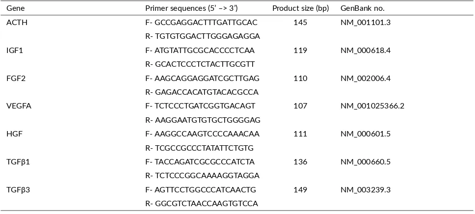
TABLE 1Primer sequences used for gene expression analysis in human.
Gene Primer sequences (5’ –> 3’) Product size (bp) GenBank no.
ACTH F- GCCGAGGACTTTGATTGCAC 145 NM_001101.3
R- TGTGTGGACTTGGGAGAGGA
IGF1 F- ATGTATTGCGCACCCCTCAA 119 NM_000618.4
R- GCACTCCCTCTACTTGCGTT
FGF2 F- AAGCAGGAGGATCGCTTGAG 110 NM_002006.4
R- GAGACCACATGTACACGCCA
VEGFA F- TCTCCCTGATCGGTGACAGT 107 NM_001025366.2
R- AAGGAATGTGTGCTGGGGAG
HGF F- AAGGCCAAGTCCCCAAACAA 111 NM_000601.5
R- TCGCCGCCCTATATTCTGTG
TGFβ1 F- TACCAGATCGCGCCCATCTA 136 NM_000660.5
R- TCTCCCGGCAAAAGGTAGGA
TGFβ3 F- AGTTCCTGGCCCATCAACTG 149 NM_003239.3
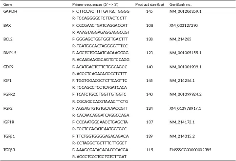
TABLE 2Primer sequences used for gene expression analysis in pigs.
Gene Primer sequences (5’ –> 3’) Product size (bp) GenBank no.
GAPDH F: CTTCCACTTTTGATGCTGGGG 145 NM_001206359.1
R: TCCAGGGGCTCTTACTCCTT
BAX F: CCCGAACTGATCAGGACCAT 108 XM_003127290
R: AAAGTAGGAGAGGAGGCCGT
BCL2 F: GGGAGCTGGTGGTTGACTTT 138 NM_214285
R: TGATGGCACTAGGGGTTTCC
BMP15 F: AGCTCTGGAATCACAAGGGG 123 NM_001005155.1
R: ACAAGAAGGCAGTGTCCAGG
GDF9 F: ACATGACTCTTCTGGCAGCC 140 NM_001001909.1
R: ACCCTCAGACAGCCCTCTTT
IGF1 F: TGGTGGACGCTCTTCAGTTC 145 NM_214256.1
R: TCCAGCCTCCTCAGATCACA
FGFR2 F: TCATCTGCCTGGTTGTGGTC 140 NM_001099924.2
R: CGCAGCCACGTAAACTTCTG
FGF2 F: AGGAGTGTGTGCAAACCGTT 124 XM_013978917.1
R: CACAACAGGATCAGGCCAGA
IGF1R F: CCCAATGGCAACCTGAGCTA 137 NM_214172.1
R: TCCTCGACATCAATGGTGCC
TGFβ1 F: TTCTGGTGGGGAGACAGACA 139 NM_214015.2
R: CCTAGGCTGCTTTCTTGGCT
TGFβ3 F: AAAGCGATACACAGCCACGA 115 ENSSSCG00000002385
R: AGCCTCCCTCCTGTCTTGAT
sequences in human and porcine. The primers for hu-man (BAX, BCl2, IGF1, FGF2, VEGFA, HGF, TGFβ1, TGFβ3 and β-actin) and porcine (BAX, BCl2, BMP15, GDF9, IGF1, IGF1R, FGF2, FGFR2, TGFβ1, TGFβ3 and GAPDH) genes were designed from sequences which ob-tained from NCBI; all primer sequences were standard-ized using a standard curve and are listed in Tables 1
and 2. Real-time PCR was performed using an ABI 7300 Real Time PCR System (Applied Biosystems, Fos-ter City, USA) according to the manufacturer’s instruc-tions with minor modification. The total volume PCR re-action mixture was 20 μL in a real-time PCR plate (Mi-croAmp optical 96-well reaction plate, Singapore) and the mixture was composed of 2 μL cDNA, 0.4 μL for-ward primer, 0.4 μL reverse primer, 10 μL SYBR Green interaction dye (Takara Bio USA Inc., Mountain View, USA) and 7.2 μL diethyl pyrocarbonate. The expres-sion of each target gene was quantified relative to that of the internal control gene (ACTB) using the equation
R = 2−[∆Ct sample−∆Ct control], as previously described
bySetyawan et al.(2016).
2.5. Sta s cal methods
All data were analyzed by one-way ANOVA then fol-lowed by Tukey’s multiple comparison test using Graph-Pad Prism 5.0 (GraphGraph-Pad, San Diego, USA) (Setyawan
et al. 2016). Values are means ± standard error of the mean. Probability values less than 0.05 were considered to be sta-tistically significant.
3. Results
3.1. The expression of signaling genes in human ASCs
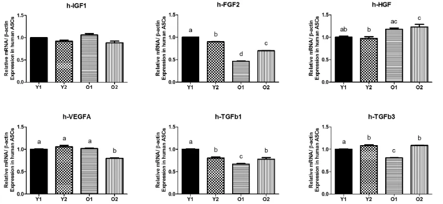
The IGF1, FGF2, VEGFA, HGF, TGFβ1 and h-TGFβ3 gene expression levels in four groups of human ASCs were analyzed (Y1, young donor 1; Y2, young donor 2; O1, old donor 1 and O2, old donor 2). There was no significant difference in levels of expression in IGF1 among the all groups (Figure 1). The Y1 and Y2 groups showed higher FGF2 gene expression levels com-pared with the O1 and O2 groups but HGF levels seemed higher in O2 than Y1 and Y2. The O2 expressed the lowest expression in VEGF than other groups. The O1 showed the lowest expression in TGFβ1 and TGFβ3 compared with the other three groups.
3.2. The expression of genes related to oocytes matu-ra on in porcine cumulus cells
FIGURE 1The expression of growth factor signaling genes from human adipose-derived stem cells (ASCs). Y1, young donor 1; Y2, young donor 2; O1, old donor 1; O2, old donor 2.
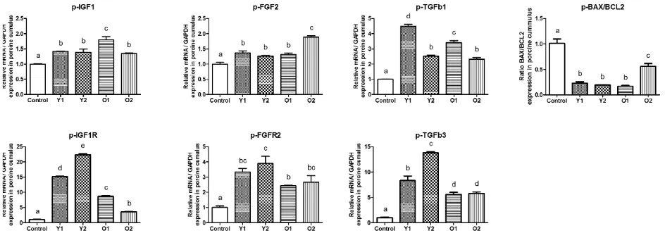
ratio of apoptotic gene expression (p-BAX / p-BCL2) in co-cultured groups showed significantly lower than con-trol. The co-cultured groups also significantly up regu-lated p-IGF1, p-IGF1R, p-FGF2, p-FGFR2, p-TGFβ1 and p-TGFβ3 expression compared with control group. The old donor groups showed lower expression in p-IGF1R and p-TGFβ3 than young donor groups. The other gene expressions among co-cultured groups exhibited different levels but still higher than control (Figure2).
3.3. The expression of genes related to matura on in porcine oocytes
The relative gene expression levels of SMAD2/3 and GDF9 in matured porcine oocytes derived from each group were analyzed. The expression of p-IGF1 among co-cultured groups were not different but significantly higher than control (P>0.05). Its receptor (p-IGF1R) also showed higher expression in co-cultured group than con-trol but exhibited different level of expression among co-cultured groups (Figure3). The expression of p-FGF2 and p-FGFR2 showed similar pattern that the significant differ-ences only found in young groups (Y1 and Y2) but there were no different between O1, O2 and control groups. The high expression in young groups were also showed in p-TGFβ1 which significantly different with old groups and all co-cultured groups were higher than control. Inter-estingly, the expression of p- TGFβ3 in control and co-cultured groups were not significantly different.
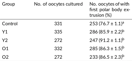
3.4. The effect of human ASCs co-culture on porcine oocyte matura on
The effects of ASCs co-culture on the nuclear maturation of porcine oocytes was observed as shown in Table3. The nuclear maturation rates in young ASCs groups (Y1 = 85.9 ± 2.2% and Y2=91.2 ± 1.1%) and old ASCs groups (O1
= 86.3 ± 1.5% and O2 = 86.5 ± 2.3%) were significantly higher than those in the control group (76.7 ± 1.1%, p < 0.05). There were not differences between Y1, Y2, O1 and O2 groups in the oocyte maturation rate.
4. Discussion
Optimum condition of IVM system is required for suc-cessful and reliable oocyte maturation which would sig-nificantly improve the effectiveness of further develop-ment. Several studies have indicated that growth factors are beneficial for oocyte maturation in vitro because of the paracrine pathway (Shimada et al. 2006). Aging is known to have a negative impact on the regenerative capacity of most tissues and human ASCs are susceptible to biologic aging (Wu et al. 2013), including changes in differentia-tion potential, proliferadifferentia-tion ability, and gene expression (Pandey et al. 2011). Herein we demonstrated that the expression of growth factors signaling gene in co-culture system using young and old donor of human ASCs during IVM could improve the porcine oocyte maturation.
The expression of signaling genes in human ASCs (Figure1) were in line with the expression in human and murine bone marrow derived stem cells that older donor MSCs have decreased gene expression compared with younger donor (Wilson et al. 2010). Higher expression of HGF in O2 and similar expression value in some groups expressed that there were individual variation between donors but mostly younger donor seems a little higher than older donor. Those various different expression in RNA levels between young and old ASCs were also reported by
FIGURE 2The expression of growth factor signalling genes from porcine oocytes a er in vitro matura on (IVM). Control, without human adipose-derived stem cells (ASCs); Y1, young donor 1; Y2, young donor 2; O1, old donor 1; O2, old donor 2.
FIGURE 3The expression of growth factor signaling genes from porcine cumulus a er in vitro matura on (IVM). Control, without human adipose-derived stem cells (ASCs); Y1, young donor 1; Y2, young donor 2; O1, old donor 1; O2, old donor 2.
and cumulus in order to enhance nuclear and cytoplasmic maturation in oocytes (Appeltant et al. 2015). The exoge-nous growth factors addition also reported in rats which affected in modulating cumulus cell expansion, stimulat-ing proliferation and inhibitstimulat-ing apoptosis (Arat et al. 2016). The exact role of cumulus during the initiation of meiotic resumption in pigs has been a matter of debate that the signal coming from cumulus as a key factor for triggering meiosis in oocytes (Gilchrist 2011) and others suggested that a cumulus-secreted soluble factor(s) may be responsi-ble for the reinitiation of nuclear progression in oocyte dur-ing IVM (Appeltant and others 2015). Our result revealed that, the higher activity of cumulus that showed by higher gene expression in co-culture IVM system with any donor age of ASCs could support oocytes maturation more than control group. Those high activities could be supported by growth factors and other bioactive molecules from ASCs via extracellular vesicles (Machtinger et al. 2016).
The growth factors from human ASCs played many roles in oocytes maturation and the high expression of those signaling gene in oocytes (Figure 3) showed their activities in several pathways. citemachtinger2016 re-ported that growth factors could escalate oocyte matu-ration in mammals by accelerate several mechanism re-lated with wingless signaling pathway (WNT),
transform-ing growth factor beta (TGFb), mitogen-activated protein kinase (MAPK), neurotrophin, epidermal growth factor re-ceptor (ErbB) pathways and ubiquitin-mediated pathways (Hull and Harvey 2014). Our result revealed that the per-centage of oocytes reached the MII stage under the co-culture IVM system with any donor age of ASCs showed a significantly higher compared with control. Those re-sult suggested that the growth factors and other bioactive molecules from ASCs supported cumulus and also directly to oocytes activities which would give the optimum meio-sis and nuclear progression in oocytes during IVM.
TABLE 3Evalua on of in vitro matura on rate of porcine oocytes co-cultured with adipose-derived stem cells (ASC) young and old donors.
Group No. of oocytes cultured No. oocytes of with first polar body
ex-a, bWithin a column, values with different superscript le ers are significantly different (p < 0.05). Experiment was replicated at least seven mes.
In the present study, the contributions of young and old donor of ASCs to oocyte maturation and gene expression were analyzed under same culture conditions between co-culture groups with control. Our study demonstrated that co-culturing with young and old donor of ASCs were es-sential for oocytes to achieve higher nuclear and cytoplas-mic maturation rates, and this would allow their develop-ment competence to improve in the next stages.
The expression of IGF1 in co-culture groups were sig-nificantly increased when compared with control but there were no significant different between treatment groups. The respond in oocytes showed higher level than in cu-mulus compared with control after co-culture with ASCs (Figure1). The IGF1R expression in oocytes and cumu-lus also showed the similar pattern that co-culture groups expressed more than control but cumulus showed higher expression than oocytes compare with each control. Inter-estingly, Young donor (Y1 and Y2) resulted higher level than old donor (O1 and O2) which were showed in Figure
2and Figure3. These result demonstrated that COCs have more IGF1 and IGF1R activities during co-cultured with human ASCs which could support maturation rate. This mechanism already reported that IGF1 have an important role in the nuclear maturation and cytoplasmic maturation of oocytes, exert its effect through cumulus cells by regula-tion of porcine cumulus proliferaregula-tion and suppressed apop-tosis (Toori et al. 2014). The IGF1R is activated by locally-produced or circulating IGF1 which leads to autophospho-rylation of the tyrosine kinase domain, with ensuing ac-tivation of the Ras-Raf-MAP kinase and PI3K-PKB/Akt signaling pathways and exert its mitogenic and antiapop-totic activities (Bentov and Werner 2011).
The FGF2 and FGFR2 expressions from oocytes and cumulus in all treatment groups were significantly higher than each control group. Young donor in oocytes showed more expression value of FGF2 and FGFR2 compared with both expressions in cumulus (Figure3). Higher ex-pression also founded in BPM15 and GDF9 from oocytes and only GDF9 showed that young donor expressed higher level than old donor. The expression of FGF2 and FGFR2
in all treatment groups from cumulus also showed signif-icant difference compared to each control group. Those growth factors and receptors together modulated the mat-uration process in IVM. The FGF2 activity enhances the maturation of mammalian cumulus-oocyte complexes (COCs) by increasing the expression of cumulus cell expansion-related genes in pigs (Son et al. 2017). The FGF2 receptors (FGFR2) can be utilized between cumu-lus cells and oocytes (Pomini Pinto et al. 2015). BMP15 and GDF9 plays a role in oocyte maturation by bind-ing with various TGF-beta receptors leadbind-ing to activate MAPK pathways for cumulus expansion/oocyte activa-tion and FGF) cooperate with BMP15 promote glycoly-sis in cumulus cell (Son et al. 2017). BMP15 belongs to the TGF-β superfamily and is known as a granulosa cell mitosis and proliferation inducer. The production of ade-quate amounts of BMP15 protein by the oocyte is neces-sary to promote cumulus cells expansion. The expression of BMP15 mRNA was also found in cumulus cells and it decreased along with the oocyte maturation and cumulus expansion in vitro (Bogacki et al. 2014).
The TGFβ1 in oocytes showed that young donor ex-pressed higher value than old donor groups and exhibited various expression in cumulus but still higher than control group. The expression of TGFβ3 in oocytes (Figure2) showed similar value but the high respond was founded in cumulus (Figure 3) that young donor group exhibited significant different compared with old donor groups and all treatment groups had significant different with control group. Both growth factors could activate MAPK path-ways which crucially involved in the processes of oocyte development and TGFβ1 have more influence than TGFβ3 in cumulus expansion and oocytes activation (Machtinger et al. 2016). Hepatocyte growth factor (HGF) promote anti-apoptotic on porcine cumulus cells (Uzumcu et al. 2006). Vascular endothelial growth factor A (VEGFA) improves quality of matured porcine oocytes (Arat et al. 2016) and contributes in regulation of crucial processes for prematurasional growth, differentiation, and develop-ment of oocytes (Kranc et al. 2017).
The co-culture system can create a microenvironment through secretion of autocrine and paracrine factors to the media (Saadeldin et al. 2014). In this study, we found that co-culture with human ASCs can support and improve the oocytes maturation when compared to the control group. Even though there were various expression in growth fac-tor signaling genes between young and old donor ASCs, the growth factor secretions from both donors could in-crease the mRNA profiles in oocytes and cumulus which would contribute in nuclear and cytoplasmic maturation.
5. Conclusions
control group indicated that the co-culture IVM system us-ing human ASCs could enhance the interaction between oocytes and cumulus which increase the oocytes matu-ration rate. The expression of growth factors signaling genes from young and old donor ASCs seems various be-tween samples and not affect the maturation rate among co-culture treatment groups. There is a critical need for further studies in human ASCs to provide conclusive ex-perimental evidence in cellular communication mediated by extracellular vesicles which might be contributed to oocyte maturation and embryo development.
Acknowledgments
This study was supported by Korea IPET (#114059-03-3-SB010), Nature Cell (#550-20150030), Research Institute for Veterinary Science, and the BK21 plus program.
Authors’ contribu ons
EMNS, HJO, MJK, BCL designed the study. EMNS, SHL, YBC, KHR carried out the laboratory work. EMNS, MJK, GAK, BCL analyzed the data. PK, BTF, GH, MJ, DW wrote the manuscript. All authors read and approved the final version of the manuscript.
Compe ng interests
None of the authors have any competing interests.
References
Appeltant R, Somfai T, Kikuchi K, Maes D, Van Soom A. 2016. Influence of co-culture with denuded oocytes during in vitro maturation on fertilization and developmental competence of cumulus-enclosed porcine oocytes in a defined system: co-culture with denuded oocytes. Anim Sci J. 87(4):503–510. doi:10.1111/asj.12459.
Appeltant R, Somfai T, Nakai M, Bodó S, Maes D, Kikuchi K, Van Soom A. 2015. Interac-tions between oocytes and cumulus cells during in vitro maturation of porcine cumulus-oocyte com-plexes in a chemically defined medium: effect of denuded oocytes on cumulus expansion and oocyte maturation. Theriogenology 83(4):567–576. doi:10.1016/j.theriogenology.2014.10.026.
Arat S, Caputcu AT, Cevik M, Akkoc T, Cetinkaya G, Bagis H. 2016. Effect of growth factors on oocyte maturation and allocations of inner cell mass and tro-phectoderm cells of cloned bovine embryos. Zygote 24(04):554–562. doi:10.1017/S0967199415000519. Bentov Y, Werner H. 2011. IGF1R (insulin-like growth
factor 1 receptor). Atlas Genet Cytogenet Oncol Haematol. (8). doi:10.4267/2042/44533.
Bogacki M, Wasielak M, Kitewska A, Bogacka I, Jalali B. 2014. The effect of hormonal estrus induction on ma-ternal effect and apoptosis-related genes expression in
porcine cumulus-oocyte complexes. Reprod Biol En-docrinol. 12(1):32. doi:10.1186/1477-7827-12-32. Coticchio G, Dal Canto M, Mignini Renzini M, Guglielmo
MC, Brambillasca F, Turchi D, Novara PV, Fadini R. 2015. Oocyte maturation: gamete-somatic cells in-teractions, meiotic resumption, cytoskeletal dynamics and cytoplasmic reorganization. Hum Reprod Update 21(4):427–454. doi:10.1093/humupd/dmv011. da Silva Meirelles L, Fontes AM, Covas DT,
Ca-plan AI. 2009. Mechanisms involved in the therapeutic properties of mesenchymal stem cells. Cytokine Growth Factor Rev. 20(5-6):419–427. doi:10.1016/j.cytogfr.2009.10.002.
da Silveira JC, Carnevale EM, Winger QA, Bouma GJ. 2014. Regulation of ACVR1 and ID2 by cell-secreted exosomes during follicle maturation in the mare. Re-prod Biol Endocrinol. 12(1):44. doi: 10.1186/1477-7827-12-44.
Fu X, He Y, Xie C, Liu W. 2008. Bone marrow mes-enchymal stem cell transplantation improves ovar-ian function and structure in rats with chemotherapy-induced ovarian damage. Cytotherapy 10(4):353–363. doi:10.1080/14653240802035926.
Gilchrist RB. 2011. Recent insights into oocyte - follicle cell interactions provide opportunities for the develop-ment of new approaches to in vitro maturation. Reprod Fertil Dev. 23(1):23. doi:10.1071/RD10225.
Hull KL, Harvey S. 2014. Growth hormone and reproduction: A review of endocrine and au-tocrine/paracrine interactions. Int J Endocrinol. 2014:1–24. doi:10.1155/2014/234014.
Jin JX, Lee S, Khoirinaya C, Oh A, Kim GA, Lee BC. 2016. Supplementation with spermine during in vitro maturation of porcine oocytes improves early embry-onic development after parthenogenetic activation and somatic cell nuclear transfer1. J Anim Sci. 94(3):963– 970. doi:10.2527/jas.2015-9761.
Kranc W, Budna J, Chachuła A, Borys S, Bryja A, Ryb-ska M, Ciesiółka S, Sumelka E, Jeseta M, Brüssow KP, Bukowska D, Antosik P, Bruska M, Nowicki M, Zabel M, Kempisty B. 2017. “Cell migration” is the ontology group differentially expressed in porcine oocytes before and after in vitro maturation: a mi-croarray approach. DNA Cell Biol. 36(4):273–282. doi:10.1089/dna.2016.3425.
Machtinger R, Laurent LC, Baccarelli AA. 2016. Extra-cellular vesicles: roles in gamete maturation, fertiliza-tion and embryo implantafertiliza-tion. Hum Reprod Update 22(2):182–193. doi:10.1093/humupd/dmv055. Overman J, Helder M, ten Bruggenkate C, Schulten E,
Pandey AC, Semon JA, Kaushal D, O’Sullivan RP, Glowacki J, Gimble JM, Bunnell BA. 2011. Mi-croRNA profiling reveals age-dependent differential expression of nuclear factor κB and mitogen-activated protein kinase in adipose and bone marrow-derived hu-man mesenchymal stem cells. Stem Cell Res Ther. 2(6):49. doi:10.1186/scrt90.
Pereira G, Lorenzo P, Carneiro G, Ball B, Bilodeau-Goeseels S, Kastelic J, Pegoraro L, Pimentel C, Esteller-Vico A, Illera J, Granado G, Casey P, Liu I. 2013. The involvement of growth hor-mone in equine oocyte maturation, receptor local-ization and steroid production by cumulus–oocyte complexes in vitro. Res Vet Sci. 95(2):667–674. doi:10.1016/j.rvsc.2013.06.024.
Pomini Pinto RF, Fontes PK, Loureiro B, Sousa Castilho AC, Sousa Ticianelli J, Montanari Razza E, Satrapa RA, Buratini J, Moraes Barros C. 2015. Effects of FGF10 on bovine oocyte meiosis progression, apop-tosis, embryo development and relative abundance of developmentally important genes in vitro. Reprod Domest Anim. 50(1):84–90. doi:10.1111/rda.12452. Ra JC, Shin IS, Kim SH, Kang SK, Kang BC, Lee
HY, Kim YJ, Jo JY, Yoon EJ, Choi HJ, Kwon E. 2011. Safety of intravenous infusion of human adi-pose tissue-derived mesenchymal stem cells in ani-mals and humans. Stem Cells Dev. 20(8):1297–1308. doi:10.1089/scd.2010.0466.
Saadeldin IM, Kim SJ, Choi YB, Lee BC. 2014. Improvement of cloned embryos development by co-culturing with parthenotes: a possible role of exosomes/microvesicles for embryos paracrine communication. Cell Reprogram 16(3):223–234. doi:10.1089/cell.2014.0003.
Scruggs BA, Semon JA, Zhang X, Zhang S, Bowles AC, Pandey AC, Imhof KM, Kalueff AV, Gimble JM, Bunnell BA. 2013. Age of the donor reduces the ability of human adipose-derived stem cells to alle-viate symptoms in the experimental autoimmune en-cephalomyelitis mouse model. Stem Cells Transl Med. 2(10):797–807. doi:10.5966/sctm.2013-0026. Setyawan EMN, Kim MJ, Oh HJ, Kim GA, Jo YK,
Lee SH, Choi YB, Lee BC. 2016. Spermine re-duces reactive oxygen species levels and decreases cryocapacitation in canine sperm cryopreservation. Biochem Biophys Res Commun. 479(4):927–932. doi:10.1016/j.bbrc.2016.08.091.
Shimada M, Hernandez-Gonzalez I, Gonzalez-Robayna I, Richards JS. 2006. Paracrine and autocrine regulation of epidermal growth factor-like factors in cumulus oocyte complexes and granulosa cells: key roles for prostaglandin synthase 2 and proges-terone receptor. Mol Endocrinol. 20(6):1352–1365. doi:10.1210/me.2005-0504.
Son YJ, Lee SE, Hyun H, Shin MY, Park YG, Jeong
SG, Kim EY, Park SP. 2017. Fibroblast growth factor 10 markedly improves in vitro maturation of porcine cumulus-oocyte complexes. Mol Reprod Dev. 84(1):67–75. doi:10.1002/mrd.22756.
Toori MA, Mosavi E, Nikseresht M, Barmak MJ, Mah-moudi R. 2014. Influence of insulin-like growth factor-I on maturation and fertilization rate of imma-ture oocyte and embryo development in NMRI mouse with TCM199 and α-MEM medium. J Clin Diagn Res. 8(12):AC05–08. doi:10.7860/JCDR/2014/9129.5242. Uzumcu M, Pan Z, Chu Y, Kuhn PE, Zachow R. 2006. Immunolocalization of the hepatocyte growth factor (HGF) system in the rat ovary and the anti-apoptotic effect of HGF in rat ovarian granu-losa cells in vitro. Reproduction 132(2):291–299. doi:10.1530/rep.1.00989.
Wilson A, Shehadeh LA, Yu H, Webster KA. 2010. Age-related molecular genetic changes of murine bone mar-row mesenchymal stem cells. BMC Genomics 11:229. doi:10.1186/1471-2164-11-229.
Woods DC, Tilly JL. 2012. The next (re)generation of ovarian biology and fertility in women: is current sci-ence tomorrow’s practice? Fertil Steril. 98(1):3–10. doi:10.1016/j.fertnstert.2012.05.005.