Citation: Mohammed SY, Nimir AHH and Ahmed AA. Identiication of Fungi from Police Dogs in Borri, Khartoum,
Sudan. Austin J Vet Sci & Anim Husb. 2017; 4(1): 1033. Austin J Vet Sci & Anim Husb - Volume 4 Issue 1 - 2017
ISSN: 2472-3371 | www.austinpublishinggroup.com Ahmed et al. © All rights are reserved
Austin Journal of Veterinary Science &
Animal Husbandry
Open Access
Abstract
The dermatophytes are a group of closely related fungi that have the capacity to invade keratinized tissue (skin, hair, and nails) of humans and other animals to produce an infection, dermatophytosis, commonly referred to as ringworm.
Dogs can suffer a dermatophyte infection at any age, but a ringworm infection is more frequent in the young. Microsporum canis is considered highly contagious and potentially patho genic for people. Cats are considered the reservoirs of M.canis. Isolation and identiication of fungi in German Shepherd dogs have not been reported, yet. In this study (81) hair samples, (2) skin scrapings were collected from apparently healthy and clinically infected dogs of German-Shepherd dogs breed, of both sexes and different ages. The skin samples were taken from two dogs with cutaneous lesions. Determinations of dermatophytes, as well as the possible involvement of other fungi in dermatomycosis in dogs were studied. Mycological investigations were conducted by direct microscopy and by fungal culture on Sabouraud Dextrose Agar supplemented with 0.05% Chloramphenicol and 0.5% Cycloheximide to study the presence of fungi based on the colonial morphology and pigmentation. Two hair samples (2.40%) yielded growth suggestive of Dermatophytes; 76 hair samples (91.5%) yielded growth of non-dermatophyte fungi (Aspergillus, Penicillium and Alternaria ), while 5 samples (6.02%) - three hair samples and 2 skin scrapings did not show any fungal growth. The cultures from the two hair samples which were positive for Dermatophytes gave pure cultures of Microsporum canis and M.gypseum. Microsporum canis was isolated from one hair sample (1.2%) and so was M.gypseum. Along with Dermatophytes, saprobic fungi were the most isolated fungi in this study especially Aspergillus spp(72%), Penicillium spp(12%) and Altenaria spp (7.2%). The study reveald that pathogenic Dermatophytes, in addition to saprobic fungi, may be the causative agents of Dermatophytosis (Ringworm) in Police dogs department.
Keywords: Dermatophytes; German shepherd; Ringworm
are T.mentagrophytes and M.gypseum. hese three species comprise approximately 96% of the isolated dermatophytes from dogs.
In this present study we obtained hair and skin scraping for mycological investigation to perform the most frequent dermatophytes existed in dogs.
Ma te ria ls a n d Me th o d s
Animal of study
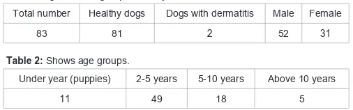
his study included 81 healthy dogs 2 with dermatitis. he samples were collected between January 2011 and May 2011 with collaboration of veterinary clinic located in Borri-Khartoum-Sudan. Breed was German shepherd dogs. Age sex and groups and clinical manifestation were arranged in Table 1 and 2.
Collection of samples
he dogs with lesions were sampled by plucking hairs with sterile forceps and by scraping epidermal scales with a sterile surgical blade from the afected areas. he samples from each dog were placed in separate sterile Petri dishes. Each animal (with or without lesions) was sampled by brushing its fur with a sterilized nailbrush using the following standard protocol. he animals were brushed from
In tro d u ctio n
Dermatophytosis is the most common fungal infections in dogs [1,2]. he dermatophytes have a high ainity for keratin, an important component of fur, skin and nails, which are the primary sites of fungal infection [3].
Several reports have stated that Microsporum canis, a typical zoophilic species, is the most common dermatophyte isolated from dogs worldwide [1,4-6].
Epidemiological studies on the isolation of dermatophytes from dogs with suspected lesions of dermatophytosis have been reported by diferent authors [7,8]. he proportion of positive samples in relation to the number of samples examined from cases of Dermatophytosis varied considerably from one study to another. he relatively low prevalence of Dermatophytes in dogs with suspected lesions of Dermatophytosis is well documented. It ranges between 4% and 10% and few studies show higher prevalence [8,9].
With few exceptions, M.canis was the most common species isolated, showing a high variability in its percentages of isolation (40-90%). Other dermatophytes less commonly isolated from dogs
Research Article
Identiication of Fungi from Police Dogs in Borri,
Khartoum, Sudan
Mo h a m m e d S Y1, N im ir AH H2 a n d Ah m e d AA3*
1Department of Veterinary Hospital, Ministry of Animal
Resources, South Darfur State, Sudan
2Department of Microbiology, Faculty of Veterinary
Medicine, University of Khartoum, Sudan
3Department of Physiology and Biochemistry, Faculty of
Veterinary Science, University of Nyala, Sudan
*Co rre s p o n d in g a u th o r: Abdelkareem Abdallah
Ahmed. Department of Physiology and Biochemistry, Faculty of Veterinary Science, University of Nyala, Nyala, P.O Box: 155 Nyala, Sudan
Austin J Vet Sci & Anim Husb 4(1): id1033 (2017) - Page - 02
Ahmed AA Au s tin Pu blis h in g Gro u p
Submit your Manuscript | www.austinpublishinggroup.com
the head down to the tail and down the lanks and the legs. Ater specimen collection, the nailbrushes were placed in sterile Petri dishes for transport to the laboratory. Samples collected by brushing were inoculated on plates of Sabouraud dextrose agar (Difco) with chloramphenicol (400 ppm) (sca) supplemented with 0.5% cycloheximide.
Plucked hairs and scraped scales, originating from dogs with lesions, were examined for fungal elements by direct microscopy in 20% potassium hydroxide, covered with cover-slip and heated mildly above the lame (3min), ater 10min they were diagnosed microscopically by potassium hydroxide mounts and by culture on the above mentioned media. he plates were incubated at 28°C and regularly examined for a month. Taxonomic identiication of all mycelial colonies considered diferent was based on thorough macroscopic and microscopic studies. Shape dimensions, arrangement of macroconidia and other parts of isolated cultures. Suspected dermatophytes were identiied to species level, and most of the remaining fungi were identiied to genus level.
Potassium hydroxide mounts
A drop of 20% KOH was added on a clean glass slide, the sample (hair and skin) was placed in KOH drop and slide passed through a burner lame to hasten keratolysis. When keratolysis sotened the sample, a clean glass cover slip was slowly placed at inclined angle on the sample and slightly pressed, preventing the formation of the air bubbles. he sample was kept in 20% KOH for a variable duration ranging from 5 minutes to 20 minutes, depending upon the thickness of the scales and examined every 5 minutes. Each slide was thoroughly examined for the presence of ilamentous, septate, branched hyphae with or without arthrospores crossing the margins of the squamous epithelial cells of the skin, in case of hair, type and arrangement of the spore was noticed.
Processing of samples
he skin scraping and the hair were collected in the sterile plastic Petri-dishes to prevent contamination in transit to the laboratory.
he samples were kept at temperature in the laboratory room. he agar plates were arranged around the lame, and then with help of inoculation straight wire all the agar plates were inoculated.
Agar plates were then inoculated upside down at 28°C degrees centigrade for 4 weeks before each was discarded.
Agar plates were frequently removed from incubator and observed (checked) for fungal (i.e. dermatophytes) growth.
Fungal (dermatophyte) growth was checked for colonial characteristics and pigmentation.
Results were recorded straight on the data sheet.
Inoculation of culture media
prior to inoculation , the media were dried in oven for about 15 minutes, ater which the plates were arranged around the lame and lamed straight wire was used to inoculate all the Petri dishes aseptically in order to prevent contamination of cultures, specimens and safety of personal as well as the environment. All sterile technique measures were strictly observed.
he temperature of the incubator was adjusted at 28°C (degrees centigrade) where all the cultures were maintained for one month before they are discarded or subcultured.
Microscopic examination procedure
During microscopic procedure two diferent techniques were used for the determination of fungal morphology, spores, shapes, size, chlamydospores and irregular hyphae.
Needle mount technique
Clean sterile glass slide and cover slip lactophenol cotton blue (LPCB) stain.
Positive pure agar plates were arranged around the lame on the work bench.
Needle wire was sterilized by heating until it becomes hot red.
Few drops of lactophenol cotton blue (LPCB) stain was added onto glass slide.
Needle wire was used to pick few colonies from the culture plates then placed onto glass slide with lactophenole cotton blue (LPCB) stain.
he cover slip was gently placed at inclined angle on the glass slide, in order to prevent the formation of air bubbles.
Microscopic observation was carried out using l ower power of 10x and 40x objectives to determine the colony morphology, size,
shape, chlamydospores and irregular hyphae.
Result was inserted straight onto data sheet.
Slide culture technique
About 6-8 mm square block of Sabouraud dextrose agar (SDA) medium was on to a sterile slide and sub-inoculated at four sides with the pure culture, the inoculated block is then covered with sterile cover glass and placed in a petri dish supported on glass rod. Small amount of sterile water was added to a ilter paper at the bottom of dish to that prevented drying of the agar media. hen the preparation was for a week at 28°C ater which the cover slip was removed and mounted in the drop of lactophenol cotton blue (LPCB).
he block was then discarded into disinfectant. Few drop of lactophenol cotton blue (LPCB) was added on to the growth on the slide and it was covered with a cover glass. Where both preparations were then examined microscopically using the 10x and 40x objectives with the condenser iris diaphragm adjusted to give maximum contrast.
Re s u lts
he study was carried out on 83 dogs in the police dogs Department in Borri-Khartoum-Sudan. Dogs were examined to detect dogs with
Total number Healthy dogs Dogs with dermatitis Male Female
83 81 2 52 31
Table 1: Age sex and groups of study.
Under year (puppies) 2-5 years 5-10 years Above 10 years
11 49 18 5
Austin J Vet Sci & Anim Husb 4(1): id1033 (2017) - Page - 03
Ahmed AA Au s tin Pu blis h in g Gro u p
Submit your Manuscript | www.austinpublishinggroup.com
suspected Dermatophytosis. Only a few dogs (n=2) presented skin lesions with focal or difuse hair loss and/or scaling. A representative number of dogs without visible skin lesions (n=81), were used to carry out the Dermatophyte of their fur. he approximate age was between (1-12) years, breed was German-Shepherd and genders of the dogs were male (70) and female (13).
Out of (83) samples (76) of hair samples (91.5%) yielded positive growth for fungal colony, while (5) samples (6%) - 3 hair samples and 2 skin scrapings were negative for fungal growth in (SDA) with Cycloheximide. he species isolated were from hair samples, and were able to grow in (SDA) supplemented with (0.05%) Chloramphenicol and (0.5% Cycloheximide) at 28°C. he isolated species were in two hair samples Microsporum-canis was in (one samples), and M.gypseum (one sample).
Microscopic diagnostic results were positive for 2 samples (2.40%). and negative result were 76 samples (91.5%) from the number of the colonies tested, while 5 samples (6%) didn’t show any fungal growth.
he most common fungi isolated from skin of dogs with susceptible Mycoses were saprobe, especially Aspergillus spp (72%), Penicillium spp (12%), and Altenaria spp (7.2%). Cultural Characteristic of Isolated Species is arranged in Table 3.
D is cu s s io n
Most cases of der matophytosis in pet are caused by Microsporum canis. According to Kaplan and Ivens (1961) cats are considered to be reservoirs of infection.
In this study we investigate the prevalence of suspected Dermatophytosis in the sample of clinically examined dogs (n=83), only 2 samples (2.40%) had visible skin lesions but none of them had a dermatophytosis.
Of the 81 samples from dogs without visible lesions, 2 animals (2.40%) had positive cultures for dermatophytes which are usually considered causal agents of dermatophytosis (M.canis and M.gypseum).
his research showed that M.canis and M.gepseum were the only species isolated. Saprobic fungi were isolated as follows: Aspergillus (72%), Penecillum (12%) and Altrnaria (6%).
In the last 10 years several studies on the prevalence of Dermatophytes in the fur of asymptomatic dogs have been published [9-11], in those studies the positive samples for Dermatophytes varied between 4.4% and 11.9%.
Comparing with the previous studies the number of animals examined was more than 100 in all of them. Microsporum canis was generally the most frequent dermatophyte isolated (>60% of the total isolates). However in our study M.canis and M.gypseum are equally isolated.
he present research showed a particularly low proportion of positive results (2.40%). this inding is probably due to low number of samples examined (<100 samples) of clinical specimen’s collection.
his research showed that M.canis and M.gepseum were the only species isolated. Not as in the most other studies of canine Dermatophytosis [12-14], which showed that M.canis was the most frequently Dermatophyte isolated, followed by T.mentagrophytes. So, these data are not totally in agreement with other studies of the literature.
Regarding the saprophytic fungi Our indings were similar to those reported by these authors Morielo and DeBoer, (1991). hey found Aspergillus, Alternaria, Cladosporium and Penicillium spp.
Co n clu s io n
We conclude that pathogenic Dermatophytes (M.canis and M.gepseum), were isolated in two cases (2.40%), however it is strongly suggested its role in contributing in the frequent occurring of dermatomycosis.
he most isolated fungi were saprobe with the alarming number of positive result of saprobe in our study: Aspegillus (72%), Penecillum (12%) and Altrnaria (6%); indicates the involvement of saprobic fungi as pathogenic agents of mycoses in dogs especially in immuno compromised and diabetic dogs is probable.
During this study, it revealed that certain factors might be the main reason of repeated cases of dermatomycosis: mal hygiene, administration of drugs without laboratory diagnosis, consideration of all cases as bacterial infection and focusing on treating allergic reaction rather than the main causative agent.
Hygienic administration of dogs stables by washing it daily by antiseptic (Dettol) and then clean it by water to prevent leaking it with dogs.
Surveillance of dogs and test them daily by veterinarians and dogs trainers.
Trainers should be aware about risk of skin conditions and probability of transmission to humans. And they should Use gloves.
Ackn o w le d gm e n t
animals in Iran. Mycoses. 2003; 46: 222–225.
2. Simpanya MF & Baxter M. Isolation of fungi from pelage of cats and dogs
using the hairbrush technique. Mycopathologia. 1996; 134: 129–133.
3. Borgers M, Degreef H & Cauwenbergh G. Fungal infections of the skin:
infection process and antimycotic therapy Current Drug Targets Dermatol. 2005; 6: 849–862.
Species Growth rate Colonial appearance Microscopic appearance
Microsporum canis 4-7 days white and luffy center; border closely spaced radial grooves knob end and spiny with a rough, thick wall 6-15 cells
Microsporum gypseum 4-9 days buff (yellowish-brown) with white border rapidly spreading
mycelium many, spiny thin wall with 6 to 15 cells, rounded ends
Austin J Vet Sci & Anim Husb 4(1): id1033 (2017) - Page - 04
Ahmed AA Au s tin Pu blis h in g Gro u p
Submit your Manuscript | www.austinpublishinggroup.com
4. Brilhante RS, Cavalcante CS, Soares-Junior FA, Cordeiro RA, Sidrim JJ and
Rocha MF. High rate of Microsporum canis feline and canine dermatophytoses
in Northeast Brazil: epidemiological and diagnostic features. Mycopathologia. 2003; 156: 303–308.
5. Cafarchia C, Romito D, Sasanelli M, Lia R, Capelli G and Otranto D. The epidemiology of canine and feline dermatophytoses in southern Italy. Mycoses. 2004; 47: 508–513.
6. Segundo C, Martinez A, Arenas R, Fernandez R and Cervantes RA. Supericial infections caused by Microsporum canis in humans and animals.
Rev Iberoam Micol. 2004; 21: 39–41.
7. Pepin GA, Austwick PKC. II - Skin disease. mycological origin. Vet Rec. 1968;
82: 208-214.
8. Cabañes FJ, Abarca ML, Bragulat MR. Dermatophytes isolated from domestic animals in Barcelona, Spain. Mycopathologia. 1997; 137: 107-113. 9. Faggi E, Saponetto N, Sagone M. Dermatophytes isolés des carnivore
domestiques a Florence (Italie): enquêt épidémiologique. Bull Soc Fr Mycol Med. 1987; 16: 297-301.
10. Costa EO, Diniz LSM, Benites NR and et al. Surtos interespecíicosde
dermatomicoses por Microsporum canis e Microsporum gypseum. Rev.
Saúde Publ. 1994; 28:337-340.
11. Cabañes FJ. Dermatoitosis animals. Recientes avances. Rev.Iberoam.
Micol. 2000; 17: S8-S12
12. Gambale W, Correa B, Paula CR and et al. Occurrence of fungi in supericial
lesions of dogs in city São Paulo, Brazil. Rev. Fac Med Vet Zootec. USP. 1987; 24: 187-191.
13. Caretta G, Mancianti F, Ajello L. Keratinophilic fungi and dermatophytes in
cats and dogs. Mycoses. 1989; 32: 620-626.
14. Romano C, Valenti L, Barbara R. Dermatophytes isolated from asymptomatic
stray cats. Mycoses. 1997; 40: 471-472.
Citation: Mohammed SY, Nimir AHH and Ahmed AA. Identiication of Fungi from Police Dogs in Borri, Khartoum, Sudan. Austin J Vet Sci & Anim Husb. 2017; 4(1): 1033.