Indo. J. Chem., 2011, 11 (2), 131 - 134
Anissa A. Putri et al.
131
* Corresponding author. Tel/Fax : +62-274-545188 Email address : [email protected]
HYDROTHERMAL SYNTHESIS OF ROD AND CHRYSANTHEMUM LIKE
NANOSTRUCTURED ZnO
Anissa A. Putri
1, Tutik D. Wahyuningsih
2, and Indriana Kartini
2,3,*
1Chemistry Graduate School, Faculty of Mathematics and Natural Sciences, Universitas Gadjah Mada, Sekip Utara, Yogyakarta, Indonesia, 55281
2
Department of Chemistry, Faculty of Mathematics and Natural Sciences, Universitas Gadjah Mada, Sekip Utara, Yogyakarta, Indonesia, 55281
3
Coating based Functional Material Group, Department of Chemistry, Faculty of Mathematics and Natural Sciences, Universitas Gadjah Mada, Sekip Utara, Yogyakarta, Indonesia, 55281
Received February 10, 2011; Accepted July 19, 2011
ABSTRACT
Rod and chrysanthemum-like nanostructured ZnO thin film have been prepared hydrothermally in NaOH and NH3 solution utilizing zinc nitrate hexahydrate as the precursor. ZnO thin films were grown on ITO glass substrate
with the seed layer of zinc oxide. Perpendicularly aligned arrays of corrugated ZnO nanorods were grown in NaOH solution, while the chrysanthemum like structure was obtained in ammonia solution. X-Ray diffraction patterns confirmed that both morphologies presenting wurtzite crystal of ZnO. The nanorod showed strong (002) orientation of ZnO.
Keywords:Semiconductor, ZnO, Hydrothermal, Nanostructures
INTRODUCTION
Semiconductor thin films with dimension in the nanometer scale were still in a research stages. The research has been increased significantly because of their scientific importance likes electrical, optical and chemical properties of the resulted semiconductor materials that can be transformed by changing their particle size [1]. Since carbon nanotubes were discovered by Iijima [2], nanostructure semiconductor material like ZnO has been successfully synthesized to many shape and structure by various routes. ZnO is one of semiconductor material which has many functions in optoelectronics application. ZnO has the wide bandgap (3.37 eV) with large binding exciton energy (60 eV) at room temperature. Various chemical synthetic methods such as hydrothermal, sol gel, precipitation, electrochemical deposition, microwave assisted, and aqueous solution method at low temperature have been developed to prepare ZnO nanoparticles like nanorods [3-4,7], nanobelts [5], nanoflowers [4-5], nanodiscs [4], nanoribbons [8], nanocombs [8], nanotubes [9], and nanowires [4].
Generally, the particle properties such as morphology and size can be controlled via hydrothermal process by adjusting the reaction temperature, time, and concentration of precursors. Hydrothermal synthesis of ZnO powders has four advantages; powders with nanometer-size can be obtained, the reaction condition was moderate, powders with different morphologies can
be obtained by adjusting the reaction conditions, and the as-prepared powders have different properties from that of the bulk [5].
Most hydrothermal synthesis of ZnO nanorods relies on the use of methenamine (C6H12N4) as capping
agent and metal zinc precursor [1]. Weak base as an effective capping agent in ZnO nanorod synthesis, template free in the process of ZnO nanorod synthesis, and easy to get were to be the several advantages of methenamine [1]. But the usage of methenamine made the cost of ZnO nanorod synthesis increases significantly. Although non toxic to aquatic organisms, it is dangerous to the environment. In this paper, we tried to replace methenamine with NaOH and NH3as a base
in ZnO nanorod synthesis. Consequently, NaOH and NH3 could be used as a good mineralizer due to its
alkalinity. As the illustration, Yu et al. [6] confirmed that Li et al. have studied well aligned ZnO nanorods with diameters of 200–400 nm, which were synthesized via an aqueous solution route in a constant equimolar aqueous solution of ZnNO3·xH2O and C6H12N4at 95 °C
Indo. J. Chem., 2011, 11 (2), 131 - 134
Anissa A. Putri et al.
132
utilizing zinc nitrate hexahydrate (Zn(NO3)2.6H2O) with
ammonia (NH3) and sodium hydroxide (NaOH) as its
capping agent in modified ITO substrate by ZnO seed layer from Zinc Acetate Hexahydrate (Zn(CH3COO)2.6H2O) in ethanol system. As we know,
NH3 and NaOH are lower cost and easier to get than
methenamine. ZnO nanorod films will be prepared as Lee et al. [11] that fabricate ZnO nanorod as a template to prepare vertically-alligned TiO2 nanotubes by etching
and deposition process in liquid phase deposition (LPD) [10]. Although we tried with the same method as Lee et al. [11], we got different results that will be discussed in this paper. It will be shown that different treatment and condition may result different shapes nanostructured ZnO. We got reproducible and uniform rod and chrysanthemum like ZnO nanostructures in our method.
EXPERIMENTAL SECTION
Materials
Zinc nitrate hexahydrate (Zn(NO3)2.6H2O), zinc
acetate hexahydrate (Zn(CH3COO)2.6H2O), ammonia
(NH3), and sodium hydroxide (NaOH) were from Merck.
Tin-doped indium oxide (ITO) glasses were used as film substrates in this study.
Instrumentation
X-Ray Diffraction (XRD, Shimadzu XRD-6000 Elmer) and Scanning Electron Microscope (SEM, JEOL JSM-6360 LA) were used to analyze crystal phase and morphology of ZnO films, respectively.
Procedure
Firstly, ZnO seed layer was prepared by dissolving of 0.47 g Zn(CH3COO)2.6H2O in 100 mL of ethanol at
room temperature. Pre-cleaned ITO glass (2x1 cm) was immersed in the resultant solution. The system was packed in Teflon bottle. ZnO seed layer will stick on ITO glass substrate after heating process in Teflon line bottle at 130 °C for 2 h. Then, modified ITO glass substrate was dried in the air for a while.
ZnO rod- and chrysanthemum like structure were prepared by dipping of modified ITO glass substrate in aqueous solution of 0.8 M Zn(NO3)2.6H2O and 0.8 M
NH3, and in aqueous solution of 0.04 M Zn(NO3)2.6H2O
and 0.4 M NaOH, respectively. The methods were modified from Yu’s and Yoshikawa’s methods [7,10]. ZnO will grow as chrysanthemum like structure by heating the first mixture at 90 °C for 4 h. While, rod-like ZnO structure was obtained by heating the second mixture at 110 °C for 20 min. After heating process, each
film was washed by double distilled water into neutral pH condition to remove any residual salt or hydroxide complex and then dried in air. The films were characterized using XRD and the morphology of films was investigated by SEM.
RESULT AND DISCUSSION
ZnO seed layer as the layer of modified ITO glass substrate was prepared by heating of ITO glass substrate in ethanolic solution of (Zn(CH3COO)2.6H2O.
The existence of ZnO seed layer decreased interfacial energy between crystal cores and substrate, so facilitated the nucleation process.
From Fig. 1, it is shown that the highest diffraction peak of ZnO seed layer at 36° corresponds to (101) diffraction peak with interplanar distance of 0.2473 nm. Peak at 34° corresponds to (100) diffraction peak (d=0.2805 nm), and the one at 32° corresponds to (002) diffraction pattern (d=02603 nm). The crystal type of ZnO seed layer was identified as wurtzite.
Jung et al. [12] reports that hydrothermal synthesis of ZnO may result in various morphologies such as rod, flower, disk, cup, and sphere like structure. It was also shown that the XRD patterns of the resulted ZnO have strong relationship with the resulted morphologies. ZnO in one dimensional structure like rod shape has diffraction pattern with (002) diffraction peak as the highest peak. While the three dimensional morphology has a specific order and ratio of peaks intensity of wurtzite crystal, i.e (100), (101), and (002) diffraction peaks. Flower, cup, or sphere morphologies have diffraction patterns with sequence intensity of d[101]>d[100]>d[002] [10]. While disk
morphology has diffraction pattern with intensity sequence of d[101]>d[100]=d[002]. Therefore, we assume
that ZnO seed layer which has diffraction pattern as in Fig. 1 will exist in flower, cup, or sphere shape as evidenced by their diffraction peaks sequence of d[101]>d[100]>d[002]. The existence of flower, cup, or
sphere morphology may cause the crystal grow in 3D as directed by the crystal axis. Thus, reducing the intensity of d[002]as the signature of rod-like ZnO [12].
Indo. J. Chem., 2011, 11 (2), 131 - 134
Anissa A. Putri et al.
133
Fig 2.Diffraction pattern of chrysanthemum like ZnO
Fig 3. SEM images of ZnO thin film from NH3: (a) top
view; (b) cross sectional view
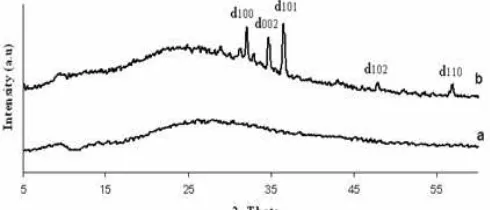
Fig 4.XRD patterns of rod like structure ZnO
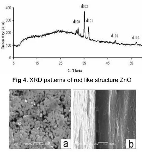
Fig 5.SEM images of ZnO thin film from NaOH: (a) top view; (b) cross sectional view
After ZnO seed layer has been successfully formed on ITO substrate, the modified substrate was then applied as a substrate in preparation of ZnO thin film nanostructure utilizing Zn(NO3)2and NH3with molar ratio
of 1:10. The XRD pattern and SEM images of the resulted nanostructured ZnO were shown in Fig. 2 and Fig. 3, respectively.
From Fig. 2, it is clear that the sequence of diffraction peaks is d[101]>d[100]>d[002]. ZnO thin film
obtained hydrothermally from Zn(NO3)2 with NH3as the
alkaline solvent did not exist in single rod shape, but as a bunch of flower-like structure. Each bunch has one central point in the centre. It is predicted that the rod started to grow from the centre to all directions composing chrysanthemum like structure. The size of a bunch of chrysanthemum was up to 1 μm. It was acquired from the approximately as estimated from the SEM image (Fig. 3). Such morphology was often called chrysanthemum like structure [13].
We also used the modified ITO glass as film substrate in the preparation of nanostructured ZnO using the same zinc nitrate precursor with NaOH as the alkaline solvent. By this route, we got white solid which was strongly adhered on the surface of the ITO glass substrate. The film formed was thinner than that of ZnO chrysanthemum like. Fig. 4 showed the diffraction pattern of the white thin layer. It is clear that (002) diffraction pattern was the highest peak indicating the presence of highly oriented ZnO in its c-axis. The growth of rod was along the c-axis. The morphology of ZnO thin
film from NaOH solution was then evaluated by SEM as shown in Fig. 5.
The top view image displayed rounded base of rods which are aligned vertically on the ITO glass substrate. Unfortunately, the cross section view did not clearly show the rows of the vertical rods. It may be due to fine morphology of the nanorods. The rod diameter is about 125 nm as calculated from the SEM image. The rod length reached 0.75 m. This dimension is smaller than that reported by Jung et al. [4], which was 180 nm in the diameter and 1.5 m length. Hydrothermal treatment in NaOH solution was effectively suppress the growth of multiple rods as occurred in NH3 alkaline solution resulted vertically
oriented rod like nanostructured ZnO.
CONCLUSION
In summary, we have shown that different alkaline solution as hydroxide anion generating agent generated different ZnO nanostructures after hydrothermal treatment. Rod like ZnO nanostructure was obtained by using NaOH as hydroxide anion generating agent, while chrysanthemum like was obtained by using NH3. Short hydrothermal treatment
Indo. J. Chem., 2011, 11 (2), 131 - 134
Anissa A. Putri et al.
134
ACKNOWLEDGEMENT
The authors acknowledge financial support from Universitas Gadjah Mada under Competitive Research Grant of Science and Technology Research Cluster 2009 (MAK. 2311.0154.521219).
REFERENCES
1. Vayssieres, L., and Keis, K., 2001, Chem. Mater., 13, 12, 4395–4398.
2. Iijima, S., 2002,Phys B, 323, 1–5.
3. Guo, L., Liang Ji, Y., and Xu, H., 2002, J. Am. Chem. Soc., 124, 50, 14864–14865.
4. Jung, S.H., Oh, E., Lee, K.H., Yang, Y., and Park, C.G., 2007,Cryst. Growth Des., 8, 1, 265–269. 5. Tang, J., and Yang, X., 2006,Mater. Lett., 60, 29-30,
3487–3491.
6. Aneesh., P.M., Vanaja., K.A., and Jayaraj., M.K., 2007, Synthesis ZnO nanoparticles by hydrothermal method,Proc. Of SPIE, vol. 6639
7. Yu, K., Jin, Z., Liu, X., Liu, Z., and Fu, Y., 2006, Mater. Lett., 61, 13, 2775–2778.
8. Leung, Y.H., Djurišić, A.B., and Xie, M.H., 2005, Synthesis and Properties of ZnO Nano-ribbon and Comb Structures, A I P Conference Proceedings, 772, 879–880.
9. Öztürk, S., Taşaltin, N., Kilinç, N., and Öztürk, Z.Z.,
J. Optoelectron. Biomed. Mater., 1, 1, 15–19. 10. Rattanavoravipa, T., Sagawa, T., and Yoshikawa,
D., 2008, Sol. Energy Mater. Sol. Cells, 92, 11, 1445–1449.
11. Lee, J.H., Leu, I.C., Hsu, M.C., Chung, W.Y., and Hon, M.H., 2005, J. Phys. Chem. B, 109, 27, 13056–13059.
12. Tonto, P., Mekasuwandumrong, O., Phatanasri, S., Pavarajarn, V., and Praserthdam, P., 2006,Ceram. Int., 34, 1, 57–62.