TROPICAL AGRICULTURAL SCIENCE
Journal homepage: http://www.pertanika.upm.edu.my/
ARTICLE INFO
Article history:
Received: 14 September 2016 Accepted: 14 August 2017
E-mail addresses:
[email protected]; [email protected] (Sianipar, N. F.), [email protected] (Vidianty, M.),
[email protected] (Chelen), [email protected] (Abbas, B. S.) * Corresponding author
Review Article
Micropropagation of Rodent Tuber Plant (Typhonium flagelliforme
Lodd.) from Medan by Organogenesis
Sianipar, N. F.1*, Vidianty, M.2, Chelen3 and Abbas, B. S.4
1Food Technology Department, Engineering Faculty, Research Interest Group Food Biotechnology,
Bina Nusantara University, Jl. Jalur Sutera Barat Kav. 21, Alam Sutera, 15325 Tangerang, Indonesia
2Master of Management, Graduate Program, Bina Nusantara University, Jl. Jalur Sutera Barat Kav. 21,
Alam Sutera, 15325 Tangerang, Indonesia
3IBM Indonesia, The Plaza Office Tower, Jl. MH. Thamrin Kav. 28-30, 10350 Jakarta Pusat, Indonesia 4Industrial Engineering Department, Engineering Faculty, Bina Nusantara University,
Jl. K H. Syahdan No. 9. Kemanggisan – Palmerah, 11480 Jakarta Barat,, Indonesia
ABSTRACT
Rodent Tuber is an anticancer herbal plant from Araceae family which is very sensitive to environmental condition and has a low plantlet reproduction rate. This research was aimed to an obtain effective method of micropropagation on Rodent Tuber plant with high rate multiplication factors. The source of explants used the mother plant originating from Medan (Indonesia). MS medium supplemented with the combination of 0.5 mg/L of BAP and various concentrations of NAA was used. Explants were successfully induced in medium containing 0.5 mg/L of BAP and 0.5 mg/L of NAA. Growing media for plant multiplication were ½
MS and MSO. In the treatment media, BAP was given in five different concentrations, i.e.
0.5, 1, 1.5, 2, and 2.5 mg/L. The result showed that, ½ MS medium added with 1.5 mg/L of BAP was effective in inducing the production of 4.20 ± 1.03 plantlets. In vitro root induction of Rodent Tuber was achieved in MSO medium supplemented with 0.5, 1, 1.5, 2, and 2.5 mg/L of NAA. MSO medium supplemented with 1.5 mg/L of NAA could induce the formation of 43.20 ± 21.21 new roots. Viability percentages of Rodent Tuber from Medan acclimatization was 100%. The usage of MS basal media enriched with BAP and NAA is able to effectively increase the production of new plants and roots of Rodent Tuber plant.
Keywords: BAP, NAA, organogenesis, Rodent Tuber
plant,Typhonium flagelliforme Lodd
INTRODUCTION
Rodent Tuber (Typhonium flagelliforme Lodd.) is an Indonesian herbal plant that belongs to the Araceae family (Surachman, 2009). This plant can be found in several countries such as India (Mankaran et al.,
2013), Indonesia (Essai, 1986), Malaysia (Lin, 2005), Sri Lanka and Australia (Sai et al., 2000). Rodent Tuber lives 1-300 m above sea level (Essai, 1986) in humid and shady areas (Nobakht et al., 2009).
Rodent Tuber is able to reach 26 cm in
height. This plant has flowers that resemble a rat’s tail. The flower of the rodent tuber
plant is sterile i.e. it cannot reproduce sexually (Nobakht et al., 2009). Rodent Tuber is commonly propagated by the vegetative method i.e. by separating buds produced from the mother plant’s tuber (Syahid & Kristina, 2007).
According to phytochemical analysis, this plant contains several bioactive
compounds such as alkaloids, flavonoids
(Mankaran et al., 2013), terpenoids, steroids (Nobakht et al., 2010), Ribosome Inactivating Proteins (RIP) (Indrayudha et al., 2006; 2011), antioxidants (Sukardi, 2011) and antibacterial compounds (Mohan et al., 2008). Besides that, Rodent Tuber extract has been found to be successful in inducing apoptosis of breast cancer cells T47D (Norrochmand et al., 2011) and MCF-7 (Putra et al., 2011). It was also found to inhibit the proliferation of human T4-lymphoblastoids (Mohan et al., 2008; Mohan et al., 2010) and NCI-H23 non-small cell lung carcinoma (Lai et al., 2008).
The ability of Rodent Tuber to inhibit and kill cancer cells has made this plant one of the plant commodities used as a raw material in complementary and alternative medicine (Mohan et al., 2011; Mankaran et al., 2013).
Rodent Tuber has a low reproduction rate and is very sensitive to enviromental factors. Therefore, the number of Rodent Tubers in the plant’s natural habitat is very low. However, demand for this plant is very high due to its biological activity. Vegetative propagation in in vitro culture is an appropriate method for overcoming this problem of low numbers because this method is able to produce many plants in a short period of time (Tiwar et al., 2011). The success of propagation
in tissue culture is influenced by several
factors i.e. genotype of plant, formulation media and physiological conditions of the mother plant. Same plants from different locations will show different results in the
same formulation media. The influence
of climate and conditions of the physical environment can also have an effect on the physiological conditions of the mother plant (George & Sherrington, 1984; Wattimena et al., 1992).
to induce the production of plants in in vitro
culture is 6-benzylamniopurin (BAP). BAP is a cytokinin-type PGR that is effective in inducing the production and propagation of buds from bud eyes (Gunawan, 1987). The supplementation of BAP in a Murashige-Skoog (MS) medium could induce the production of buds from plants such as
Amygdalus communis L.cv. Yaltsinki
(Akbas et al., 2009), Psoralea corylifolia
Linn (Pandey et al., 2013), Vinca rosea L. (Haq et al., 2013) and Potulaca grandiflora Hook (Jain et al., 2010).
In Indonesia, Rodent Tuber has been found in several regions such as Bogor, Pekalongan and Medan. Laurent et al.,
(2013) identified the genetic differences
between Rodent Tubers from three different regions by analysing their RAPD molecular
marker profiles. Rodent Tuber from Bogor
was multiplicated in MS medium to which was added 1 mg/L of NAA and 0.5 mg/L of BAP (Sianipar et al., 2011). Besides that, Rodent Tuber from Pekalongan has also been propagated in vitro in MS medium supplemented with 0.5 mg/L of NAA and 0.5 mg/L of BAP (Sianipar et al., 2015). Rodent Tuber from Medan is one of the plant accessions in Indonesia. The regeneration of this plant in in vitro
culture can be done in two ways i.e. by somatic embryogenesis and organogenesis. In our previous research (Sianipar et al., 2011), the somatic embryogenesis micropropagation method was performed on Rodent Tuber from Bogor. However, plants from different accessions usually have different optimal micropropagation
conditions as well. Therefore, this research
aimed to formulate a new and efficient
micropropagation method for propagating Rodent Tuber plants from Medan by applying different concentrations of BAP as PGR.
MATERIALS AND METHOD
Sterilisation and Initiation of Explant
This research used bud eyes and tubers of Rodent Tuber from Medan as the initial plant materials/explant source. The explants were washed in running tap water to remove any soil particles attached to them. The explants were submerged in 200 ml solution containing 0.25 g of bactericide and fungicide for 30 min, and 2.25 mg/ml of rifampicin for 4 h. The explants were then submerged in 2% Clorox® bleach for 15
min, 1.5% Clorox®bleach for 15 min, 1%
Clorox®bleach for 10 min and 0.1% HgCl
2
for 10 min. The explants were washed twice with sterile water and then cultured in growth media for initiating buds. There were three types of growth media for initiating buds, each with a different PGR composition. The media used were all Murashige-Skoog (MS) with 30 g sucrose and 100 mL coconut water added for a
one-Litre media. The first medium formulation
contained 0.5 mg/L Benzylaminopurine
(BAP) and NAA in three different concentrations i.e. 0.5, 1.0 and 1.5 mg/L. The explants were stored in an incubation room with light intensity 2000 lux for 16 h with a room temperature of about 24 ± 10C
Shoot Multiplication
The shoot multiplication media were ½ MS and MSO, and to each was added
BAP in five different concentrations
i.e. 0.5, 1.0, 1.5, 2.0 and 2.5 mg/L. Each treatment was replicated 10 times. The variables observed were the number of shoots in the 10th week. The explants
were then subcultured in the optimal medium.
Root Induction
The growth medium for root induction was an MSO basal medium supplemented with
NAA in five different concentrations (0.5,
1.0, 1.5, 2.0 and 2.5 mg/L). The experiment was repeated 10 times. The variables observed were the length and number of roots in the 10th week.
Acclimatisation
Plantlets which had many roots were acclimated after the 10th week. The
plantlets, especially their roots, were washed with water and submerged in 1 g/L of Agrimicyn bacteriocide and 0.25 g Benlate fungicide solutions for 3 min each. The plantlets were then acclimated in a medium composed of husk and compost in the ratio of 1:1 in plastic bottles in a greenhouse. Watering was done twice a day to maintain humidity. The variable observed was the viability percentage of plantlets during four weeks of pre-acclimatisation. Plants which could withstand the pre-acclimatisation period
were planted in media containing soil and compost in the ratio of 1:1. These plantlets were maintained in a greenhouse. The parameters observed were the plantlet’s viability percentages for a period of four weeks.
Research Design and Statistical Analysis
The experimental design of this experiment was a completely random design with eight replications. The statistical analysis of the average number of shoots was done at the 10th week. The statistical analyses used were the normality test Kolmogorov-Smirnov, followed by an analysis of variance (ANOVA) test for normally distributed data.
RESULTS AND DISCUSSION
Induction and Initiation of Explant
Rodent Tuber explants from Medan, Indonesia were cultured in MS medium supplemented with BAP and NAA. The part of explant used for initiating buds formation was the bud on the mother plant’s tuber. Buds were used as explants for micropropagation because they contain meristematic somatic cells. According to Nobakht et al. (2009), the bud eye of Rodent Tuber is a potential explant for micropropagating Rodent Tuber in in vitro
culture.
An in vitro culture medium of Rodent
of NAA i.e. 0.5, 1.0 and 1.5 mg/L. The percentages of sterile and viable explants in different media were supplemented with
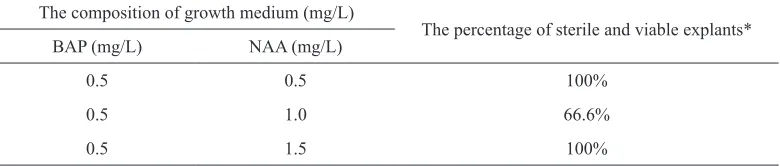
0.5 mg/L BAP and NAA and 0.5 mg/L BAP and 1.5 mg/L NAA, both at 100% (Table 1).
Table 1
Percentage of sterile and viable explants in initiation media
The composition of growth medium (mg/L)
The percentage of sterile and viable explants*
BAP (mg/L) NAA (mg/L)
0.5 0.5 100%
0.5 1.0 66.6%
0.5 1.5 100%
* The number of explants initiated for each treatment was three explants
Sterile and viable explants were obtained from all of the three types of media during the initiation stage. The percentage of sterile explants in medium containing 1.0 mg/L NAA was quite low i.e. 66.6% because the explants were contaminated with fungi.
At the bud induction and initiation stages, the percentage of sterile and viable explants was determined by the type, concentration and time of sterilisation. Organic and inorganic components of the medium were the determining factors of differentiation and de-differentiation (for example, formation of meristem-interfascicular cambium and cork cambium from fully differentiated parenchyma cells) processes. PGRs largerly contributed to the plant’s morphogenesis (George & Sherrington, 1984). In this research, the MS medium to which had been added sucrose and coconut water was able to
fulfil the explant’s requirements of macro-
and micronutrients. The addition of BAP
is important for mitosis cell division and inducing bud formation, while NAA is for cell division and root induction. Roots will be induced if the ratio of NAA to BAP is higher than one (George & Sherrington, 1984). According to Nobakht et al. (2009), the application of BAP as cytokinin, either alone or combined with NAA as auxin, is effective in increasing the proliferation rate of buds and the fresh weight of Rodent Tuber explant (Syahid & Kristina, 2007).
Multiplication
Figure 1. The increase in the number of shoots in various ½MS media supplemented with BAP
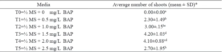
The highest number of shoots i.e. 4.20 was achieved on ½MS medium supplemented with 1.5 mg/L of BAP. A Duncan test at the accuracy level of 5% showed that the number of shoots in ½MS medium supplemented with 1.5 mg/L
BAP was significantly different from that
produced by the control, ½MS with 0.5
mg/L BAP, ½MS with 1.0 mg/L BAP and ½MS with 2.5 mg/L BAP. Meanwhile, the average number of shoots in the ½MS medium supplemented with 1.5 mg/L
of BAP was not significantly different
from that produced by the ½MS medium supplemented with 2.0 mg/L of BAP (Table 2 and Figure 4(c)).
Table 2
Multiplication of Rodent Tuber from Medan in ½MSO medium
Media Average number of shoots (mean ± SD)*
T0=½ MS + 0 mg/L BAP 0.00±0.00a
T1=½ MS + 0.5 mg/L BAP 2.30±1.49b
T2=½ MS + 1.0 mg/L BAP 3.00±.15bc
T3=½ MS + 1.5 mg/L BAP 4.20±1.03d
T4=½ MS + 2.0 mg/L BAP 4.10±0.88cd
T5=½ MS + 2.5 mg/L BAP 2.70±1.95b
* Each treatment was replicated 10 times. Average numbers with the same letters indicated that there were
no significant differences at p-value ≤0.05 according to the Duncan analysis.
The increasing concentration of BAP up to 2.5 mg/L lowered the number of shoots. This showed that a relatively low concentration of PGR will greatly affect the differentiation process. Like the ½MS treatment, the MSO medium supplemented
Figure 2.The number of shoots of Rodent Tuber from Medan in MS medium supplemented with BAP
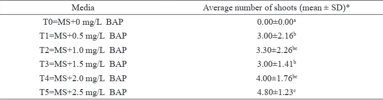
Among the MS medium treatments, the highest average number of shoots was obtained by the MS medium supplemented with 2.5 mg/L of BAP. The average number of shoots in that medium reached 4.8. A Duncan test with an accuracy level
of 5% showed that there were significant
differences in the number of shoots between the MS medium to which had been added
BAP 2.5 mg/L and the control medium, as well as between the MS medium to which had been added 0.5 mg/L of BAP and the MS medium to which had been added 1.5 mg/L of BAP. The average number of shoots between the MS medium supplemented with 1.0 mg/L of BAP and the MS medium supplemented with 2.0 mg/L of BAP was
not significantly different.
Table 3
Multiplication of Rodent Tuber from Medan in MS medium
Media Average number of shoots (mean ± SD)*
T0=MS+0 mg/L BAP 0.00±0.00a
T1=MS+0.5 mg/L BAP 3.00±2.16b
T2=MS+1.0 mg/L BAP 3.30±2.26bc
T3=MS+1.5 mg/L BAP 3.00±1.41b
T4=MS+2.0 mg/L BAP 4.00±1.76bc
T5=MS+2.5 mg/L BAP 4.80±1.23c
* Each treatment was replicated 10 times. Average numbers with the same letters indicated that there were
The ½MS and MSO basal media were able to support the growth of the plant cultures based on the observation of the number of heathly shoots produced. However, the application of the ½MS and MS without the addition of BAP was not able to increase the number of shoots. BAP is important for inducing shoot multiplication (Wattimena et al., 1992) and it can induce cell division and differentiation to produce new buds, either directly or indirectly. The same concentration of BAP can also induce shoot production of Musa
acuminata cv. Berangan plant (Jafari et al.,
2011). The application of PGR outside the safe concentration range will destroy plant tissue and inhibit bud production and cell enlargement, so plant growth will also be inhibited (George & Sherrington, 1984; Sharman et al., 2012). The same applied
to Melissa officinalis (Tavares et al.,
1996) and Hedeoma multifolium (Koroch, 1997). The ½MS medium to which had been added 1.5 mg/L BAP produced the highest number of shoots compared to the
other treatment with ½MS, while the MSO medium to which had been added 2.5 mg/L BAP was the optimum treatment compared to the other treatments with MSO.
Root Induction
Root induction of in vitro shoots was achieved in the MS medium supplemented
with five different concentrations of NAA
i.e. 0.5 mg/L, 1.0 mg/L, 1.5 mg/L, 2.0 mg/L and 2.5 mg/L. Root length and average number of roots were observed in the 10th week. Root length was not increased
significantly in the medium supplemented
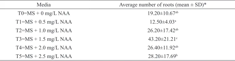
with NAA. The longest root was obtained in the MSO medium i.e. 7.79 cm (Table 4). NAA was important for root induction, according to the analysis of the average number of roots (Table 5). The highest average number roots was obtained by the culture in the MS medium supplemented with 1.5 mg/L NAA i.e. 43.20 (Figure 4D). The Duncan test with an accuracy level
of 5% showed that there were significant
differences between the treatments.
Table 4
Average root length of Rodent Tuber from Medan
Media Average length (cm) (mean±SD)*
T0=MS+0 mg/L NAA 7.79±2.97b
T1=MS+0.5 mg/L NAA 5.66±0.57a
T2=MS+1.0 mg/L NAA 6.11±1.39a
T3=MS+1.5 mg/L NAA 6.14±1.56a
T4=MS+2.0 mg/L NAA 5.14±0.92a
T5=MS+2.5 mg/L NAA 5.36±0.58a
* Each treatment was replicated 10 times. Average numbers with the same letters indicated that there were
Table 5
The average number of roots of Rodent Tuber from Medan
Media Average number of roots (mean ± SD)*
T0=MS + 0 mg/L NAA 19.20±10.67ab
T1=MS + 0.5 mg/L NAA 12.50±4.03a
T2=MS + 1.0 mg/L NAA 26.20±17.42ab
T3=MS + 1.5 mg/L NAA 43.20±21.21c
T4=MS + 2.0 mg/L NAA 26.40±11.92ab
T5=MS + 2.5 mg/L NAA 28.20±17.69b
* Each treatment was replicated 10 times. Average numbers with the same letters indicated that there were
no significant differences at p-value ≤0.05 according to the Duncan analysis.
The addition of NAA in the MS medium affected the production of the roots of the Rodent Tuber plant from Medan but did not
significantly affect the elongation of roots.
NAA is a synthetic auxin that is able to stimulate cell growth, cell division and the formation of fruit and roots (Wattimena et al., 1992). Auxin that has been absorbed will be metabolised through the auxin transport method and is important for stimulating the elongation of roots, formation of adventive shoots and root hairs and determining the roots’ growth direction (Teale et al., 2004). The application of exogenous auxin in the culture medium was essential for lateral root formation (Thimann, 1936).
The application of exogenous auxin in a culture medium is important for initiating lateral root formation (Chhun et al., 2003). NAA in plant tissue culture medium was able to induce the formation of lateral roots of red betel plant (Sianipar et al., 2016), rice mutant Lrt 1 (Chhun et al., 2003) and Mellissa officinalis (Sevik &
Guney, 2013). This research generated the micropropagation method of Rodent Tuber
from Medan, Nort Sumatra (Indonesia) up to the phase of acclimatisation. Although Nobakht et al. (2009) conducted micropropagation research on the same plant with a mother plant originating from Malaysia, the different climatic conditions and the physical environment had a huge effect on the physiological conditions of the mother plant as a source of the explants. This phenomenon is a problem that is common with tissue culture even in the same location as a different isolation of plants (dry/wet climate) will affect the success of micropropagation (totipotency cells) (George & Sherrington, 1984; Wattimena et al., 1992).
Acclimatisation
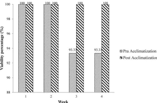
Figure 3. Acclimatisation of the plantlets of Rodent Tuber from Medan by plant tissue culture method
The viability percentage of the plantlets during pre-acclimatisation was very high i.e. reaching 93.33%. This has also been observed by Sianipar et al (2015), who showed a viability percentage of 100% from Rodent Tuber from Pekalongan. The viability percentage of Rodent Tuber from Malaysia documented by Nobakht et al. (2009) reached 90%. During post-acclimatisation, the viability percentage of Rodent Tuber from Medan even reached 100%. This was even higher than the viability percentage of Rodent Tuber from Pekalongan, which was only 58%. This result showed that different accessions/genotypes
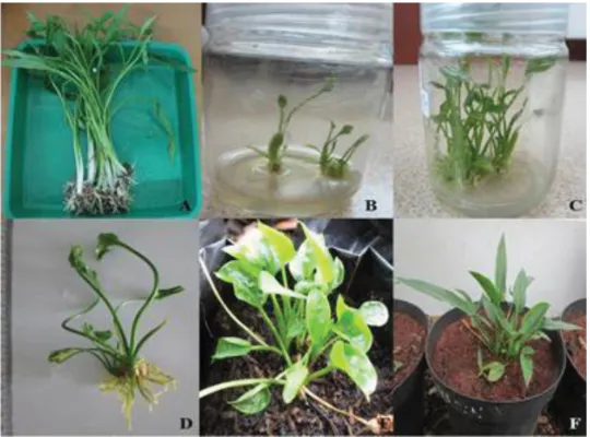
Figure 4. Micropropagation of Rodent Tuber from Medan
Note: A- Rodent Tuber from Medan used as the explant mother plant; B- Initiation in MS medium to which had been added 0.5 mg/L BAP and 0.5 mg/L NAA; C: Multiplication of shoots in the 10th week in ½MS
medium to which had been added 1.5 mg/L BAP; D: Root induction in MS medium to which had been added 1.5 mg/L NAA; E: Pre-acclimatisation of plantlets; and F: Post-acclimatisation of the plant that had been propagated in in vitro culture previously.
CONCLUSION
Rodent Tuber from Medan was able to multiply and produce a high number of shoots i.e. 4.20 ± 1.03 shoots in a ½MS medium supplemented with 1.5 mg/L BAP in the 10th week. Rodent Tuber from
Medan is unique because it can grow optimally in a ½MSO medium. The highest number of shoots i.e. 4.80 was obtained from the MSO medium supplemented with 2.5 mg/L BAP. The highest number of roots i.e. 43.20 lateral roots was achieved from the MSO medium supplemented with 1.5 mg/L NAA. Rodent Tuber from Medan has good adaptability to environmental conditions, with a viability percentage of 93.33% during pre-acclimatisation and 100% during acclimatisation.
ACKNOWLEDGEMENT
The author would like to thank The Directorate General of Higher Education, Indonesia for a competitive grant funding and Prof. Ika Mariska for reviewing this manuscript.
REFERENCES
Akbas, F., Isikalan, C., Namli, S., & Ak, B. (2009). Effect of plant growth regulators on in vitro shoot multiplication of Amygdalus communis L. cv. Yaltsinki. African Journal of Biotechnology,
8(22), 6168–6174.
Essai. (1986). Medicinal herbs index in Indonesia. Jakarta: PT Essai Indonesia.
George, E. F., & Sherrington, P. D. (1984). Plant propagation by tissue culture. Basingstoke, England: Exegetics Ltd.
Gunawan, L. W. (1987). Teknik kultur jaringan. Bogor, Indonesia: Laboratorium Kultur Jaringan Tanaman, Pusat Antar Universitas Bioteknologi Institut Pertanian Bogor.
Haq, R., Naz, S., Aslam, F., & Manzoor, F. (2013). Comparison of in vitro response of micropopragation and callogenesis of medicinal plant, L. Journal of Agricultural Research, 51(1), 9–17.
Indrayudha, P., Wijaya, A., & Iravati, S. (2006). Uji aktivitas ekstrak daun dewandaru dan daun keladi tikus terhadap pemotongan dna superkoil untai ganda. Jurnal Farmasi Indonesia, 3(2), 63–70.
Indrayudha, P., Wijayanti, N., & Sismindari. (2011). Antiangiogenesis activity of protein fraction containing MJ C acidic ribosome inactivating protein of Mirabilis jalapa L. Jurnal Bahan Alam Indonesia, 7, 277–281.
Jafari, N., Othman, R. Y., & Khalid, N. (2011). Effect of benzylaminopurine (BAP) pulsing on in vitro shoot multiplication of Musa acuminate (banana) cv. Berangan. African Journal of Biotechnology, 10(13), 2446–2450.
Jain, A. K., & Bashir, M. (2010). In-vitro propagation of a medicinal plant Portulaca grandiflora. Hook. World Journal of Agricultural Sciences, 6(3), 327–330.
Koroch, A. R., Juliani, H. R., Juliani, H. R., Jr., & Trippi, V. S. (1997). Micropropagation and acclimatization of Hedeoma multiform. Plant Cell Organ Culture: Journal of Plant
Biotechnology, 48(3), 213–217.
Lai, C. S., Mas R. H., Nair, N. K., Majid, M. I. A., Mansor, S. M., & Navaratnam, V. (2008).
Typhonium flagelliforme inhibits cancer cell growth in vitro and induces apoptosis: An evaluation by the bioactivity guided approach. Journal of Ethnopharmacology, 118(1), 14–20.
Laurent, D., Sianipar, N. F., Chelen, L., & Wantho, A. (2013). Analysis of genetic diversity of Indonesian rodent tuber (Typhonium
flagelliforme Lodd.) cultivars based on RAPD marker. Presented at the International Conference on Biological Science, the Faculty
of Biology, University of Gadjah Mada. Yogyakarta, Indonesia: AIP Publishing.
Lin, K. (2005). Ethanobotanical study of medical plants used by the Jah Hut People in Malaysia. Indian Journal of Medical Sciences, 59(4), 156-161.
Mankaran, S., Dinesh, K., Deepak, S., & Gurmeet,
S. (2013). Typhonium flagelliforme: A
multipurpose plant. International Research Journal of Pharmacy, 4(3), 45-48.
Mohan, S., Abdul, A. B., Abdelwahab, S., Zubairi, A., Sukari, M., Abdullah, R., … & Syam, S. (2010). Typhonium flagelliforme induce apoptosis in CEMs cells via activation of caspase-9, PARP cleavage and cytochrome c release: Its activation coupled with G0/G1 phase cell cycle arrest. Journal of Ethnopharmacology, 131(3), 592–600.
Mohan, S., Bustamam, A., Ibrahim, S., Zubairi, A., & Aspollah, M. (2008). Anticancerous effect of Typhonium flagelliforme on human T4-Lymphoblastoid cell line CEM-ss. Journal of Pharmacology, 3(6), 449–458.
Mohan, S., Bustamam, A., Ibrahim, S., Al-Zubairi, A., Aspollah, M., Abdullah, R., & Elhassan, M. M. (2011). In vitro ultramorphological assessment of apoptosis CEMs induced by linoleic acid-rich fraction from Typhonium flagelliforme tuber. Evidence-Based and Alternative Medicine,
Mustafa, N. S., & Taha, R. (2012). Influence of
plant growth regulator and subculturing on in vitro multiplication of some fig (Ficus Carcia) cultivars. Journal of Applied Science Research,
8(8), 4038–4044.
Nobakht, G. M., Kadir, M. A., & Stanslas, J. (2009). In vitro mass propagation of Typhonium
flagelliforme as affected by plant growth regulators. African Journal of Biotechnology,
8(24), 6840–6843.
Nobakht, G. M., Kadir, M. A., & Stanslas, J. (2010). Analysis of preliminary phytochemical screening of Typhonium flagelliforme. African Journal of Biotechnology, 9(11), 1655–1657.
Nurrochmad, A., Lukitaningsih, E., & Meiyanto, E. (2011). Anti-cancer activity of rodent tuber (Typhonium flagelliforme Lodd.) blume on human breast cancer t47d cells. International Journal of Phytomedicine, 3(1), 138–146.
Pandey, P., Mehta, R., & Upadhyay, R. (2013). In vitro propagation of an endangered medical plant Psoralea corylifolia Linn. Asian Journal of Pharmaceutical and Clinical Research, 6(3), 115–118.
Putra, A., Tjahjono, & Winarto. (2011). Ekstrak rodent tuber (Typhonium flagelliforme) fraksi diklorometanolik dan ekspresi caspase-3 dan p21 cell-Line kanker payudara MCF-7. Media
Medika Indonesiana, 45(2), 95–104.
Sai, S., Keng, C., Pargini, N., & Teo, C. (2000). In vitro propagation of Typhonium flagelliforme
(Lodd) blume. In vitro Cellular and
Developmental Biology – Plant, 36(5), 402–406.
Sevik, H., & Guney, K. (2013). Effect of IAA, IBA, NAA and GA3 on rooting and morphological features of Melissa officinalis L. stem cuttings.
The Scientific World Journal, 2013, 1–5.
Sharman, J., Khan, S., & Varman, R. (2012). In vitro culture establishment and shoot induction of mallotus philippenesis (Lam.) M. ARG. Pharmacie Globale International Journal of Comprehensive Pharmacy, 3, 1–5.
Sianipar, N. F., Laurent, D., & Tanty, H. (2015, September). Induction, multiplication, and acclimatization of rodent tuber (Typhonium
flagelliforme Lodd.) plant from Indonesia by in
vitro organogenesis. In International Conference on Technology, Informatics, Management,
Engineering & Environment (TIME-E), 2015
(pp. 59-63). IEEE.
Sianipar, N. F., Rustikawati, Maarisit, W., Wantho, A., & Sidabutar, D. N. R. (2011). Embryogenic calli induction, proliferation and regeneration of
rodent tuber plant (Thyphonium flagelliforme
Lodd.) by single node culture. Paper presented at the International Conference on Biological Science, the Faculty of Biology, University
of Gadjah Mada BIO-UGM. Yogyakarta, Indonesia: AIP Publishing.
Sianipar, N. F., Verlina & Rosaria. (2016). Induction, multiplication, and acclimatization of red betel plant (Piper crocatum Ruiz and Pav.) by in vitro organogenesis. Jurnal Teknologi, 78(5-6), 35–40.
Sukardi. (2011). Identifikasi dan karakterisasi umbi
Rodent Tuber sebagai zat antioksidan alami. Gamma, 6(2), 143–151.
Surachman, D. (2009). Penggunaan beberapa taraf konsentrasi paklobutrazol dalam media konservasi rodent tuber (Typhonium
flagelliforme Lodd.) in vitro. Buletin Teknik Pertanian, 14(1), 31–33.
Syahid, S., & Kristina, N. (2007). Induksi dan regenerasi kalus Rodent Tuber (Typhonium
Tavares, A. C., Pimenta, M. C., & Goncalves, M. T. (1996). Micropropagation of Melissa officinalis L. through proliferation of axillary shoots. Plant Cell Respiratory, 15(6), 441–444.
Teale, W. D., Papanov, I. A., Ditengou, F., & Palme, K. (2004). Auxin and the developing root of Arabidopsis thaliana. Physiologia Plantarum, 123(2), 130–138.
Thimann, K. V. (1936). Auxins and the growth of roots. American Journal of Botany, 23(8), 561– 569.
Tiwar, S., & Arnold, R. (2011). Micropropagation as an advanced technology for the conservation of plants. International Journal of Drug Discovery
and Herbal Research, 1(2), 95–99.