Polymorphism of
PXR
gene associated with the increased risk of drug-induced
liver injury in Indonesian pulmonary tuberculosis patients
Z. Zazuli* MPharm, M. I. Barliana†PhD, U. A. Mulyani‡MSc, D. A. Perwitasari§PhD, H. Ng* BPharm and R. Abdulah* PhD
*Department of Pharmacology and Clinical Pharmacy, Faculty of Pharmacy, Universitas Padjadjaran, Jatinangor,†Department of Biological Pharmacy, Faculty of Pharmacy, Universitas Padjadjaran, Jatinangor,‡Center for Applied Health Technology and Clinical Epidemiology, National Institute of Health Research and Development, Ministry of Health Republic of Indonesia, Jakarta, and§Faculty of Pharmacy, Universitas Ahmad Dahlan, Yogyakarta, Indonesia
Received 25 December 2014, Accepted 25 August 2015
Keywords:antituberculosis drugs, drug-induced hepatotoxicity, drug-induced liver injury, polymorphism, PXR
SUMMARY
What is known and objective:Tuberculosis is still a major infectious disease in Indonesia. Patients are treated mostly using fixed-dose combination treatment in primary public health facilities. The incidence of antituberculosis drug-induced liver injury (AT-DILI) is approximately 10% among Indonesian tuberculosis patients who used standard fixed combination regimens during the intensive phase of treatment. However, information regarding genetic polymorphism associated with the increase risk of drug-induced liver injury is still limited. The aim of this study was to investigate pregnane X receptor (PXR) gene polymorphisms as one of the risk factors of AT-DILI.
Methods:In this prospective cohort study, we recruited 106 adult patients diagnosed with pulmonary tuberculosis and treated with category I FDC (fixed-dose combination). The identification of SNP -25385C>T (rs3814055) was conducted by ARMS (ampli-fication refractory mutation system). Hepatotoxicity was defined as ALT and/or AST levels above the normal threshold on the second, fourth and sixth months of monitoring during tubercu-losis treatment.
Results and discussion:The logistic regression analysis showed that patients with the TT genotype of PXR gene (rs3814055) significantly had a greater risk of AT-DILI (OR 889; 95% CI 136–5793, P<005), compared with those of wild-type CC genotype.
What is new and conclusion:The result suggests that in Indonesian patients with tuberculosis, the risk of having AT-DILI was associated with TT genotype of thePXRgene.
WHAT IS KNOWN AND OBJECTIVE
Tuberculosis (TB) is an infectious disease that is still a major problem in developing countries, including Indonesia. In 2014, the World Health Organization categorized Indonesia as a high-TB-, multi-drug resistant (MDR)-TB-, and human immunodeficiency virus (HIV)-burdened country. There are an estimated 90 million incident cases of TB and 15 million people died of the disease each year (11 million deaths were HIV negative, and 360 000 were HIV positive). The correspondingfigures for Indonesia are 325 582, and 7964 cases of TB have been identified as new and relapse cases,
respectively.1Given this high burden of disease, the government of Indonesia is focusing on the control and elimination of TB. One of the efforts is through the directly observed treatment, short-course strategy (DOTS) programme.
One of the main concerns in antituberculosis drugs (ATDs) is drug-induced liver injury (DILI) or hepatotoxicity caused by drugs.2 Of the first-line anti-TB drugs, isoniazid, pyrazinamide and rifampicin can all cause liver damage (drug-induced hepati-tis).3 In addition, rifampicin can cause asymptomatic jaundice without evidence of hepatitis.3A previous publication has shown that the incidence of antituberculosis drug-induced liver injury (AT-DILI) is around 10% among Indonesian TB patients treated with standard fixed combination regimens during the intensive phase of treatment.4Many researchers have reported on specific gene polymorphisms as risk factors for those hepatotoxic effects. Some of the reported genes are N-acetyltransferase 2 (NAT2),5–10 cytochrome P450 2E1 (CYP2E1),9,11,12 glutathione S-transferase mu-1 (GSTM1)9,10,13and glutathione S-transferase theta (GSTT).9,13 Other genes also reported to be potential predictors of ATDs-DILI are pregnane X receptor (PXR), glutathione S-transferase alpha-1 (GSTA1), manganese superoxide dismutase (MnSOD, SOD2), UDP-glucuronosyltransferase (UGT), nitric oxide synthase 2A (NOS2A), BTB and CNC homology 1 (BACH1), Maf basic leucine zipper protein (MAFK), and human leucocyte antigen (HLA).14–18 We examinedPXRgene polymorphism because ligand forma-tion between rifampicin and PXR could trigger the expression of various genes, including various cytochromes and car-boxylesterases that may be relevant to the metabolism of isoniazid in producing hepatotoxic metabolites. PXR plays a role in the regulation of a number of hepatic and intestine genes involved in detoxification and elimination of xenobiotics.19 The polymor-phisms are generally located in the 50-flanking region of the target genes.20 PXR activates various genes through binding and heterodimer formation with the retinoid X receptor (RXR). PXR ligands stimulates the expression of genes involved in xenobiotics oxidation (phase I), conjugation (phase II) and transport (phase III) in the liver. PXR is involved in phase I metabolism through the expression of CYP2B6, CYP2C8, CYP2C9, CYP2B9 and CYP2C19.21–25Moreover, PXR is involved in phase II metabolism through the expression of glutathione S-transferase (GST), sulfotransferase, UDP-glucuronosyltransferase and carboxyles-terase.26–31
PXR is involved in phase III through the expression of organic anion transporting polypeptide 2(OATP2) and multiple drug resistance protein 2 (MRP2).32–34
This study aimed to investigate possible associations ofPXRgene polymorphisms with the increased risk of ATDs-DILI.
METHODS
Patients
A total of 106 patients with active lung TB who visited selected primary public health facilities in Bandar Lampung and Yogya-karta were enrolled consecutively and followed up in this prospective cohort study. Inclusion criteria were as follows: (i) adult patients (over 18 years of age) newly diagnosed with active lung TB, (ii) treated withfixed-dose combination of antitubercu-losis drugs (FDC-ATD) category I using directly observed treat-ment, short-course strategy (DOTS) and (iii) have agreed to the terms in the written informed consent. Patients with any of the following conditions were excluded from the study: (i) positive HIV/AIDS, (ii) abnormal serum ALT and AST levels over twice upper limit of normal (ULN) value before treatment with ATD, (iii) with hepatitis or have a history of hepatitis, (iv) haemoglobin serum<8 mg/dL, (v) not having ATDs for over 2 weeks, (vi) have a history of kidney disease and (vii) refusal to blood collection.
All patients received FDC-ATD category I intensive phase (rifampicin 150 mg, isoniazid 75 mg, pyrazinamide 400 mg and ethambutol 275 mg per tablet) and FDC-ATD category I contin-uation phase (rifampicin 150 mg and isoniazid 150 mg) in 6 months of therapy. The dosage of ATDs was selected according to the patients’weight. ALT and AST serum levels were obtained five times: before ATD treatment, after completing the second, the fourth, the sixth months of ATD therapy and 1 month after the patient finished the last ATDs therapy. Antituberculosis drug-induced liver disease (ATD-DILI) was designated as an increase in serum ALT and AST levels above the ULN after ATDs treatment, according to the criteria of drug-induced liver injuries developed by Drug-Induced Liver Injury Network (DILIN), National Institute of Diabetes and Digestive and Kidney Diseases (NIDDK), and Division of Specialized Information Services of the National Library of Medicine (NLM), and National Institutes of Health and Common Toxicity Criteria for Adverse Events 4.0 version (CTCAEv.4.0, Bethesda, MD, USA).35
The written informed consent was obtained from each patient enrolled in this study. This study protocol conformed to the ethical guidelines of the 1975 Declaration of Helsinki and was approved by the Ethics Committee for Health Research, National Institute of Health Research and Development, Ministry of Health, Republic of Indonesia.
Single Nucleotide Polymorphism genotyping
DNA was extracted from the peripheral blood sample collected from each patient using the standard proteinase K digestion and GeneJET Genomic DNA Purification Kit (Thermo Scientificâ
, Walthman, MA, USA.). Polymerase chain reaction–amplification refractory mutation system (PCRARMS) was used to detect the -25385C>T (rs3814055) PXR polymorphisms located in the 50UTR
promoter. The two primer sets were as follows: forward 50
-TTTTGGCAATCCCAGGTTT-30 to detect PXR polymorphic gene
(F-primer–1) and 50-TTTTGGCAATCCCAGGTTC-30 to confirm normal fragment gene (F-primer–2); reverse 50
-CGAATGTGGTG-GATACCAG-30. PCR was carried out using 5x FIREPolâ
Master Mix Ready from Solis BioDyneâ
(Tartu, Estonia), according to the manufacturer’s instruction. Initial denaturation at 95°C for 4 min was followed by 40 cycles of denaturation at 95°C for 30 s, annealing at 597°C, extension at 72°C for 1 min, with final extension at 72°C for 7 min. The products obtained were
electrophoresed on 2% agarose gels. Polymorphic gene TT showed 219-bp PCR product only when it was identified using F-1 forward primer, non-polymorphic gene CC showed 219 bp only when it was identified using F-2 reverse primer, and heterozygote gene CT showed 219 bp when it was identified using both forward primers.
Statistical analysis
Allele frequencies were calculated, and the agreement between the allele frequencies and Hardy–Weinberg equilibrium was tested using a chi-square test (d.f.=1) for each locus. Odds ratios (ORs) and 95% confidence intervals (95% CIs) were calculated using binomial logistic regression analysis, adjusting for sex and age. The baseline ALT and AST differences among genotype groups were determined using the Kruskal–Wallis test. Statistical analyses were performed using a data analysis freeware.
RESULTS AND DISCUSSION
Results
The result of UV gel visualization is shown in Fig. 1. Homozygotes TT and CC showed single bands on F-primer 1 and F-primer 2, respectively. Heterozygote CT showed double bands with the F-primer 1 and F-F-primer 2.
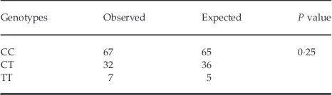
Seven patients (66%) were TT homozygotes, 32 patients (3019%) were CT heterozygotes, and most patients (67; 6321%) were wild-type CC homozygotes. The genotype frequencies were not different from those predicted by the Hardy–Weinberg equation (Table 1).
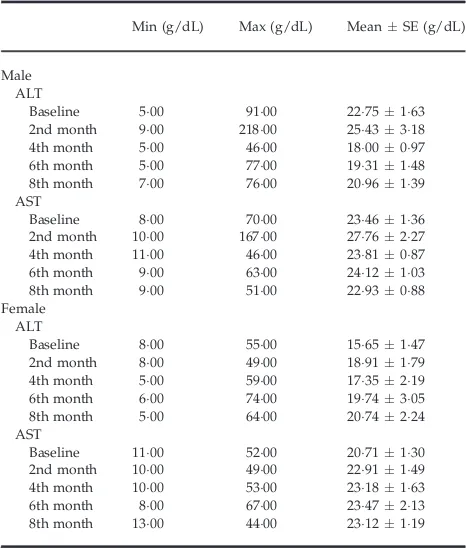
Table 2 shows that there was no statistical difference in ALT and AST baseline levels among both male and female patients with different genotypes (P>005). The mean of ALT and AST levels increased at the end of thefirst 2 months of ATD therapy or after completion of the intensive phase and decreased during the continuous phase (Table 3). Most of the patients (71 patients; 6698%) experienced no hepatotoxicity, but 35 patients showed elevated transaminase levels. The transaminase elevation was seen in 33 patients (3113%) with mild hepatotoxicity (1+) and 2 patients (189%) with moderate hepatotoxicity (2+) (Table 4).
219 bp Sample 1 (TT) Sample 2 (CC) Sample 3 (CT)
Fig. 1. UV visualization result of 25385C>T (rs3814055) polymor-phisms.
Table 1. Comparison of observed and expected genotype frequency based on Hardy–Weinberg equilibrium
Genotypes Observed Expected Pvalue
CC 67 65 025
CT 32 36
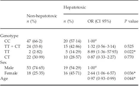
The risk of having ATD-DILI was significantly associated with the TT genotype. For this genotype, the risk of having an ATD-DILI was 889-fold (OR: 889; 95% CI: 136–5793;P=0022) that of
the wild-type (CC genotype) (Table 5). The results also show that the combined (CT+TT) genotype had a tendency for higher incidence of ATD-DILI in comparison with the wild-type CC genotype (OR: 132; 95% CI: 056–314;P=0525). We also found that the risk of an ATD-DILI in females was 264-fold (OR: 264; 95% CI: 106–657;P=0036) that of males.
Discussion
We found that there was a significant association between the variant TT homozygote ofPXR (-25385C>T; rs3814055) and the risk of AT-DILI. This result is consistent with results reported by Andrewset al.36who reported that a differentPXRpolymorphism at the same location was significantly associated with liver injury caused byflucloxacillin, a PXR agonist. Andrewset al.concluded that the CC genotype was associated with the risk offl ucloxacillin-induced liver injury. However, a previous study conducted in Europe and South Asia reported no association between ATDs and PXR gene polymorphism in patients.37
As genetic polymorphism is only one of the many factors that may contribute to drug-induced hepatotoxicity, its significance requires further testing.38 Rifampicin is a PXR agonist. PXR is involved in the regulation of bile acid and cholesterol homeostasis. Table 2. Characteristics of patients according to genotype
Genotype
Pvalue
TT CT CC
n(%) MeanSD n(%) MeanSD n(%) MeanSD
Sex
Male 5 (7143) 18 (5625) 49 (7313) Female 2 (2857) 14 (4375) 18 (2687) Total 7 (10000) 32 (10000) 67 (10000) Age group (years)
18–45 4 (5714) 23 (7188) 46 (6866) 46–55 1 (1429) 6 (1875) 13 (1940)
>55 2 (2857) 3 (938) 8 (1194) Total 7 (10000) 32 (10000) 67 (10000) ALT baseline
Male 3120855 2456287 2122196 0195 Female 1200200 1707313 1494138 0767 AST baseline
Male 3360870 2600300 2149135 0053 Female 1750250 2121263 2067140 0754
Table 3. Serum aminotransferase level cohort data according to sex
Min (g/dL) Max (g/dL) MeanSE (g/dL)
Male ALT
Baseline 500 9100 2275163 2nd month 900 21800 2543318 4th month 500 4600 1800097 6th month 500 7700 1931148 8th month 700 7600 2096139 AST
Baseline 800 7000 2346136 2nd month 1000 16700 2776227 4th month 1100 4600 2381087 6th month 900 6300 2412103 8th month 900 5100 2293088 Female
ALT
Baseline 800 5500 1565147 2nd month 800 4900 1891179 4th month 500 5900 1735219 6th month 600 7400 1974305 8th month 500 6400 2074224 AST
Baseline 1100 5200 2071130 2nd month 1000 4900 2291149 4th month 1000 5300 2318163 6th month 800 6700 2347213 8th month 1300 4400 2312119
Table 4. Frequency of hepatotoxicity according to severity of drug-induced hepatotoxicity
Drug-induced Hepatotoxicity Level n(%)
1+, Mild (<259ULN) 33 (3113) 2+, Moderate (25–59ULN) 2 (189) No hepatotoxicity 71 (6698)
Some of the bile acids are toxic. Therefore, the regulation by PXR of the expression of proteins involved in both phase I and phase II drug metabolism and transport requires further investigation.39 Murine studies with the humanized pregnane X receptor (hPXR) suggested that rifampicin and isoniazid combination therapy leads to accumulation of protoporphyrin IX, an endogenous hepatotoxin through a mechanism involving PXR’s effect on the haem biosynthesis pathway.40
This study also indicates that females are significantly more likely to show ATD-DILI than males. This is consistent with some previous studies which showed that female patients were more likely to show hepatotoxicity.41–43 They also reportedly show a higher risk of hepatotoxicity with nitrofurantoin, erythromycin, flucloxacillin, minocycline and isoniazid.44 Drug-induced hepatotoxicity in females tends to be hepatocellular with vascular damage on the centrilobular vein.44,45The presence of genetic abnormality in the mitochondria is also said to be a
major determinant of idiosyncratic drug-induced hepatotoxicity especially in women and the elderly.46
Our study has some limitations because we did not use other liver function parameters such as total serum bilirubin, serum ALP, INR and GGT. Their measurements would have been useful in defining liver function and the severity of hepatotoxicity better. We were also not able to assess other confounding factors, such as exposure to hepatotoxic agents including commonly encountered items such as acetaminophen, potentially hepatotoxic herbal and food products, and other chemicals in the patient’s environment. The small number of PXR T homozygotes calls for a larger confirmatory study.
WHAT IS NEW AND CONCLUSION
We found a significant association between thePXRTT variant (-25385C>T; rs3814055) and the AT-DILI. Whether testing forPXR genotype will provide reliable predictions of AT-DILI requires further validation.
ACKNOWLEDGEMENT
The authors thank Jarir At Thobari, Anas Subarnas, Keri Lestari, Tiana Milanda, Iswandi Darwis, Soraya Rahmanisa, Syachroni and Muhammad Khairuman for their guidance and technical support in this study.
CONFLICT OF INTEREST
None declared.
SOURCE OF FUNDING
The study was funded by the Centre for Applied Health Technology and Clinical Epidemiology, The National Institute of Health Research and Development, Ministry of Health Republic of Indonesia.
REFERENCES
1. Organization WH. 2014. Global tuberculosis report 2014. Available at: http://www.who. int/tb/publications/global_report/en/ (acc essed 30 October 2014).
2. Saukkonen JJ, Cohn DL, Jasmer RMet al. An official ATS statement: hepatotoxicity of antituberculosis therapy.Am J Respir Crit Care Med, 2006;174:935–952.
3. Organization WH. 2010.Treatment of
tuber-culosis guideline. Available at: http://
www.who.int/tb/publications/2010/978 9241547833/en/ (accessed 28 March 2014). 4. Atthobari J, Mulyani UA, Perwitasari DA, Darwis I. 2013.Early drug induced liver injury after intensive phase of tb treatment in Indone-sia: Primary care centers and lung hospital study. In The 13th International Society of Pharmacovigilance Annual Meeting Pisa: Drug Safety, 36:864.
5. Huang YS, Chern HD, Su WJ et al. Poly-morphism of the n-acetyltransferase 2 gene
as a susceptibility risk factor for antituber-culosis drug-induced hepatitis.Hepatology, 2002;35:883–889.
6. Sun F, Chen Y, Xiang Y, Zhan S. Drug-metabolising enzyme polymorphisms and predisposition to anti-tuberculosis drug-in-duced liver injury: a meta-analysis. Int J Tuberc Lung Dis, 2008;12:994–1002. 7. Ben Mahmoud L, Ghozzi H, Kamoun A
et al. Polymorphism of the
n-acetyltrans-ferase 2 gene as a susceptibility risk factor for antituberculosis drug-induced hepato-toxicity in tunisian patients with tuberculo-sis.Pathol Biol (Paris), 2012;60:324–330. 8. Wang PY, Xie SY, Hao Q, Zhang C, Jiang
BF. Nat2 polymorphisms and susceptibility to anti-tuberculosis drug-induced liver injury: a meta-analysis. Int J Tuberc Lung Dis, 2012;16:589–595.
9. Forestiero FJ, Cecon L, Hirata MH, de Melo FF, Cardoso RF, Cerda A, Hirata
RD. Relationship of nat2, cyp2e1 and gstm1/gstt1 polymorphisms with mild elevation of liver enzymes in Brazilian individuals under anti-tuberculosis drug therapy. Clin Chim Acta, 2013;415: 215–219.
10. Rana SV, Sharma SK, Ola RP, Kamboj JK, Malik A, Morya RK, Sinha SK. N-acetyl-transferase 2, cytochrome p4502e1 and glutathione s-transferase genotypes in anti-tubercular treatment-induced hepatotoxicity in North Indians. J Clin Pharm Ther, 2014;39:91–96.
11. Lee SW, Chung LS, Huang HH, Chuang TY, Liou YH, Wu LS. Nat2 and cyp2e1 poly-morphisms and susceptibility to first-line anti-tuberculosis drug-induced hepatitis.Int J Tuberc Lung Dis, 2010;14:622–626. 12. Deng R, Yang T, Wang Y, Tang N. Cyp2e1
rsai/psti polymorphism and risk of anti-tuberculosis drug-induced liver injury: a Table 5. Results of logistic regression analysis
Non-hepatotoxic
n(%)
Hepatotoxic
n(%) OR (CI 95%) Pvalue
Genotype
CC 47 (662) 20 (5714) 100a
TT+CT 24 (338) 15 (4286) 132 (056–314) 0525 TT 2 (282) 5 (1429) 889 (136–5793) 0022*
CT 22 (3099) 10 (2857) 087 (033–227) 0770 Sex
Male 53 (7465) 19 (5429) 100a
Female 18 (2535) 16 (4571) 264 (106–657) 0036*
Age 097 (093–099) 0044*
aReference category.
meta-analysis. Int J Tuberc Lung Dis, 2012;16:1574–1581.
13. Tang SW, Lv XZ, Zhang Y et al.Cyp2e1, gstm1 and gstt1 genetic polymorphisms and susceptibility to antituberculosis drug-induced hepatotoxicity: a nested case– con-trol study. J Clin Pharm Ther, 2012;37: 588–593.
14. Liddle C, Goodwin B. Regulation of hepatic drug metabolism: role of the nuclear
recep-tors pxr and car. Semin Liver Dis,
2002;22:115–122.
15. Brind AM, Hurlstone A, Edrisinghe D, Gilmore I, Fisher N, Pirmohamed M, Fryer AA. The role of polymorphisms of glu-tathione s-transferases gstm1, m3, p1, t1 and a1 in susceptibility to alcoholic liver disease. Alcohol Alcohol, 2004;39:478–483.
16. Zhang B, Xie W, Krasowski MD. Pxr: a xenobiotic receptor of diverse function implicated in pharmacogenetics. Pharma-cogenomics, 2008;9:1695–1709.
17. Padda MS, Sanchez M, Akhtar AJ, Boyer JL. Drug-induced cholestasis. Hepatology, 2011;53:1377–1387.
18. Huang YS. Recent progress in genetic vari-ation and risk of antituberculosis drug-induced liver injury. J Chin Med Assoc, 2014;77:169–173.
19. Kliewer SA, Goodwin B, Willson TM. The nuclear pregnane x receptor: a key regulator of xenobiotic metabolism. Endocr Rev, 2002;23:687–702.
20. Wang H, LeCluyse EL. Role of orphan nuclear receptors in the regulation of drug-metabolising enzymes.Clin Pharmacokinet, 2003;42:1331–1357.
21. Xie W, Barwick JL, Simon CMet al. Recip-rocal activation of xenobiotic response genes by nuclear receptors sxr/pxr and car.Genes Dev, 2000;14:3014–3023. 22. Gerbal-Chaloin S, Pascussi JM,
Pichard-Garcia Let al.Induction of cyp2c genes in human hepatocytes in primary culture. Drug Metab Dispos, 2001;29:242–251. 23. Goodwin B, Moore LB, Stoltz CM, McKee
DD, Kliewer SA. Regulation of the human cyp2b6 gene by the nuclear pregnane x receptor.Mol Pharmacol, 2001;60:427–431. 24. Synold TW, Dussault I, Forman BM. The
orphan nuclear receptor sxr coordinately
regulates drug metabolism and efflux.Nat Med, 2001;7:584–590.
25. Gerbal-Chaloin S, Daujat M, Pascussi JM, Pichard-Garcia L, Vilarem MJ, Maurel P. Transcriptional regulation of cyp2c9 gene. Role of glucocorticoid receptor and consti-tutive androstane receptor. J Biol Chem, 2002;277:209–217.
26. Madhu C, Klaassen CD. Protective effect of pregnenolone-16 alpha-carbonitrile on aceta-minophen-induced hepatotoxicity in ham-sters.Toxicol Appl Pharmacol, 1991;109:305–313. 27. Hosokawa M, Hattori K, Satoh T. Differen-tial responses of rat hepatic microsomal carboxylesterase isozymes to glucocorti-coids and pregnenolone 16 alpha-carboni-trile.Biochem Pharmacol, 1993;45:2317–2322. 28. Liu L, Klaassen CD. Regulation of hepatic sulfotransferases by steroidal chemicals in rats.Drug Metab Dispos, 1996;24:854–858. 29. Dunn RT 2nd, Gleason BA, Hartley DP,
Klaassen CD. Postnatal ontogeny and hor-monal regulation of sulfotransferase sult1b1 in male and female rats. J Pharmacol Exp Ther, 1999;290:319–324.
30. Runge-Morris M, Wu W, Kocarek TA. Regulation of rat hepatic hydroxysteroid sulfotransferase (sult2-40/41) gene expres-sion by glucocorticoids: evidence for a dual mechanism of transcriptional control.Mol Pharmacol, 1999;56:1198–1206.
31. Falkner KC, Pinaire JA, Xiao GH, Geoghe-gan TE, Prough RA. Regulation of the rat glutathione s-transferase a2 gene by corticoids: involvement of both the gluco-corticoid and pregnane x receptors. Mol Pharmacol, 2001;60:611–619.
32. Courtois A, Payen L, Guillouzo A, Fardel O. Up-regulation of multidrug resistance-asso-ciated protein 2 (mrp2) expression in rat hepatocytes by dexamethasone. FEBS Lett, 1999;459:381–385.
33. Fromm MF, Kauffmann HM, Fritz Pet al. The effect of rifampin treatment on intesti-nal expression of human mrp transporters. Am J Pathol, 2000;157:1575–1580.
34. Staudinger JL, Goodwin B, Jones SAet al. The nuclear receptor pxr is a lithocholic acid sensor that protects against liver toxi-city. Proc Natl Acad Sci USA, 2001;98: 3369–3374.
35. Health NIo. 2014. Severity grading in drug induced liver injury. Available at: http:// livertox.nih.gov/Severity.html (accessed 5 October 2014).
36. Andrews E, Armstrong M, Tugwood Jet al. A role for the pregnane x receptor in flucloxacillin-induced liver injury.
Hepatol-ogy, 2010;51:1656–1664.
37. Ng CS. A study of genetic polymorphism underlying idiosyncratic hepatotoxicity due to anti-tuberculosis medications. PhD thesis, Newcastle University, UK, 2012.
38. Russmann S, Kullak-Ublick GA, Gratta-gliano I. Current concepts of mechanisms in drug-induced hepatotoxicity. Curr Med Chem, 2009;16:3041–3053.
39. Xie W, Radominska-Pandya A, Shi Yet al. An essential role for nuclear receptors sxr/ pxr in detoxification of cholestatic bile acids. Proc Natl Acad Sci USA, 2001;98:3375–3380. 40. Li F, Lu J, Cheng J et al. Human pxr modulates hepatotoxicity associated with rifampicin and isoniazid co-therapy. Nat Med, 2013;19:418–420.
41. Nolan CM, Goldberg SV, Buskin SE. Hepa-totoxicity associated with isoniazid preven-tive therapy: a 7-year survey from a public health tuberculosis clinic.JAMA, 1999;281: 1014–1018.
42. Chamorro JG, Castagnino JP, Musella RMet al.Sex, ethnicity, and slow acetylator profile are the major causes of hepatotoxi-city induced by antituberculosis drugs. J Gastroenterol Hepatol, 2013;28:323–328. 43. Shu CC, Lee CH, Lee MC, Wang JY, Yu CJ,
Lee LN. Hepatotoxicity due tofirst-line anti-tuberculosis drugs: afive-year experience in a taiwan medical centre.Int J Tuberc Lung Dis, 2013;17:934–939.
44. Leise MD, Poterucha JJ, Talwalkar JA. Drug-induced liver injury. Mayo Clin Proc, 2014;89:95–106.
45. Grattagliano I, Bonfrate L, Diogo CV, Wang HH, Wang DQ, Portincasa P. Biochemical mechanisms in drug-induced liver injury: certainties and doubts.World J Gastroenterol, 2009;15:4865–4876.