Complex equilibrium study of some hydroxy aromatic ligands with
beryllium ion
Shella Permatasari Santoso
a, Artik Elisa Angkawijaya
a, Felycia Edi Soetaredjo
b, Suryadi Ismadji
b, Yi-Hsu Ju
a,⁎
aChemical Engineering Department, National Taiwan University of Science and Technology, Taipei 106-07, Taiwan
bDepartment of Chemical Engineering, Widya Mandala Surabaya Catholic University, Kalijudan 37, Surabaya 60114, Indonesia
a b s t r a c t
a r t i c l e
i n f o
Article history:
Received 15 May 2015
Received in revised form 5 August 2015 Accepted 15 September 2015
Equilibrium studies of beryllium with 2,3-dihydroxybenzoic acid, 3,4-dihydroxybenzoic acid and gallic acid in an aqueous solution at 310.15 K, an ionic strength of 0.15 mol·dm−3NaCl and pH 2.5 to 11.0 were investigated by the pH-potentiometric method. Stability constants of the complexes were determined by HyperQuad2008 and presented as logβ. Contributing binding sites of these ligands were evaluated by comparing its logβwith some structurally related ligands such as catechol and salicylic acid. Spectrophotometric measurements were done to confirm the formation of the complex species. Geometry optimization and frequency analysis of the complexes were performed by using Gaussian09W program to verify the contributing binding sites. The results indicate that the ability of ligands in preventing the hydrolysis of Be2+follows the order: 3,4-dihydroxybenzoic acid
N 2,3-dihydroxybenzoic acidNgallic acid.
© 2015 Elsevier B.V. All rights reserved.
1. Introduction
Beryllium is a considerably toxic element which may induce abnor-mal cell growth in the lung, known as chronic beryllium disease (CBD). Exposure to this metal may cause fatal health effect[1–3]. Moreover, be-ryllium is known as an amphoteric species which is reactive in both acidic and basic environment, thus greatly increases the possibility of its exposure. Studies about the chemistry of Be2+in an aqueous solution
showed that Be2+is able to form various hydroxo species. For example [Be2(OH)]3 +and Be(OH)2are formed during hydrolysis of beryllium
[4,5], while [Be(OH)4]2−and [Be2(OH)7]3−can be found in highly acidic
and basic environment, respectively[6]. The addition of organic ligands into beryllium solution may prevent the formation of these hydroxo species, where the active donor atom of the ligand will bind the Be2+
ion instead of hydroxo ions[7–12].
Based on Pearson's Hard Soft Acid Base (HSAB) theory, hard acid ion such as Be2+tends to form strong chelate complex with hard base
ligand [13]. Thus in this work, hydroxy aromatic ligands 2,3-dihydroxybenzoic acid (DA), 3,4-2,3-dihydroxybenzoic acid (PA) and gallic acid (GA) were used as the ligand to form complex with Be2+. These
compounds are categorized as hard bases which possess at least three oxygen donor atoms in their carboxyl (−COOH) and hydroxyl (−OH) functional groups. These natural phytochemicals can be found in aquatic ferns, tea leaves, gallnuts, oak barks and many other plants. They are
biologically significant for humans due to their antioxidant, antimicro-bial and anti-inflammatory properties[14–16].
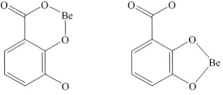
During complexation, these antioxidants may coordinate with Be2+ in several possible binding modes, in particular DA may form complex in salicylate or catecholate manner as depicted inFig. 1 [17,18]. It was an interesting subject to study the ligand possible binding modes to-wards metal ion. Therefore to gain an insight into the contributing bind-ing sites of the studied ligands, the complexation properties of catechol (CAT) and salicylic acid (SAL) were also studied. The aim of this work was to determine the complexing ability of DA, PA and GA along with CAT and SAL towards Be2+by the potentiometric method at 310.15 K
and an ionic strength of 0.15 mol·dm−3NaCl in an aqueous solution. These complexing ability properties were determined by using HyperQuad2008 program and quantitatively presented as stability con-stants (logβ), while qualitatively the complex formation was observed by using spectrophotometric measurement. The species distribution diagrams in the pH range of 2.5 to 11.0 obtained from HySS2009 were graphically presented. The complex structures were verified by the den-sity functional theory calculation method using Gaussian09W program.
2. Experimental section
2.1. Materials and solutions
Beryllium sulfate tetrahydrate (BeSO4·4H2O, 98% purity),
2,3-dihydroxybenzoic acid (C7H6O4, 99% purity) and 3,4-dihydroxybenzoic
acid (C7H6O4, 97% purity) were obtained from Alfa Aesar (Lancashire,
UK). Gallic acid (C7H6O5, 97.5% purity) was acquired from Sigma Aldrich
–
⁎ Corresponding author.
E-mail address:[email protected](Y.-H. Ju).
http://dx.doi.org/10.1016/j.molliq.2015.09.026
0167-7322/© 2015 Elsevier B.V. All rights reserved.
Contents lists available atScienceDirect
Journal of Molecular Liquids
(Steinheim, Germany). Catechol (C6H6O2, 99% purity) and hydrochloric
acid (HCl, 37.6%) were purchased from Fisher Scientific (Waltham, MA). Salicylic acid (C7H6O3, 99% purity) was supplied by Shimakyu (Osaka,
Japan), Carbonate-free sodium hydroxide (NaOH, 96% purity) and sodium chloride (NaCl, 99.5% purity) were obtained from Yakuri Pure Chemical (Kyoto, Japan) and Showa (Tokyo, Japan), respectively. All solutions in this study were prepared freshly before use in distilled deionized water (resistance 18.3 MΩ·cm). The beryllium sulfate tetrahydrate solution, NaOH and HCl were standardized before use.
2.2. Potentiometric method
All titrations were carried out in a 150 cm3glass vessel which was
connected to a thermostated circulating bath to maintain the tempera-ture at 310.15 K. The determination of stability constants of metal-ligand complexes was done by titrating a mixture of solution containing 0.0004–0.001 mol·dm−3of Be2+salt (T
M) + 0.001–0.0012 mol·dm−3
of ligand DA/PA/GA/CAT/SAL (TL), where the TMto TLratios used were
Fig. 1.DA binding mode in salicylate (left) and catecholate (right) manner.
Fig. 2.Titration curve of the Be2+
1:1, 1:2, 1:2.5 and 1:3. The ionic strength of each solution was main-tained with 0.15 mol·dm−3NaCl and the solutions were initially
acidi-fied by adding 0.003 mol·dm−3HCl. The solutions were titrated against standardized carbonate-free NaOH under nitrogen atmosphere.
All titrations were performed using a Metrohm 888 Titrando with 805 dosimat and equipped with a 802 rod stirrer and an Ecotrode Plus pH-glass electrode with pH unit readability up to third digit decimal. Calibration of the electrode was evaluated by means of strong acid-strong base titration using GLEE program. This potentiometer was connected to a personal computer equipped with Tiamo 2.3 titration software to record the titration data. Each set of titration was repeated at least 3 times with repeatability of ±0.02 in pH unit. The data then were used for the determination of stability constants from refinement using HyperQuad2008 program[19]. The stability constants were presented as logβpqrvalues, which can be expressed as follows:
pBeþqLþrH⇌BepLqHr; logβpqr¼
species formation, respectively. After the stability constants of various complex species were obtained, species distribution of each system was graphically presented by using the HySS2009 program[20].
2.3. Spectrophotometric measurement
The formation of the complexes was confirmed spectrophotometri-cally. The measurements were performed by using a double-beam Jasco V-550 spectrophotometer and a standard 10 mm quartz cell. The range of wavelength used in the measurement is 200 to 500 nm. The solutions were prepared (each 50 cm3in total) as follow: (a) 0.0004 mol·dm−3 ligand solution and (b) 0.0004 mol·dm−3 ligand + 0.00016 mol·dm−3metal solution.
2.4. Molecular modeling
The complex structures were optimized by the density functional theory (DFT) method with B3LYP exchange-correlation function and 6-311++G(d) basis set. Along with geometry optimization, frequency calculations were also carried out using Gaussian09W program[21]. For simplification purpose, (a) the addition of salt and base in the system was not included in the calculation, (b) the calculations were only intended for the BeL–core species and (c) the complex structures were evaluated from their free energy values.
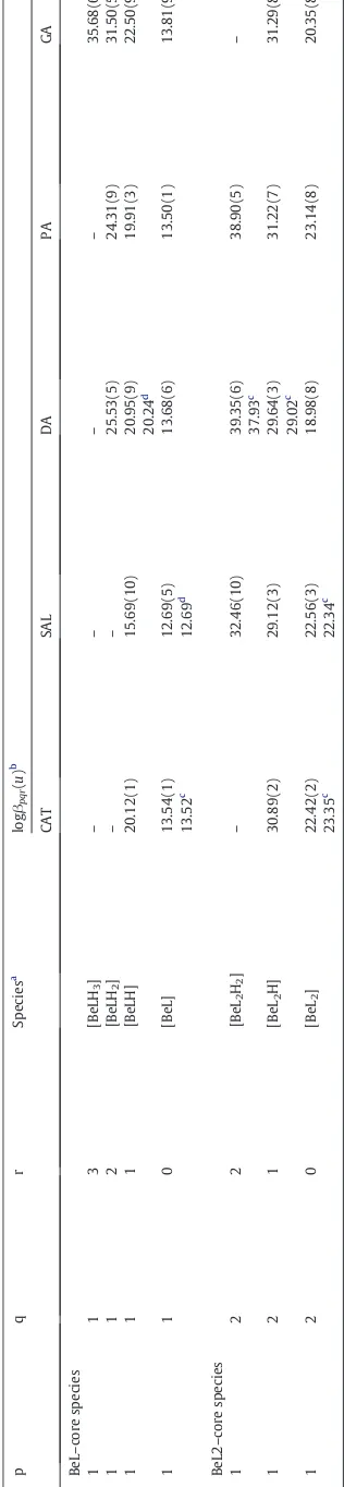
Table 2
Stepwise formation constants of Be2+complexes.
Ligand logKMLH(n−1)
Values of logKMLH(n−1)were obtained by the following equation:
CAT or SAL:logKMLHðn−1Þ¼logβ111−pKa2;n¼2 DA or PA:logKMLHðn−1Þ¼logβ112−pKa2−pKa3;n¼3 GA:logKMLHnð−1Þ¼logβ113−pKa2−pKa3−pKa4;n¼4
b Values of logK
3. Results and discussion
3.1. Stability constant of beryllium complexes
The ligands used in this work are the hydroxy aromatic ligands (DA, PA, GA, CAT and SAL) which contain both carboxylic (−COOH) and hydroxyl (−OH) donor groups, except for CAT which only has–OH groups. The protonation constants of the ligands were examined and compared with those reported in the literatures[22,23]and the results show good agreement. In the case of protonation constants which have valueN11, the values obtained from the literatures[22,23]were input-ted into the HyperQuad2008 for the determination of stability con-stants. The hydrolysis constants of Be2 +species such as Be(OH)+,
Be(OH)2, Be2(OH)3 +and Be3(OH)33 +were also examined and
com-pared with those reported in the literature[5].
The formation of the metal complex indicated by the inflection point which occur at approximatelym= 4 for all systems as shown in the ti-tration curves inFig. 2. The species observed in the system of Be2+with hydroxy aromatic ligands were the BeL–core species (BeLH3, BeLH2,
BeLH and BeL) and the BeL2–core species (BeL2H2, BeL2H and BeL2).
The refined stability constants from HyperQuad2008 are presented in Table 1.
The stepwise formation constants of the species were calculated and these values are given inTable 2. Stepwise formation constant (logK) corresponds to stepwise addition of one proton/ligand at a time, while stability constant (logβ) corresponds to the addition of protons/ligands
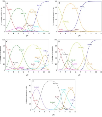
Fig. 3.Species distribution diagram of Be2+
all at once or known as overall formation constant. logKMLH(n−1) repre-sents the stepwise formation constant of the earliest species which is formed from the binding of Be2 +via the
–COOH group of ligand, following the steps described below:
Be2þ
þLH−⇌BeLHþ ;L¼CAT or SAL ð1Þ
Be2þ þLH−
2⇌BeLH−2 ;L¼DA or PA ð2Þ
Be2þ þLH−
3⇌BeLH þ
3 ;L¼GA: ð3Þ
These species have similar logKMLH(n−1)values of 2.69, 2.55, 2.59 and 2.98 for SAL, DA, PA and GA, respectively which indicate that the same contributing donor atom was bound to the metal ion, in this case the
–COOH group (except for CAT which has a logKMLH(n−1)of 7.12 and bound through one–OH group). The species distribution diagrams in
Fig. 3also prove this analysis, where complexation took place in more acidic pH in which only the–COOH group has higher possibility to dissociate.
At higher pH more species occurred, as can be observed in the species distribution diagram. These species occurred due to further dissociation of ligand. For example in the complexation of Be2+with
DA, species were formed according toEqs. (4) and (5).
Be2þþDAH2−⇌BeDAH ð4Þ
Be2þ
þDA3−⇌BeDA− ð5Þ
The latter species formed was [BeL] which was formed by the bind-ing of Be2+with the fully dissociated ligand. The stepwise formation constant of these species expressed as logKML, and the values are
13.54, 12.69, 13.68, 13.50 and 13.81 for CAT, SAL, DA, PA and GA,
respectively. These species have higher stability constant value than other BeL–core species, indicating more stable species. A fully dissociat-ed ligand has more negative charge hence it attract metal ion stronger. The [BeL] species of systems with DA, PA and GA have similar logKML
values as the one with CAT. This indicates that [BeL] species are coordi-nated via the same binding group as that of CAT (chelate complex via two–OH groups or catecholate type).
It is expected that [BeL] will form a 5-membered chelate ring as catecholate-like complex rather than a 6-membered chelate ring salicylate-like complex, since 5-membered chelate ring has more stable and rigid structure than 6-membered chelate ring. Moreover, the 5-membered ring of the observed [BeL] complex involves only single bond atom resulting in more stable binding, while the 6-membered ring involves one single bond atom and one double bond atom. A 6-membered chelate ring will be more stable if all the atoms are double bonded[25,26]. The structures of [BeL] species were discussed further in the molecular modeling section later.
On the other hand, BeL2–core species were formed by the ability of
metal ions to bind multiple ligands to form ML2species. As presented
inTable 1, the logβ120values of chelate complex between Be2 +ion
and two fully dissociated ligands or [BeL2] species are 22.42, 22.56,
18.98, 23.14 and 20.35 for CAT, SAL, DA, PA and GA, respectively. The stepwise formation constant of [BeL2] species are represented as
logKML2, where this value shows the strength of the second ligand to bind with the complex. It was shown that logKML2is much lower than logKML, indicating that the second ligand was bound not as strong as
thefirst ligand. This phenomenon often occurred in the formation of [BeL2] species due to the bulkier species that caused a steric interaction
between the ligands attached. However, overall [BeL2] species are more
stable than [BeL] species as indicated by the higher logβ120values. The
species distribution diagrams inFig. 3show that BeL2species is more
likely to form at high pH. It is due to the fact that at high pH dissociation is easier to occur; hence the chance of chelation is higher.
As presented inFig. 3, the hydrolysis of Be2+happened in the CAT,
DA and PA systems; where the Be(OH)+species existed at acidic pH (b7) and the neutral Be(OH)2species was predominantly found in the
physiological pH (7.4). However, these two hydrolysis species of Be2+
were not found in the SAL and PA systems. This suggests that in the physiological pH, SAL and PA are able to form stronger binding with Be2+. The other hydrolyzates such as Be
2(OH)3+and Be3(OH)33+were
not observed in the system indicating that the ligands were capable of inhibiting further hydrolysis of Be2+.
3.2. Spectrophotometric measurement
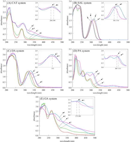
The formation of the complex species were confirmed qualitatively by using UV–Visible spectrum measurement at wavelength 200 to 500 nm. The spectrum measurement were done for all the systems at pH 7 and 11. As shown inFig. 4, the absorbance at range 200 to 250 occurred due to the aromatic ring of ligand. The shifting of the Be2+–
ligand spectrum compared to ligand spectrum was clearer observed at the basic pH 11. The shift indicated that the complex formation occurred in the system. Specifically, for (a) CAT system, initially the peak of ligand is showed at 284 nm, the formation of Be2+complex cause shifting to
290 nm; (b) SAL system, the shifting is occur at 301 nm to 314 nm; (c) DA system, the formation of complex species caused the increasing of absorbance at 331 nm; (d) PA system, the shifting is occur at 305 nm to 313 nm; and (e) GA system, the shifting is occur at 273 nm to 285 nm.
3.3. Effectiveness of ligands
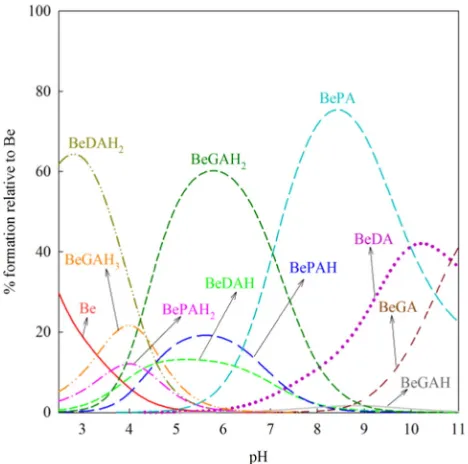
The effectiveness of the investigated ligand (DA, PA and GA) to bind Be2+was analyzed from the ability of the ligand to form [BeL] species.
When all three ligands are present in a system, they will compete with each other to bind the metal ion. Ligand which possesses stronger activity to attract and bind to the metal ion will form the most dominant species. As presented in competition diagram inFig. 5, DA was thefirst ligand that bound Be2 +, followed by PA and GA to form species that
were attached via–COOH group. This result is expected since the–
COOH group of DA was tend to dissociate at more acidic pH compared to PA and GA[23]. While among these three ligands, PA is the most effective ligand to bind Be2 +since its species ([BePA]) was found in
the highest concentration in the system. The maximum concentration of [BePA] species formed was 75.40% at pH 8.41, while the maximum concentration of [BeDA] and [BeGA] species was 42.08% at pH 10.24 and 41.03% at pH 11.0, respectively as shown inTable 3.
Fig. 5.Competition diagrams of DA, PA and GA as ligand to bind Be2+
in the mixed system, with a metal to ligand molar ratio of 1:2.5.
Table 3
Percentage of species formed in the mixed DA, PA and GA system.
Species %max pH Species %max pH
Free Be 29.70 2.50 BeGAH3 21.59 3.99
BeDAH2 64.28 2.80 BeGAH2 60.20 5.77
BeDAH 13.14 5.14 BeGAH 1.62 8.83
BeDA 42.08 10.24 BeGA 41.03 11.00
BePAH2 12.06 3.99
BePAH 19.15 5.60
BePA 75.40 8.41
Table 4
Gibb's free energy (ΔrG) values of BeL–core complex calculated by DFT method with 6-311++G(d) basis seta
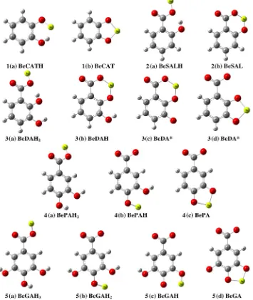
This table is related toFig. 6; for example ligand number1(CAT) and model number (a)withΔrG−0.1372 represent model1(a)inFig. 6.
b
3.4. Molecular modeling
The molecular structure of BeL–core species ([BeLH3], [BeLH2],
[BeLH] and [BeL]) were calculated using density functional theory (DFT) method with B3LYP correlation and basis set of 6-311++G(d) by the Gaussian09W program, represented as Gibb's free energy of reaction (ΔrG).ΔrG value was calculated by the following
equation:
ΔrG¼ΣGproduct−ΣGreactant: ð6Þ
The calculatedΔrG are shown inTable 4and optimized model of the
species are shown inFig. 6where each ligand is symbolized by a number (1= CAT,2= SAL,3= DA,4= PA and5= GA).
ΔrG values inTable 4are proportional to the logKvalues obtained
from potentiometric result. In all ligand systems, species with lower logKMLH(n−1)yielded less negativeΔrG (−0.1372,−0.1129,−0.1118, −0.1245 and −0.1240 for CAT, SAL, DA, PA and GA, respectively where the structures are shown inFig. 6by models1(a),2(a),3(a),
4(a)and5(a)); compare to [ML] species which have higher logKML
and yielded more negative ΔrG (−0.2582, −0.2489, −0.2797,
−0.2628 and −0.2936 for CAT, SAL, DA, PA and GA, respectively
where the structures are shown in Fig. 6 by model 1(b), 2(b),
3(d),4(c)and5(d)). Based on the result for DA, the [BeDA] species prefer the catecholate manner than the salicylate manner (Fig. 6, model3(c)for salicylate manner and3(d)for catecholate manner), in which the catecholate manner complexes have more negativeΔrG
values.
4. Conclusion
The complexation of hydroxy aromatic antioxidants (DA, PA and GA) with Be2 +was investigated potentiometrically atT= 310.15 K
andI= 0.15 mol·dm−3NaCl. These antioxidants were capable of forming stable complexes with Be2+. When the antioxidant (ligand)
was fully dissociated, [BeL] species were formed by binding Be2 +
through two OH groups in catecholate type with a logKMLvalue of
13.68, 13.50 and 13.81 for DA, PA and GA, respectively. The DFT calcula-tions well supported the [BeL] structure in catecholate binding mode with aΔrG value of−0.2797,−0.2628 and−0.2936 for DA, PA and GA, respectively. Among the three ligands, PA was shown to be the most effective ligand in preventing the hydrolysis of Be2+especially
in the physiological pH (7.4), followed by DA and then GA.
Acknowledgment
This work was supported by the National Taiwan University of Science and Technology through the project 103 M47006.
References
[1] T. Gordon, D. Bowser, Beryllium: genotoxicity and carcinogenicity, Mutat. Res.
Fundam. Mol. Mech. Mutagen. 533 (2003) 99–105.
[2] P.F. Wambach, J.C. Laul, Beryllium health effects, exposure limits and regulatory
requirements, J. Jchas (2008) 1–12.
[3] F. Cecconi, C.A. Ghilardi, A. Ienco, P. Mariani, C. Mealli, S. Midollini, A. Orlandini, A. Vacca, Different complexation properties of some hydroxy keto heterocycles toward beryllium(II) in aqueous solutions: experimental and theoretical studies, Inorg.
Chem. 41 (2002) 4006–4017.
[4] H. Kakihana, L.G. Sillen, Studies on the hydrolysis of metal ions: the hydrolysis of the beryllium ion, Be2+
, Acta Chem. Scand. 10 (1956) 985–1005.
[5] J. Bruno, Beryllium(II) hydrolysis in 3.0 mol dm−3perchlorate, J. Chem. Soc. Dalton
Trans. (1987) 2431–2437.
[6] H. Schmidbaur, Recent contributions to the aqueous coordination chemistry of
beryllium, Coord. Chem. Rev. 215 (2001) 223–242.
[7] M.W. Luczak, A. Zhitkovich, Role of direct reactivity with metals in chemoprotection byN-acetylcysteine against chromium(VI), cadmium(II), and cobalt(II), Free Radic. Biol. Med. 65 (2013) 262–269.
[8] F. Borges, C. Guimaraes, J.L.F.C. Lima, I. Pinto, S. Reis, Potentiometric studies on the complexation of copper(II) by phenolic acids as discrete ligand models of humic substances, Talanta 66 (2005) 670–673.
[9] N. Turkel, M. Berker, U. Ozer, Potentiometric and spectroscopic studies on
aluminium(III) complexes of some catechol derivatives, Chem. Pharm. Bull. 52 (8) (2004) 929–934.
[10] T. Kiss, H. Kozlowski, G. Micera, L.S. Erre, Copper(II) complexes of
2,3-dihydroxybenzoic acid, J. Coord. Chem. 20 (1989) 49–56.
[11]L.H.J. Lajunen, R. Portanova, J. Piispanen, M. Tolazzi, Stability constants foralpha -hydroxycarboxylic acid complexes with protons and metal ions and the
accompa-nying enthalpy changes: part I: aromaticortho-hydroxycarboxylic acids, Pure
Appl. Chem. 69 (1997) 329–381.
[12] T.S. Keizer, N.N. Sauer, T.M. McCleskey, Designer ligands for beryllium, J. Am. Chem. Soc. 126 (2004) 9484–9485.
[13] R.G. Pearson, Hard and soft acids and bases, J. Am. Chem. Soc. 85 (1953) 3533–3539.
[14]S.M. Fiuza, C. Gomes, L.J. Teixeira, M.T.G.d. Cruz, M.N.D.S. Cordeiro, N. Milhazes, F. Borges, M.P.M. Marques, Phenolic acid derivatives with potential anticancer
properties—a structure–activity relationship study. Part 1: methyl, propyl and
octyl esters of caffeic and gallic acids, Bioorg. Med. Chem. 12 (2004) 3581–3589.
[15] M.I. Choudhary, N. Naheed, A. Abbaskhan, S.G. Musharraf, H. Siddiqui,
Atta-ur-Rahman, Phenolic and other constituents of fresh water fern Salvinia molesta,
J. Phytochem. 69 (2007) 1018–1023.
[16] M. Grootveld, B. Halliwell, 2,3-Dihydroxybenzoic acid is a product of human aspirin
metabolism, Biochem. Pharmacol. 37 (1988) 271–280.
[17] T. Cam, G. Irez, R. Aydin, Determination of stability constants of mixed ligand
complexes of the lanthanum(III) ion and identification of structures, J. Chem. Eng.
Data 56 (2011) 1813–1820.
[18] S. Giroux, S. Aury, P. Rubini, S. Parant, J.-R. Desmurs, M. Dury, A spectroscopic inves-tigation of the complexing ability of catecholate or salicylate derivatives towards
aluminium(III), Polyhedron 23 (2004) 2393–2404.
[19] P. Gans, A. Sabatini, A. Vacca, Investigation of equilibria in solution. Determination of equilibrium constants with the HYPERQUAD suite of programs, Talanta 43 (1996) 1739–1753.
[20] L. Alderighi, P. Gans, A. Ienco, D. Peters, A. Sabatini, A. Vacca, Hyperquad simulation and speciation (HySS): a utility program for the investigation of equilibria involving soluble and partially soluble species, Coord. Chem. Rev. 184 (1999) 311–318.
[21] Frisch, M.J., G.W. Trucks, H.B. Schlegel, G.E. Scuseria, M.A. Robb, J.R. Cheeseman, G. Scalmani, V. Barone, B. Mennucci, G.A. Petersson, H. Nakatsuji, M. Caricato, X. Li, H.P. Hratchian, A.F. Izmaylov, J. Bloino, G. Zheng, J.L. Sonnenberg, M. Hada, M. Ehara, K. Toyota, R. Fukuda, J. Hasegawa, M. Ishida, T. Nakajima, Y. Honda, O. Kitao, H. Nakai, T. Vreven, J. J. A. Montgomery, J.E. Peralta, F. Ogliaro, M. Bearpark, J.J. Heyd, E. Brothers, K.N. Kudin, V.N. Staroverov, R. Kobayashi, J. Normand, K. Raghavachari, A. Rendell, J.C. Burant, S.S. Iyengar, J. Tomasi, M. Cossi, N. Rega, J.M. Millam, M. Klene, J.E. Knox, J.B. Cross, V. Bakken, C. Adamo, J. Jaramillo, R. Gomperts, R.E. Stratmann, O. Yazyev, A.J. Austin, R. Cammi, C. Pomelli, J.W. Ochterski, R.L. Martin, K. Morokuma, V.G. Zakrzewski, G.A. Voth, P. Salvador, J.J. Dannenberg, S. Dapprich, A.D. Daniels, O. Farkas, J.B. Foresman, J.V. Ortiz, J. Cioslowski, and D.J. Fox, Gaussian, Inc., Wallingford CT, (2009)
[22] M. Aplincourt, A. Debras-Bee, C. Gerard, R.P. Hugel, Modelling of the interactions of metal cations with soil organic matter. I: Thermodynamic stability of copper (II)
complexes with dihydroxybenzoic acids, J. Chem. Res. (S) 4 (1986) 134–135.
[23] L.D. Pettit, K.J. Powell, A Comprehensive Database of Published Data on Equilibrium Constants of Metal Complexes and Ligands, IUPAC and Academic Software, 2001.
[24] S.A. Abbasi, Binary and ternary complexes of interest to environmental systems.
Part-II: interaction of beryllium(II) with a mixture of ligands and failure in forming mixed ligand complexes, J. Indian Chem. Soc. 61 (1984) 125–127.
[25]S.J.S. Flora, V. Pachauri, Chelation in metal intoxication, Int. J. Environ. Res. Public Health 7 (2010) 2745–2788.