Nd-YAG laser irradiation damages to
Verrucaria nigrescens
M. Speranza
a,*, M. Sanz
b, M. Oujja
b, A. de los Rios
a, J. Wierzchos
a, S. Pérez-Ortega
a,
M. Castillejo
b, C. Ascaso
a,*aMuseo Nacional de Ciencias Naturales, MNCN-CSIC, Serrano 115 bis, 28006 Madrid, Spain bInstituto de Química Física Rocasolano, CSIC, Serrano 119, 28006 Madrid, Spain
a r t i c l e
i n f o
Article history:
Received 19 December 2011 Received in revised form 27 January 2012 Accepted 16 February 2012 Available online 15 October 2012
Keywords: Biodeterioration Stone Laser Algae Fungi Lichen
a b s t r a c t
Epilithic and endolithic microorganisms and lichens play an important role in stone biodeterioration. The structural and physiological damage caused by nanosecond pulsed laser of 1064 nm from Nd:YAG laser toVerrucaria nigrescenslichen as well as to endolithic algae and fungi were investigated in the present study. Ultrastructural laser effects on lichen and endolithic microorganisms were study without dis-turbing the relationship between lichen and lithic substrate by taking lichen-containing rock fragments and processing both together. SEM-BSE, LT-SEM and FM were used to determine cell integrity and ultrastructure, which reflect microorganism viability. Photobiont vitality was determined using a PAM chlorophyllfluorescence technique. The lichen thalli were completely removed by irradiation with 5 ns pulses at afluence of 2.0 J/cm2with no stone damage as showed by micro-Raman spectroscopy. The fungal and algal endolithic cells located below were completely destroyed or presented a high plas-molysis degree resulting from heating their microenvironment. The lichen and endolithic mycobiont near the irradiated zone were also damaged. Algal photosynthetic damage prevents fungal survival and lichen viability. This is thefirst report of laser removal and inactivation of lichen and lithic microor-ganisms, and thus provide an environmentally friendly and efficient method to control stone biodeterioration.
Ó2012 Elsevier Ltd. All rights reserved.
1. Introduction
Diverse groups of microorganisms such as heterotrophic bacteria, cyanobacteria, free-living algae and fungi are responsible for biodeterioration in stones used to construct heritage buildings and monuments (Caneva et al., 2008). Lichens, a symbiotic associ-ation of a fungus (the mycobiont) and a photosynthetic partner, usually an alga and/or cyanobacteria (the photobiont), are also common colonizers of monumental stone. These biological agents can colonize the surface of the lithic substrate (epilithic colonizers) as well as the internal zone of the stone (endolithic colonizers), where they develop complex interactions with the mineral substrate (Ascaso et al., 1998a, 2002; Caneva et al., 2008). Both types of colonizers play an important role in stone biodeterioration, being responsible for several chemical and physical processes that enhance weathering reactions (Ascaso et al., 1998a;De los Ríos and
Ascaso, 2005). The presence of endolithic microbial forms and their relationship with weathering effects on stone was proven by using differentin situmicroscopy techniques (Ascaso et al., 1998a,2002). Current control of lichens and microorganisms involved in stone biodeterioration processes includes chemical and physical treat-ments (Caneva et al., 2008;Scheerer et al., 2009;Doehne and Price, 2010). However, in spite of the great diversity of protocols currently in use in the field of stone conservation, methods to control biodeterioration have proven their technical and environmental limitations (Bartolini et al., 1999;Alakomi et al., 2006;Caneva et al., 2008;Scheerer et al., 2009;Doehne and Price, 2010). In this sense, further research is necessary on the effects on the substrate and the efficiency of physic methods to control stone-deteriorating micro-organisms (Scheerer et al., 2009). Furthermore, chemical methods have limitations as biocide low substrate penetration, certain side effects such as irreversible stone damage and/or low retention in stone that often limit their action (Caneva et al., 2008;Doehne and Price, 2010). Therefore, short-term applications must be repeated (deCruz et al., 2005). Scanning electron microscopy in back-scattered electron mode (SEM-BSE) technique showed that in some cases biocide action to control epilithic and endolithic microorganisms was limited when applied either alone or *Corresponding authors. Dept. Environmental Biology, National Museum of
Natural Sciences, Spanish National Research Council (CSIC), Serrano 115, 28006 Madrid, Spain. Tel.:þ34 917 452 500981010 and 981040; fax:þ34 915 640 800. E-mail addresses: [email protected] (M. Speranza), ascaso@ mncn.csic.es(C. Ascaso).
Contents lists available atSciVerse ScienceDirect
International Biodeterioration & Biodegradation
j o u r n a l h o m e p a g e : w w w . e l s e v i e r . c o m / l o c a t e / i b i o d
0964-8305/$esee front matterÓ2012 Elsevier Ltd. All rights reserved. http://dx.doi.org/10.1016/j.ibiod.2012.02.010
combined with mechanical pretreatments (Cámara et al., 2011;De los Ríos et al., 2012). In recent decades chemical biocides were banned because of toxicity-related environmental and health hazards (European-Comission-Regulation, 2007;SCENIHR, 2009). Consequently, these more restrictive environmental regulations have led to a growing need for safe, efficient, and cost-effective treatments to remove and control microorganisms and lichen that avoid the disadvantages involved by traditional management of stone biodeterioration processes (European-Comission-Regulation, 2007).
Laser irradiation is used as a successful procedure to control human pathogenic microorganisms such as pathogenic bacteria, keratinophilic, and dermatophytic fungi (Burns et al., 1993;Smijs and Schuitmaker, 2003;Vural et al., 2007;Manevitch et al., 2010). Laser treatment could be used for antifouling purposes, causing mortality to biofilm-forming microorganisms (Nandakumar et al., 2002, 2004). In the field of cultural heritage, laser cleaning is a well-established technique because it providesfine and selective removal of superficial deposits and encrustations such as biological and black crust (Cooper, 1998;Marakis et al., 2000; Maravelaki-Kalaitzaki et al., 2003;Oujja et al., 2005;Potgieter-Vermaak et al., 2005;Gomoiu et al., 2006;Tornari et al., 2006;Pouli et al., 2008; López et al., 2010). Nowadays, portable laser cleaning systems can remove contaminants from substrates with no surface damage, thus being a great solution for many conservation projects. These systems allow a cost effective use of laser technology to conserve large objects such as buildings or large sculptures (Pouli et al., 2011). However, despite the widespread use of lasers in conservation, few laser cleaning studies have been focused on the removal of biode-terioration agents such as epilithic lichen and fungi from stone (deCruz et al., 2005,2009; Sarantopoulou et al., 2006;Speranza et al., 2011). Furthermore, most of these researches mainly rely on the chemical/physical aspects of the processes (deCruz et al., 2005, 2009) or the irradiation of fungi previously isolated from deterio-rated stone parts and cultured in artificial media that differed greatly from the stone substrate (Sarantopoulou et al., 2006).
In this work, we study the degree of structural and physiological damage to mycobiont and photobiont cells from Verrucaria nigrescensthalli as well as fungi and algae inside the stone caused by nanosecond pulsed laser of 1064 nm from Nd:YAG laser. To the best of our knowledge, this is thefirst study on the effect of laser on lithic microorganisms growing on the surface and inside the stone.
2. Materials and methods
2.1. Samples collection, chlorophyll afluorescence (ChlaF) measurements, and selection of vital lichen thalli
Lichen thalli on dolostone were collected from the Redueña quarry (917 m above sea level; N 4047.660; W 333.250, Madrid, Spain).V. nigrescens thalli were identified according toClauzade et al. (1985), andNimis and Martellos (2008).
In order to select V. nigrescens thalli with active photobiont, ChlaF measurements were taken in thefield as previously described
(Schroeter et al., 1999;Tretiach et al., 2010). The photosystem II quantum yield (Fv/Fm ¼variable fluorescence/maximal fl uores-cence) is an indicator of lichen vitality and was determined with MiniPAM (Walz, GmbH, FRG) in fully-hydrated and dark-adapted thalli (Tretiach et al., 2007). Dolostone samples with healthy V. nigrescens thalli were taken to the laboratory and kept under controlled low-humidity conditions for 24 h. Samples with V. nigrescens epilithic thalli and endolithic colonization were selected for laser treatment using a stereomicroscope. Before laser irradiation (a maximum of 72 h after sample collection), ChlaF
measurements were repeated to ensure lichen vitality.
2.2. Laser laboratory treatments
Laboratory laser irradiations were performed on the selected hydrated stone samples (fourteen zones), colonized both by V. nigrescens and endolithic colonization, using a Q-switched Nd:YAG (neodymium-doped yttrium aluminum garnet) laser (Quantel Brilliant B) that delivered pulses of 5 ns full width half maximum (FWHM). The unfocussed laser beam was directed to the surface of the sample with the help of mirrors. Irradiation was per-formed at the fundamental wavelength (1064 nm) with afluence of 2.0 J/cm2at a 10 Hz repetition rate, delivering 100 pulses in an area of ca. 0.5 cm2. Pulse energy was measured by a joulemeter (Gentec ED-200). The selected laserfluence was just below the stone ablation threshold and determined in previous experiments evaluating 19 laser irradiation conditions (Speranza et al., 2011). Immediately after irradiation, the colonized-stone samples were prepared for obser-vation under the stereomicroscope and electron microscopy.
2.3. Stereomicroscopic observations
Morphological changes produced in the lichenized microor-ganisms as well as in endolithic microormicroor-ganisms after irradiation were observed with a Leica S8APO stereomicroscope equipped with a Leica EC3. The images were taken at different magnifications using LAS EZ software.
2.4. Fluorescence microscopy
Fluorescence microscopy (FM) was used to assess vitality of lichen symbionts after the different treatments. The preparations were observed using a Zeiss AxioImager D1fluorescence micro-scope (Carl Zeiss, Jena, Germany) Plan-Apochromat 63/1.40 oil-immersion objectives. A CCD Axiocam HRc Rev 2 Zeiss camera and Carl Zeiss AxioVision 4.7 software were used to capture and record fluorescence signals. Fragments were taken from sound lichen thalli, and lichen thalli next to the irradiated cleaned surface were gently separated from the rock surface using a sterile blade. After shortfixation (2 h at 4C) in 2.5% glutaraldehyde, they were stained with the SYTOX Green dead cell stain (S-7020, Molecular Probes). The SYTOX Green dye is a simple and quantitative dead-cell indicator, a high-affinity nucleic acid stain that penetrates only cells with damaged plasma membranes (i.e., dead cells) (Wierzchos et al., 2004). The original solution containing 5 mM SYTOX Green in anhydrous DMSO was water diluted 1:100 and added to fragmented lichen thallus. After 10 min counterstaining at room temperature, small pieces of lichen thallus deposited on slides were examined by fluorescent microscopy using sets of
filters: for eGFP (Zeiss Filter Set 38; Ex/Em: 450e490/500e550 nm)
and rhodamine (Zeiss Filter Set 20; Ex/Em: 540e552/567e647 nm).
Signals from both channels were recorded: a green signal (blue excitation; 38 filter set) that reveals nuclear DNA structures in damaged algal and fungal cells, and a red emission signal (green excitation; 20filter set) that reveals red auto-fluorescence emitted by the chlorophyll of algal phycobiont cells. To obtain 3D images, 15 sections (0.5
mm between them) were recorded by Z-stack capture
and multichannel image acquisition. The obtained images were 3D reconstructed using software package AXIOVISION 4.7 and repre-sented as maximum intensity projection (MIP) images.2.5. Scanning electron microscopy
Control and irradiated stones (five and seven samples respectively), colonized by lichen and microorganisms, were processed for SEM-BSE observation according to a method introduced byWierzchos and Ascaso (1994). Laser irradiated samples included: the cleaned stone surface area (removed lichen thalli) and the neighboring areas containing apparently healthyV. nigrescensthalli. Briefly, the stone samples covered byV. nigrescensand containing endolithic microorganisms were cut transversely and fixed with 3.25% glutaraldehyde in 0.05 M cacodylate buffer, and subsequently stained using a 1% OsO4solution in 0.025 M cacodylate buffer. After
fixing, the samples were dehydrated in a series of ethanol solutions, embedded in LRÔ
White resin, andfine-polished after polymeri-zation. The fine-polished surfaces of the stone sample cross-sections were carbon coated andfinally examined using a DMS 960 Zeiss SEM equipped with a four-diode, semiconductor BSE detector at ICA-CSIC facilities (Madrid, Spain). Control and irradi-ated stone surfaces colonized byV. nigrescens(four samples of each type) were air dried, carbon coated, and observed using a DMS 960 Zeiss SEM in secondary electron mode (SE).
2.6. Low temperature scanning electron microscopy (LTSEM)
Lichen thalli (three irradiated and two non-irradiated) were examined using the LTSEM technique (De los Ríos and Ascaso, 2005). Small sample fragments were mounted using OCT (Optimal Cutting Temperature) compound (Gurr), and mechan-icallyfixed onto the specimen holder of the cryotransfer system (Oxford CT1500). Subsequently, the samples were plunge-frozen into subcooled liquid nitrogen, and then transferred to the prepa-ration unit. Therefore, the frozen specimens were cryofractured and etched for 2 min at 90 C. After ice sublimation, sample surfaces were gold sputter coated and then transferred onto the cold stage of the SEM chamber. Fractured surfaces were observed under a DSM960 Zeiss SEM microscope at 135C.
2.7. Micro-Raman spectroscopy analyses
Micro-Raman spectroscopy was used to detect possible struc-tural and chemical changes on irradiated areas. Raman analyses were carried out using a Thermo Fischer DXR Raman microscope (West Palm Beach, FL 33407, USA), whose point and shoot spatial resolution capability was 1
mm. The spectra were taken with 4 cm
1 resolution and 20s exposure using the 10objective of the confocalmicroscope (spot size 2
mm). A laser emitting at 532 nm with
10 mW power was used as an excitation source. OMNIC 1.0 soft-ware package was used for system operation.3. Results
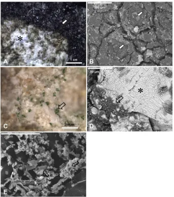
All photobiont from lichen thalli used for laser irradiation were photosynthetically active in the field and under the laboratory conditions, Fv/Fm values ranging between 0.60 and 0.79. The small dark brown crustose thallus ofV. nigrescens showed the typical aspect of this epilithic lichen (Fig. 1A and B).V. nigrescensproduces no substance responsible for stone darkening, but the natural color of the dolomite stone is masked by the thalli and, as a consequence, the substrate looks rather dark (Fig. 1A and B).
Stereomicroscope examinations proved that treatment condi-tions were very effective in removingV. nigrescensthalli (Fig. 1A). The laser irradiated area had been cleaned, and the fungi that composed the lichen basal layer had been efficiently removed (Fig. 1A, C and D). Although green color from algae is clearly visible on the surface by stereomicroscopy (Fig. 1C), remnants composed by severely damaged algal and fungal cells could be observed on the dolostone surface by SEM-SE (Fig. 1D and E).
These remnants are mainly formed by broken and empty fungal hyphae that showed altered cell walls (Fig. 1E). Several laser cleaned areas, as shown inFig. 1A and D, were analyzed using micro-Raman spectroscopy. The micro-Raman spectra of the dolomite stone acquired in control and stone areas colonized byV. nigrescensthalli before and after irradiation are shown inFig. 2, andTable 1lists the bands observed, together with their corresponding assignments. The spectrum of theV. nigrescenscontrol thalli (non irradiated) presents the characteristic bands of disordered and graphitic carbon, centered at 1340 and 1580 cm 1respectively, which are related to the pres-ence of organic compounds. WhenV. nigrescensthalli are irradiated, the spectra of the laser cleaned areas display the characteristic bands of calcium carbonate, as it can be observed in the dolomite control sample, which correspond to the vibration modes of the free CO2
3 ion of calcium carbonate. These bands are attributed to lattice vibrations at 175 and 299 cm 1, and to in-plane bending and symmetric stretch modes at 727 and 1097 cm 1, respectively. The spectrum also displays bands assigned to the hydrocarbons present in the dolomite stone at 1440 cm 1(
n
s(CeO)þn
(CeC)vibration mode), 1615 cm 1 (
n
a (CeO) vibration mode), and
1744 cm 1(
n
(C]O) stretching).
In general, irradiated stone showed similar aspect to the control, andfissures, fractures or porosity changes were not observed in the irradiated surface or internal zones of the stone (Fig. 3A, C, and E; Fig. 4AeD andFig. 5AeD) when observed by SEM-BSE and SEM-SE.
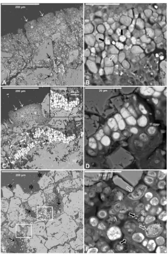
The anatomy of the epilithic lichen thalli and the cytology of pho-tobiont and mycobiont, and the microorganisms in endolithic zones before and after laser irradiation were examined by SEM-BSE and LTSEM (Figs. 3e5). Thin to moderately thickV. nigrescensthalli of 50e250
mm, including the basal layer, were observed on the
superficial layer of the dolostone (Fig. 3A). A typical thallus pres-ents a poorly differentiated upper cortex of variable width composed of thick, densely packed fungal cells (2.5e3.0
mm),
a photobiont layer formed by green algae (6.0e9.0
mm in
diam-eter), and mycobiont cells (3.0e6.0
mm in diameter) (Fig. 3B). The
mycobiont hyphae showed several small lipid globules occupying a substantial portion of the protoplast volume (Fig. 3B). InFig. 3A and C the medullary region can be seen, with abundant hyphae of 7e12
mm in diameter that have expanded through
fissures reachingdeep stone areas (up to 200
mm). The cells of fungal hyphae were
entirely occupied by lipids (oil cells). As lipids are very osmiophilic when inside a cell previously treated with osmium, a strong signal due to the lipids’osmium uptake is detected (Fig. 3C box). Other fungal clusters near the lichen thallus also showed high cyto-plasmatic lipid content (Fig. 3D).Laser irradiation left empty spaces in the dolomite surface that correspond with the spaces occupied by the lichen thalli before treatment. These can reach 80
mm from the surface (Fig. 3E
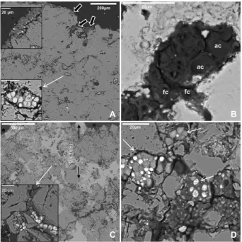
aster-isks). The microorganisms found in endolithic zones were also altered by laser surface irradiation as shown by SEM-BSE analyses (Fig. 3EeF andFig. 4). Most fungal cells near the stone surface werecompletely destroyed (Fig. 3E arrows) or showed high plasmolysis degrees (Fig. 3E box and F).
Fungal cytoplasm looked sponge-like and the lipid bodies could only be distinguished in some cells (Fig. 3F). Free fungi on the surface and endolithic colonization forms were also observed in split longitudinal sections of the dolostone using the SEM-BSE technique (Fig. 4A top box). In these samples, algal and fungal cell clusters were also frequently found randomly distributed in deep areas of the dolomite stone (Fig. 4A lower box). Fungi on the stone surface were removed by laser irradiation and the endolithic clusters were also severely affected (Fig. 4B). A similar but weaker effect was also observed in clusters of phototrophic and hetero-trophic microorganisms in deeper stone areas, down to 1000
mm
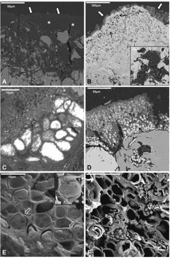
under the cleaned dolostone surface (Fig. 4C and D).Laser irradiation also affected lichen thalli and microorganisms near cleaned areas (Fig. 5). The cell damage severity observed depends on the localization of lichen thalli from the laser irradiated zone. First, on the border of the cleaned areas, lichen thalli were
observed to apparently keep their integrity (Fig. 5A). However, their algal and fungal cells had been strongly affected, as these cells were empty and only conserved their walls (Fig. 5A). Second, thalli with intermediate damage stages were detected near this area (Fig. 5B). In this case, in the lower part of the thalli some fungal cells with cytoplasm and lipid bodies remain although with evident signs of damage (Fig. 5C). The endolithic mycobiont hyphae as well as fungi and algal cell clusters under the thalli (Fig. 5B box) were also affected by the laser irradiation applied in the neighboring cleaned area. Laser-produced structural damage was evident when
3 1340
ity
1580 Verrucaria2
Verrucaria Irradiated
R
a
m
a
n
In
te
n
s
0 1
Dolomite
1440 1615 1744
175 299
727 1097
No
rm
a
liz
e
d
2000 1500 1000 500 0
Wavenumber / cm
-1Fig. 2.Micro-Raman spectra of the control dolostone sample and stone surface colo-nized byVerrucaria nigrescensbefore and after laser irradiation.
Fig. 1.Crustose thallus ofV. nigrescens(white arrows in A and B) and areoles immersed perithecia and their ostioli (arrows in B). Laser cleaned areas (asterisks in A and D) with lichen thallus remain (black arrows in A, CeE). A and C: stereomicroscopy images; B and D: SEM-SE images; E: LTSEM image.
Table 1
List of the bands and the corresponding assignments determined by micro-Raman spectroscopy in the dolomite andVerrucaria nigrescensthalli.
Wavenumbers (cm1) Vibrational assignment Origin of bands
1744 n(CaO) Hydrocarbons
1615 na(CeO) Hydrocarbons
1580 Graphitic carbon Organic compounds 1440 ns(CeO)þn(CeC) Hydrocarbons 1340 Disordered carbon Organic compounds 1097 Symmetric stretching ofðCO23 Þ CaCO3, dolomite 727 In-plane bending ofðCO2
3 Þ CaCO3, dolomite 299 Lattice vibration ofðCO2
3 Þ CaCO3, dolomite 175 Lattice vibration ofðCO2
compared to control thalli observed by SEM-BSE (Fig. 5D) and further confirmed by LTSEM (Fig. 5E and F).
Fungi and algae from control V. nigrescens thalli with well-defined cell walls and cytoplasmatic content could be observed by LTSEM (Fig. 5E). Some ultrastructural cell details in lichen pho-tobionts such as the chloroplast which often occupy most algal cell volume were also observed by LTSEM in the control sample (Fig. 5E box). However, lichen thalli close to the cleaning zone that showed
some apparent macroscopic integrity were actually formed only by algal and fungal cell walls, as their cytoplasms had been totally destroyed (Fig. 5F).
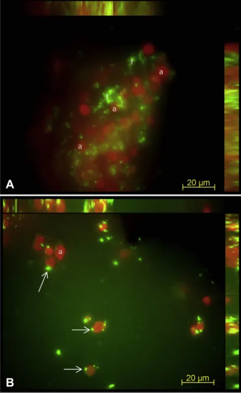
In addition to the SEM-BSE and LTSEM results, fluorescence microscopy confirms that laser irradiation also affected lichen thalli localized near cleaned areas.V. nigrescensthalli control (Fig. 6A) and thalli in the border area and near irradiated areas (Fig. 6B) were stained with the SYTOX Green Live/Dead dye. The red emission Fig. 3.SEM-BSE images ofV. nigrescensepilithic thalli and endolithic colonization of dolomite stone before (AeD) and after laser treatment (EeF). Upper cortex (thin white arrows in A and C), photobiont layer and mycobiont hyphae (white asterisks in A and C), fungal cells with several lipid globules (black arrows in B) and algae (white open arrows in B), and lichen medullar region (thin black arrows in A and C) from a controlV. nigrescensthalli. Strong signals produced by the OsO4staining of cytoplasmatic lipids in fungal hyphae from vital lichen thalli (box in C) and in clusters of fungi localized in endolithic zones (D). Empty spaces in the dolomite surface after laser removal of lichen thalli (black asterisks in E). Microorganisms localized in endolithic zones near the surface and lichen medullar region were completely destroyed (thin black arrows). Other zones showed fungal cells with a high degree of plasmolysis (white boxes in E and detail in F).
signal reveals autofluorescence signal emitted by chlorophyll in algal phototrophic cells, while the green emission signal comes from the SYTOX Green stained nucleic acids of photobiont and mycobiont cells with damaged plasma membranes (arrows inFig. 6B). While most algal cells in the control sample showed no green signal (Fig. 6A), almost all algal cells from lichen thalli near the irradiated area reveal SYTOX Green stained nucleic acids signal from cells with damaged plasma membranes (Fig. 6B).
4. Discussion
Nd:YAG laser treatment under the irradiations conditions used (1064 nm, 5 ns) was effective at removingV. nigrescenshydrated active thalli producing no chemical changes in the irradiated stone surface, as observed by micro-Raman, SEM-BSE, and SEM-SE analyses.V. nigrescensis a crustose epilithic lichen that is report-edly a widespread species on limestone heritage buildings and monuments throughout Europe (Blazquez et al., 1995;Nimis and Martellos, 2008; Smith et al., 2010) and also colonizes extensive areas of the Redueña dolostone quarry face (Madrid, Spain) (Cámara et al., 2011). In addition, severe cellular damage was produced not only in the endolithic mycobiont hyphae and neighboring lichen thalli but also in fungi and algae deep in the stone. Although identification of these microorganisms inside the stone was not carried out because it was not the main aim of the present work, these microorganisms showed similar morphologies
to those previously detected by SEM-BSE and identified by molecular biology in a nearby Redueña quarry face (Cámara et al., 2011).
The Fv/Fm values measured in the photobiont microorganisms in the laboratory were consistent with the range of values regis-tered in other crustose lichens (Schroeter et al., 1999;Tretiach et al., 2010) These values fell within the range determined in the quarry face in the autumn season when V. nigrescens is metabolically active, and confirmed the photobiont vitality of the selected samples for laser irradiation experiments.
Lichens show different sensibility to biocides related to thallus structure and physiological state, as well as to chemical biocide nature (Tretiach et al., 2010). The main problem detected during the biocide treatment of lichens encrusted in stone artworks was that considerable parts of the thalli still remained attached to the stone after several weeks of application of highly concentrated biocide with bristle brushes or poultices (deCruz et al., 2005;Caneva et al., 2008; Doehne and Price, 2010). In previous experiments, after biocide application, a mixture of dead and living microorganisms was found, these included living fungal forms infissures and under dead lichen thalli, and lichen thalli areas of no biocide action (Cámara et al., 2011;De los Ríos et al., 2012). In contrast, as shown in this and other works, laser cleaning produces complete lichen-thallus removal, even if lichen debris on the surface can be brushed away without affecting dolostone surface (Leavengood et al., 2000; deCruz et al., 2007, 2009). In addition, if the Fig. 4.SEM-BSE images of fungal epilithic and endolithic colonization of dolomite stone before (A) and after (BeD) laser treatment. Free fungi (black arrows and upper box in A) and endolithic algal and fungal cells (white thin arrow and lower box in A) in control sample. Endolithic clusters near the stone surface damaged by the laser heat effect (B). Clusters of phototrophic and heterotrophic microorganisms (white thin arrow and box in C, and white thin arrows in D) from deeper stone zones (until 1 mm) showed a weak laser effect. Empty spaces in the dolomite surface after laser removal of lichen thalli (black asterisks). fc: fungal cell; ac: algal cell.
considerable cell damage observed in lichen debris, as well as in the endolithic mycobiont cells and microorganisms in the deeper substrate areas is taken into account the possibility of stone recolonization from theses sources is rather low. The collateral damage produced in lichen thalli near cleaned areas is also important.
Laser application to lithic microorganisms and lichens without extracting them from their substrate have been scarcely studied (Leavengood et al., 2000;deCruz et al., 2007,2009). In general, the
biological component of stone crust, known as“biological crust”, is poorly characterized, and commonly used techniques enable no evaluation of laser-produced structural damage and vitality changes. In this sense, SEM-BSE microscopy allows assessing laser effect on lichen epilithic thalli and microorganisms in deep stone areas without extracting them from their microhabitat (Ascaso et al., 1998a,b, 2002; De los Ríos and Ascaso, 2005; Wierzchos and Ascaso, 1994,1996). SEM-BSE microscopy also provides infor-mation on cell-wall integrity and some ultrastructural aspects of Fig. 5.SEM-BSE (AeD) and LTSEM (EeF) images of lichen thalli and microorganisms localized near the laser cleaned areas. Lichen thalli next to the irradiated surface (white arrows in A) with empty algal and fungal cells that only conserved their walls (white asterisks in A). Fungal and algal cell clusters affected by the thermal laser (black thin arrows and box in B). Thalli from an intermediate localization (white arrows in B) showed evident fungal cell wall and lipid body damage (detail in C) compared with control thalli (D).V. nigrescens hydrated control thalli (E) showing algal cells (ac) and chloroplast (c) (white open arrows and box in E). The complete laser destruction of algal and fungal cells can be observed (F) with some fungal lipid body debris (black empty arrows).
lithic microorganisms that reflect their vitalityin situ(Ascaso et al., 1998b,2002;Cámara et al., 2011;De los Ríos and Ascaso, 2005;De los Ríos et al., 2004,2010,2012;Potgieter-Vermaak et al., 2005). In this way, the different degrees of laser-induced damage undergone by epilithic and endolithic microorganisms were determined.
Laser effect on lithic microorganisms may depend on different factors, including cellular structure, chemical composition, and location in the stone Lichen thallus anatomy, the presence of a protective upper cortex, thick mycobiont cell walls, and intimate thallusesubstrate relationship may represent certain additional
difficulties for the laser cleaning procedure. Under our experi-mental conditions, laser ablation led to complete removal of V. nigrescensepilithic hydrated thalli. However, the upper cortex of different crustose lichen, Diploicia canescens, was observed more resistant under the same laser irradiation conditions used for the removal ofV. nigrescens(Speranza et al., 2011). Higherfluence is proven necessary for thallus removal in this case. Even though Nd:YAG laser destruction of epilithic lichen and endolithic
microorganisms cannot be unequivocally ascribed to specific physical/chemical mechanisms, certain ideas based on previous research can however be pointed out. Laser-removal efficiency depends on light-material interaction, which is in turn related to wavelength,fluence, and the optical properties of the target such as light absorption and heat diffusion (Siano, 2007; deCruz et al., 2009). The influence of these factors during laser irradiation of Diploschistes scruposus(a grayish-white crustose lichen) had been proven in a recent study (deCruz et al., 2009). Moreover, the extent of laser cell damage depends on bothfluence and time of exposure to laser light. In bacteria the laser effect could be enhanced by the presence of pigments (Maravelaki-Kalaitzaki et al., 2003; Vural et al., 2007;Manevitch et al., 2010). Therefore, a rapid heat trans-fer to lichen thalli could take place whenV. nigrescensabsorbs laser light, as described for other lichens and microorganisms (Cappitelli et al., 2007; deCruz et al., 2007). In this way, the local heating induced by laser irradiation at 1064 nm could also affect the thalli near cleaned areas. This phenomenon could be responsible for cytoplasm damage of photobiont and mycobiont cells, especially in the case of alteration of fungal lipid bodies. Although several authors report diverse morphology of lichen oil cells, their function still remains unknown (Pinna et al., 1998; Gueidan et al., 2007; Kushnir and Galun, 1978).Kushnir and Galun (1978)state that oil hyphae do not occur in epilithic lichen. This fact is also supported by the interesting studies byPinna et al. (1998).
These authors describe oil-hyphae using SEM in secondary mode (SEM-SE), which allows observing the outside of the oil cells, and point out that true-oil hyphae occur beneath epilithic thalli only when these overgrow endolithic species. In our study, SEM in backscattered mode (SEM-BSE) enabled us to observe lipids inside the cells, both in the control sample and the laser-treated sample. We can perfectly describe cells with lipids (oil cells) belonging to the epilithic thalli, but it is difficult to say if there are true-oil hyphae or fungal cells with lipid inclusion (Pinna et al., 1998; Gueidan et al., 2007;Kushnir and Galun, 1978). SEM-BSE observa-tions allowed us to observe alteraobserva-tions in the lipidic integrity inside the cells of the laser-treated samples. In photobiont cells, damage to the photosynthetic system will also affect fungal survival and lichen viability.
From a practical point of view, lichen mechanical removal is unadvisable in order to not promote stone disintegrations in some circumstance. In this case, induced thermal damage of thalli could be a useful strategy to stop lichen stone biodeterioration activity, especially if the severe cytoplasmatic damage observed in the endolithic mycobiont hyphae is taken into account.
Moreover, certain resistance to the thermal laser effect was observed in mycobiont cell walls. The walls are responsible for the mechanical strength and thermal resistance of fungal cells (Osherov and Yarden, 2010). Furthermore, microenvironment heating could be responsible for the severe damage observed in microorganisms in endolithic areas. Similar microenvironment heating processes are responsible for the inactivation and death of unexposed microorganisms such as the bacteria inside the dentine microcrystalline structure during Nd:YAG laser treatments (Goodis et al., 1993). A characteristic of such a laser system is that it reaches deeper stone areas of interest when compared to UV rays and xenon lamps used against microorganisms and lichen on stone thatdin spite of their germicidal activitydalso display poor
penetration power (Goodis et al., 1993; Leavengood et al., 2000; Doehne and Price, 2010).
5. Conclusion
All results shown indicate that irradiation with a Q-switched Nd:YAG laser is a promising and environmentally friendly method Fig. 6.Fluorescence microscopy 3D reconstruction MIP images of SYTOX Green stained
lichen symbiont cells from control lichen thalli (A) and from lichen thalli located in neighbor laser cleaned zones (B). Image A reveals red autofluorescence signal of algal (a) cells and green autofluorescence of fungal cells (f). Image B show red
auto-fluorescence signal of algal (a) cells and green signal of nuclei of algal cells with damaged membranes (arrows). (For interpretation of the references to color in this
figure legend, the reader is referred to the web version of this article.)
to control lichen and microorganisms involved in stone biodeteri-oration. Although further studies are needed to elucidate the specific physical/chemical mechanisms involved, the results ob-tained extend the traditional idea that laser irradiation can only clean stone surfaces, thus proving an additional important laser effect related to the microbiota community involved in several deterioration mechanisms. The integration of SEM-BSE, LTSEM, FM, and micro-Raman analyses constitutes an effective strategy to determine the optimal laser irradiation regime to control both stone integrity and biological damage. Field experiments of laser irradiation of lichen thalli in the quarry are currently in progress and allow us to evaluate different parameters such as the biological recolonization of cleaned and irradiated surfaces.
Acknowledgments
Work funded by MICINN under Projects CTM2009-12838-CO4-O3, CTQ2010-15680 and CGL2010-16004, and CONSOLIDER CSD2007-00058, and the Programa Geomateriales (CAM, S2009/ Mat-1629). Thanks to Prof. L. G. Sancho and M. Rivas (UCM) for facilities and advice for PAM analyses. Thanks to F. Pinto and V. Souza from ICA, and T. Carnota from MNCN, for microscopy technical assistance, and to M. Castillejo and J. M. Hontoria from MNCN for sample preparation and to M. Furió Veja and A. J. García from MNCN for Raman technical assistance.
M. Speranza and S. Perez-Ortega are contract holders of the CSIC-JAE Fondo Social Europeo
References
Alakomi, H.-L., Paananen, A., Suihko, M.-L., Helander, I.M., Saarela, M., 2006. Weakening effect of cell permeabilizers on gram-negative bacteria causing biodeterioration. Applied and Environmental Microbiology, 4695e4703. Ascaso, C., Wierzchos, J., Castelló, R., 1998a. Study of the biogenic weathering of
calcareous litharenite stones caused by lichen and endolithic microorganisms. International Biodeterioration and Biodegradation 42, 29e38.
Ascaso, C., Wierzchos, J., Delgado Rodrigues, J., Aires-Barros, L., Henriques, F.M.A., Charola, A.E., 1998b. Endolithic microorganisms in the biodeterioration of the tower of Belem. International Zeitschrift für Bauinstandsetzen 4, 627e640. Ascaso, C., Wierzchos, J., Souza-Egipsy, V., De los Rios, A., Delgado Rodrigues, J.,
2002. In situ evaluation of the biodeteriorating action of microorganisms and the effects of biocides on carbonate rock of the Jeronimos Monastery (Lisbon). International Biodeterioration and Biodegradation 49, 1e12.
Bartolini, M., Pietrini, A.M., Ricci, S., 1999. Use of UV-C irradiation on artistic stone works for control of algae and cyanobacteria. In: Ciferri, O., Tiano, P., Mastromei, G. (Eds.), Microbes and Art: the Role of Microbial Communities in the Degradation and Protection of Cultural Heritage. CNR, Florence, pp. 221e227.
Blazquez, F., Calvet, F., Vendrellc, M., 1995. Lichen alteration and mineralization in calcareous monuments of northeastern Spain. Geomicrobiology Journal, 223e247.
Burns, T., Wilson, M., Pearson, G., 1993. Sensitisation of cariogenic bacteria to killing by light from a helium-neon laser. Journal of Medical Microbiology 38, 401e405.
Cámara, B., De los Ríos, A., Urizal, M., Álvarez de Buergo, M., Varas, M.J., Fort, R., Ascaso, C., 2011. Characterizing the microbial colonization of a dolostone quarry: implications for stone biodeterioration and response to biocide treat-ments. Microbial Ecology 62, 299e313.
Caneva, G., Nugari, M.P., Salvadori, O., 2008. Control of biodeterioration and bioremediation techniques. In: Caneva, G., Nugari, M.P., Salvadori, O. (Eds.), Plant Biology for Cultural Heritage: Biodeterioration and Conservation. Getty Conservation Institute, Los Angeles, pp. 309e346.
Cappitelli, F., Nosanchuk, J., Casadevall, A., Toniolo, L., Brusetti, L., Florio, S., Principi, P., Borin, S., Sorlini, C., 2007. Synthetic consolidants attacked by melanin-producing fungi: case study of the biodeterioration of Milan (Italy) Cathedral marble treated with acrylics. Applied and Environmental Microbi-ology 73, 271e277.
Clauzade, G., Roux, C., Houmeau, J.M., Raimbault, P., 1985. Likenoj de Okcidenta Europo. Ilustrita Determinlibro.
Cooper, M., 1998. Laser Cleaning in Conservation: an Introduction. Butterworth Heinemann, Oxford.
De los Ríos, A., Ascaso, C., 2005. Contributions of in situ microscopy to the current understanding of stone biodeterioration. International Microbiology 8, 181e188.
De los Ríos, A., Ascaso, C., Wierzchos, J., Eduardo, F.-V., Quesada, A., 2004. Micro-structural characterization of cyanobacterial mats from the McMurdo ice shelf, Antarctica. Applied and Environmental Microbiology 70, 569e580.
De los Ríos, A., Ascaso, C., Wierzchos, J., Sancho, L.G., 2010. Spaceflight effects on lichen ultrastructure and physiology. In: Seckbach, J., Grube, M. (Eds.), Stress and Symbiosis.
De los Ríos, A., Perez-Ortega, S., Wierzchos, J., Ascaso, C., 2012. Differential effects of biocide treatments on saxicolous communities: case study of the Segovia cathe-dral cloister (Spain). International Biodeterioration and Biodegradation 67, 64e72. deCruz, A., Palmer, R.A., Culberson, C.F., Andreotti, A., Colombini, M.P., Pinna, D., 2007. Preliminary investigation of Er:YAG laser ablation for the removal of lichen from stone. In: Joyce, H., Townsend, L.T., Francesca, C. (Eds.), Conservation Science. Archetype Publications, London, p. 308.
deCruz, A., Wolbarsht, M.L., Andreotti, A., Colombini, M.P., Pinna, D., Culberson, C.F., 2009. Investigation of the Er:YAG laser at 2.94 mm to remove lichens growing on stone. Studies in Conservation 54, 268e277.
deCruz, A., Wolbarsht, M.L., Palmer, R.A., Pierce, S.E., Adamkiewicz, E., 2005. Er:YAG laser applications on marble and limestone sculptures with polychrome and patina surfaces. In: Lasers in the Conservation of Artworks. Springer Proceed-ings in Physics.10.1007/1003-1540-27176-27177_27114.
Doehne, E., Price, C.A., 2010. Stone Conservation: an Overview of Current Research, second ed. The Getty Conservation Institute, Los Angeles, CA.
European-Commission-Regulation No. 1451/2007, 2007. The second phase of the 10-year work program referred to in Article 16(2) of Directive 98/8/EC of the European Parliament and of the Council concerning the placing of biocidal products on the market. Official Journal of the European Journal, 3e64. Gomoiu, I., Radvan, R., Sarantopoulou, E., Cefalas, A.C., 2006. Lasers in
biodeterio-ration. In: Schreiner, M., Strlic, M. (Eds.), Handbook on the Use of Lasers in Conservation and Conservation Science. COST office G7, p. 20.
Goodis, H.E., White, J.M., Marshall, S.J., Marshall, G.W., 1993. Scanning electron microscopy examination of intracanal wall dentin: hand versus laser treatment. Scanning Microscopy 7, 979e987.
Gueidan, C., Roux, C., Lutzoni, F., 2007. Using a multigene phylogenetic analysis to assess generic delineation and character evolution inVerrucariaceae (Verru-cariales, Ascomycota). Mycological Research 111, 1145e1168.
Kushnir, E.T.,A., Galun, M., 1978.“Oil hyphae”of endolithic lichens and their fatty
acid composition. Protoplasma 97, 47e60.
Leavengood, P., Twilley, J., Asmus, J.F., 2000. Lichen removal from Chinese Spirit pathfigures of marble. Journal of Cultural Heritage, s71es74.
López, A., Lamas, J., Ramil, A., Yáñez, A., Rivas, T., Silva, B., Taboada, J., 2010. Opti-mization of laser cleaning parameters for the removal of biological black crusts in granites. In: Radvan, R., Asmus, J.F., Castillejo, M., Pouli, P., Nevin, A. (Eds.), Laser in the Conservation of Artworks VIII. CRC Press, p. 227.
Manevitch, Z., Lev, D., Hochberg, M., Palhan, M., Lewis, A., Enk, C.D., 2010. Direct antifungal effect of femtosecond laser onTrichophyton rubrumonychomycosis. Photochemistry and Photobiology 86, 476e479.
Marakis, G., Maravelaki, P., Zafiropulos, V., Klein, S., Hildenhagen, J., Dickmann, K., 2000. Investigations on cleaning of black crusted sandstone using different UV-pulsed lasers. Journal of Cultural Heritage 1, s61es64.
Maravelaki-Kalaitzaki, P., Zafiropulos, V., Pouli, P., Anglos, D., Balas, C., Salimbeni, R., Siano, S., Pini, R., 2003. Short free running Nd:YAG laser to clean different encrustations on Pentelic marble: procedure and evaluation of the effects. Journal of Cultural Heritage 4, 77se82s.
Nandakumar, K., Obika, H., Shinozaki, T., Ooie, T., Utsumi, A., Yano, T., 2002. Impact of pulsed Nd:YAG laser irradiation on the growth and mortality of the biofilm forming marine bacteriumPseudoalteromonas carrageenovora. Biofouling 18, 123e127.
Nandakumar, K., Obika, H., Utsumi, A., Ooie, T., Yano, T., 2004. In vitro laser ablation of laboratory developed biofilms using an Nd:YAG laser of 532 nm wavelength. Biotechnology and Bioengineering 86, 729e736.
Nimis, P.L., Martellos, S., 2008. The Information System on Italian Lichens, Version 4.0. University of Trieste, Department of Biology, IN4.0/1.
Osherov, N., Yarden, O., 2010. The cell wall offilamentous fungi. In: Borkovich, K.A., Ebbole, D.J. (Eds.), Cellular and Molecular Biology of Filamentous Fungi. ASM Press, Washington D.C., pp. 224e237.
Oujja, M., Rebollar, E., Castillejo, M., Domingo, C., Cirujano, C., Guerra-Librero, F., 2005. Laser cleaning of terracotta decorations of the portal of Palos of the Cathedral of Seville. Journal of Cultural Heritage 6, 321e327.
Pinna, D., Salvadori, O., Tetriach, M., 1998. An anatomical investigation of calcicolous endolithic lichens from the Trieste karst (NE Italy). Plant Bio-systems 132, 183e195.
Potgieter-Vermaak, S.S., Godoi, R.H.M., Grieken, R.V., Potgieter, J.H., Oujja, M., 2005. Micro-structural characterization of black crust and laser cleaning of building stones by micro-Raman and SEM techniques. Spectrochimica Acta Part A: Molecular and Biomolecular Spectroscopy 61, 2460e2467.
Pouli, P., Fotakis, C., Hermosin, B., Saiz-Jimenez, C., Domingo, C., Oujja, M., Castillejo, M., 2008. The laser-induced discoloration of stonework; a compara-tive study on its origins and remedies. Spectrochimica Acta Part A: Molecular and Biomolecular Spectroscopy, 932e945.
Pouli, P., Oujja, M., Castillejo, M., 2011. Practical issues in laser cleaning of stone and painted artefacts: optimisation procedures and side effects. Applied Physics A: Materials Science & Processing, 1e18.
Sarantopoulou, E., Kollia, Z., Gomoiu, I., 2006. Preventing biological activity of Ulocladium sp spores in artifacts using 157-nm laser. Applied Physics A: Materials Science & Processing 83, 663e668.
SCENIHR, 2009. Assessment of the Antibiotic Resistance Effects of Biocides. Scien-tific Committee on Emerging and Newly Identified Health Risks, European Commission Health & Consumer Protection DG, Brussels, p. 87.
Scheerer, S., Ortega-Morales, O., Gaylarde, C., 2009. Microbial deterioration of stone monumentsdan updated overview. Advances in Applied Microbiology 66, 97e139 (Chapter 5).
Schroeter, B., Sancho, L.G., Valladares, F., 1999. In situ comparison of daily photo-synthetic activity patterns of saxicolous lichens and mosses in Sierra de Gua-darrama, Central Spain. The Bryologist 102, 623e633.
Siano, S., 2007. Principles of laser cleaning in conservation. In: Schreiner, M., Strlic, M., Salimbeni, R. (Eds.), Handbook on the Use of Laser in Conservation and Conservation Science. COST office, G7, Belgium.
Smijs, T.G.M., Schuitmaker, H.J., 2003. Photodynamic inactivation of the dermato-phyte Trichophyton rubrum. Photochemistry and Photobiology 5.
Smith, C.W., Aptroot, A., Coppins, B.J., Fletcher, A., Gilbert, O.L., James, P.W., Wolseley, P.A., 2010. The Lichens of Great Britain and Ireland. British Lichen Society London.
Speranza, M., Sanz, M., Oujja, M., de los Ríos, A., Pérez-Ortega, S., Ascaso, C., Cas-tillejo, M., 2011. Laser cleaning investigation to remove biological crust on stone. SPIE Optical Metrology Conference O3A: Optics for Arts, Architecture and Archeology III Technical Summaries Munich, Germany.
Tornari, V., Fotakis, C., Georgiou, S., Zafiropulos, V., Anglos, D., 2006. Laser cleaning of encrustation. In: Fotakis, C., Anglos, D., Georgiou, S., Zafiropulos, V., Tornari, V. (Eds.), Laser in the Preservation of Cultural Heritage Principles and Applications. Taylor & Francis, p. 336.
Tretiach, M., Bertuzzi, S., Salvadori, O., 2010. Chlorophyll afluorescence as a prac-tical tool for checking the effects of biocide treatments on endolithic lichens. International Biodeterioration and Biodegradation 64, 452e460.
Tretiach, M., Crisafulli, P., Imai, N., Kashiwadani, H., Heemoon, K., Wada, H., Salvadori, O., 2007. Efficacy of a biocide tested on selected lichens and its effects on their substrata. International Biodeterioration and Biodegradation 59, 44e54. Vural, E., Winfield, H.L., Shingleton, A.W., Horn, T.D., Shafirstein, G., 2007. The effects of laser irradiation onTrichophyton rubrumgrowth. Lasers in Medical Science 23, 349e353.
Wierzchos, J., Ascaso, C., 1994. Application of back-scattered electron imaging to the study of the lichen-rock interface. Journal of Microscopy 175, 54e59. Wierzchos, J., Ascaso, C., 1996. Morphological and chemical features of
bio-weathered granitic biotite induced by lichen activity. Clays and Clay Minerals 44, 652e657.