L
Journal of Experimental Marine Biology and Ecology 249 (2000) 219–233
www.elsevier.nl / locate / jembe
The synthesis of mycosporine-like amino acids (MAAs) by
cultured, symbiotic dinoflagellates
a ,1 b b ,*
Anastazia T. Banaszak, , Todd C. LaJeunesse , Robert K. Trench
a
Smithsonian Environmental Research Center, Edgewater, MD 21037, USA b
Department of Ecology, Evolution and Marine Biology, University of California at Santa Barbara, Santa Barbara, CA 93106, USA
Received 16 November 1999; received in revised form 18 February 2000; accepted 10 March 2000
Abstract
We tested the hypothesis that there is a relation between phylotypes (phylogenetic types, as determined by restriction fragment length polymorphism (RFLP) and partial sequence analysis of the small subunit ribosomal RNA gene (SSUrDNA)) and the synthesis of mycosporine-like amino acids (MAAs) by symbiotic dinoflagellates under the influence of ultraviolet radiation (UV-B /A) and photosynthetically active radiation (PAR). We exposed 27 isolates of symbiotic dinoflagellates simultaneously to UV-B /A and PAR, and subsequently determined the MAAs present in cell extracts and in the media. The algae used included 24 isolates of Symbiodinium spp. originating from jellyfishes, sea anemones, zoanthids, scleractinians, octocorals, and bivalves, and three others in the genera Gymnodinium, Gloeodinium and Amphidinium from a jellyfish, an hydrocoral and a flatworm, respectively. In this study, all of the phylotype A Symbiodinium spp. synthesized up to three identified MAAs. None of the 11 cultured phylotypes B and C Symbiodinium spp. synthesized MAAs. The three non-Symbiodinium symbionts also synthesized up to three MAAs. The results support a conclusion that phylotype A Symbiodinium spp. have a high predilection for the synthesis of MAAs, while phylotypes B and C do not. Synthesis of MAAs by symbiotic dinoflagellates in culture does not appear to relate directly to depths or to the UV exposure regimes from which the consortia were collected. 2000 Elsevier Science B.V. All rights reserved.
Keywords: Coral bleaching; Mycosporine-like amino acids (MAAs); Symbiotic dinoflagellates; Amphidinium; Gloeodinium; Symbiodinium; Ultraviolet radiation
*Corresponding author. Tel.: 11-805-893-8955; fax: 11-805-893-4724. E-mail address: [email protected] (R.K. Trench)
1
´
Present address: Estacion de Investigaciones Marinas ‘Puerto Morelos’, Instituto de Ciencias del Mar y
´ ´ ´ ´ ´
Limnologıa, Universidad Nacional Autonoma de Mexico, Apartado Postal 1152, Cancun 77500, Mexico. 0022-0981 / 00 / $ – see front matter 2000 Elsevier Science B.V. All rights reserved.
220 A. T. Banaszak, et al. / J. Exp. Mar. Biol. Ecol. 249 (2000) 219 –233
1. Introduction
In clear, tropical, shallow-water environments such as coral reefs, the fluence of ultraviolet radiation (UVR, 280–400 nm), particularly UV-B (280–320 nm), is high (Smith and Baker, 1979; Baker et al., 1980). The seminal study by Jokiel (1980) introduced the concept that UVR plays an important role in the diversity and abundance of benthic, coral-reef dwelling organisms. Jokiel and York (1982) highlighted the deleterious effects of solar UVR on the photosynthetic performance of the endo-symbiotic dinoflagellates in the coral Pocillopora damicornis, and the resulting impairment of skeletal growth. The discovery of UV-absorbing substances, ‘S-320’, by Shibata (1969), (later found to be mycosporine-like amino acids, MAAs), and the subsequent observation of an inverse relation in corals between the presence (and / or concentration) of ‘S-320 / MAAs’ and depth (Maragos, 1972; Dunlap et al., 1986; Gleason and Wellington, 1995; Kuffner et al., 1995; Shick et al., 1995; Banaszak et al., 1998) led to the concept that ‘S-320 / MAAs’ performed a photo-protective (sun-screen) function against UVR (Bandaranayake, 1998; Dunlap and Shick, 1998). That ‘S-320’ were MAAs was demonstrated by Dunlap and Chalker (1986). As the parent class of mycosporines are fungal metabolites synthesized via the shikimate pathway (Favre-Bonvin et al., 1987), and there is no known equivalent biosynthetic pathway in animals (Bentley, 1990), it was assumed that symbiotic algae synthesized MAAs and provided them to their hosts. This suggestion was sustained by the observation that the non-symbiotic dinoflagellate Alexandrium excavatum synthesized MAAs when exposed to UVR (Carreto et al., 1990a,b). Shick et al. (1999) have provided evidence for the involvement of the shikimate pathway in MAA synthesis in the stony coral Stylophora
pistillata.
Consistent with the hypothesis of symbiont synthesis of MAAs, Banaszak and Trench (1995b) demonstrated that cultured Symbiodinium microadriaticum, exposed simul-taneously to UV-B (280–320 nm) /A (320–400 nm) and photosynthetically active radiation (PAR, 400–700 nm), synthesized quantitatively more MAAs than when exposed to PAR only. Significantly, the same MAAs were detected in the cells and in the culture medium. The tissues of Cassiopeia xamachana collected at Lee Stocking Island, Bahamas, and S. microadriaticum freshly isolated from the jellyfish, both contained the same suite of three MAAs, but the aposymbiotic scyphistomae did not synthesize MAAs, even under the stimulus of UV-B /A. By contrast, the animal tissues of aposymbiotic and symbiotic Anthopleura elegantissima contained up to seven MAAs, but neither freshly isolated symbionts nor cultured S. californium demonstrated evidence of MAAs, even when exposed to UV-B /A (cf. Stochaj et al., 1994). It was suggested that the animals acquired MAAs from their diet. Analogous situations may exist in the Caribbean zoanthid Palythoa caribaeorum (Lesser et al., 1990) and the scleractinian coral Acropora microphthalma (Shick et al., 1995).
1998), and sensitivity to temperature and light, has been hypothesized for the
Montastraea annularis species complex (Rowan et al., 1997). Phylotypes A and B are
regarded as high-light / high-temperature adapted, and are likely to persist, whereas phylotype C algae are considered to be low-light / low-temperature adapted, and tend to be eliminated during ‘stressful’ events (Rowan, 1998). Baker and Rowan (1997) found
´
that all the corals they analysed from the Pacific coast of Panama harboured
Symbiodinium that conformed to phylotype C, and several of these are known to occur
in very shallow habitats. It has been shown that there is no correlation between algal phylotype and (i) photo-acclimatory capability with respect to PAR (Iglesias-Prieto and Trench, 1994, 1997a,b), or (ii) temperature tolerance (Fitt and Warner, 1995; Warner et al., 1996, 1999). Although Warner et al. (1996) did not record the genetic types of the algae in the corals they studied, data from Rowan et al. (1997) and Baker and Rowan (1997) show that the algae in Montastraea annularis (at depth, 15 m), Agaricia
lamarckii (15 m), and A. agaricites (15 m), are all phylotype C, and we have found that
the algae in Siderastrea radians (1.5 m) are also phylotype C. These algae demonstrate different temperature tolerances, the latter being quite resistant. To date, there have been no analyses attempting to test a correlation of phylotypes of Symbiodinium with the synthesis of MAAs under the stimulus of UVR. Rowan (1998) emphasized the importance of such analyses using cloned algal cultures.
We exposed 27 cultured isolates of symbiotic dinoflagellates simultaneously to UV-B /A and PAR, and analyzed the extracts of the cells, and the media in which they grew, for MAAs. Twenty-four of the symbionts are in the genus Symbiodinium representing phylotypes A, B and C from sea anemones, zoanthids, scleractinian corals, jellyfishes, octocorals and bivalve molluscs, and three others, including an Amphidinium sp. from a flatworm, Gloeodinium viscum from a hydrocoral, and Gymnodinium
linucheae from a jellyfish. The results indicate that there is a high predilection for
phylotype A Symbiodinium to synthesize MAAs under the influence of UVR and PAR, while there is a low propensity for phylotypes B and C Symbiodinium to synthesize MAAs. All the non-Symbiodinium dinoflagellates tested synthesized MAAs.
2. Materials and methods
2.1. Sources of symbiotic dinoflagellates and growth conditions
The identity and geographic origins of the symbiotic dinoflagellates, and the hosts (with the depth of collection) from which they were isolated are shown in Table 1. For routine maintenance, the algae were grown in the defined medium ASP-8A (Blank,
22 21
1987), at 75 mmol quanta m s PAR (measured in the flask with a QSL-100 meter
222 A. T. Banaszak, et al. / J. Exp. Mar. Biol. Ecol. 249 (2000) 219 –233
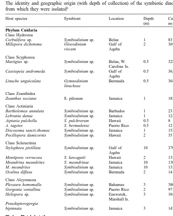
Table 1
The identity and geographic origin (with depth of collection) of the symbiotic dinoflagellates and the hosts a
from which they were isolated
Host species Symbiont Location Depth Culture
Phylo-(m) no. type
Phylum Cnidaria Class Hydrozoa
Corbulifera sp. Symbiodinium sp. Belau 1 83 B
b Millepora dichotoma Gloeodinium Gulf of 2 369 –
viscum Aqaba
Class Scyphozoa
Mastigias sp. Symbiodinium sp. Belau, W. 0.5 328 A Caroline Is.
Cassiopeia andromeda Symbiodinium sp. Gulf of 0.5 362 A Aqaba
b Linuche unguiculata Gymnodinium Bermuda 0.5 368 –
linucheae
Class Zoanthidea
Zoanthus sociatus S. pilosum Jamaica 1 185 A
Class Actiniaria
Bartholomea annulata Symbiodinium sp. Barbados 1 23 A Lebrunia danae Symbiodinium sp. Jamaica 1 125 B Aiptasia pulchella S. pulchrorum Hawaii 0.5 8 B A. tagetes S. bermudense Puerto Rico 0.5 12 B Discosoma sancti-thomae Symbiodinium sp. Jamaica 1 152 C Pocillopora damicornis Symbiodinium sp. Hawaii 2 351 B
Class Scleractinia
Stylophora pistillata Symbiodinium sp. Gulf of 10 370 A Aqaba
Monitpora verrucosa S. kawagutii Hawaii 2 135 C Meandrina meandrites S. meandrinae Jamaica 10 130 A
M. meandrites Symbiodinium sp. Jamaica 10 133 C
Oculina diffusa Symbiodinium sp. Bermuda 2 141 B
Class Alcyonacea
Plexaura homomalla Symbiodinium sp. Bahamas 3 380 A Gorgonia ventallina Symbiodinium sp. Puerto Rico 2 97 A Heliopora sp. Symbiodinium sp. Enewetok, 3 104 A
Marshall Is. Psuedopterogorgia
bipinnata Symbiodinium sp. Jamaica 3 147 B
Phylum Platyhelminthes Class Turbellaria
b Amphiscolops Amphidinium sp. Belau 3 19 – australis (?)
Phylum Mollusca Class Bivalvia
Corculum cardissa S. corculorum Belau 0.5 350 A Tridacna crocea Symbiodinium sp. GBR, 1 168 A
Australia
T. crocea Symbiodinium sp. Belau 1 220 A T. gigas Symbiodinium sp. GBR, 3 169 A
Australia
Hippopus hippopus Symbiodinium sp. Belau 10 203 C
a
The culture identification number and phylotype designation for each symbiont are also given. Phylotype designation was determined by restriction fragment length polymorphism (RFLP) analysis and partial sequencing of the small subunit ribosomal RNA gene (SSUrDNA). GBR, Great Barrier Reef.
b
22 21
were exposed to 100mmol quanta m s PAR (measured in the flask) delivered by 40 W Duro-Test Vita-Lite fluorescent tubes, and UV-B /A simultaneously, with temperature and photoperiod as above. The UV source was a G30T8 tube (Panasonic PPF, Hammond, IN, USA). A 6-mm thick UV-transparent Plexiglas shield (cut off at about 280 nm) was used to eliminate wavelengths in the UV-C (,280 nm) range (Banaszak and Trench, 1995a). The resulting spectrum, taken with a LI-COR Spectral Radiometer (LI-1800, Lincoln, Nebraska) is shown in Fig. 1. The UV flux measured beneath the Plexiglas shield but outside the flasks at 310 nm with a UVX-31 Radiometer (UVP, San
22
Gabriel, CA, USA) was 85mW cm . The cells were grown under these conditions for 1 month, after which they were harvested by centrifugation (8003g). The yield of algal
6 21
cells in all cases was approximately similar (ca. 2310 cells ml ). The cell pellets and the media were separately lyophilized, and stored under N2 gas at 2808C until analysed.
2.2. Analysis of MAAs
The lyophilized algal cells and media were extracted overnight with HPLC-grade absolute methanol at 48C. After centrifugation, the supernatants were analysed for MAAs as described by Dunlap and Chalker (1986), using reverse-phase, isocratic HPLC with a Brownlee RP-8 column. The mobile phase consisted of 25% (v / v) methanol,
21
0.1% (v / v) glacial acetic acid in H O, and the flow rate was 0.7 ml min2 . A diode array, UV absorbance detector (Beckman Gold System) was employed, along with secondary standards for mycosporine-glycine, shinorine, porphyra-334, palythine, asterina-330, palythinol, and palythene to allow for identification of peaks, both by their retention times and absorbance spectra (300–400 nm), including the wavelengths of maximum absorption. The primary standards were originally isolated by Walter Dunlap, and were kindly provided by Deneb Karentz and Michael Lesser. In many analyses there
224 A. T. Banaszak, et al. / J. Exp. Mar. Biol. Ecol. 249 (2000) 219 –233
were components (with absorption maxima at 300 nm or less) that could not be identified, but these do not include any of the samples that were scored as ‘non-synthesizers’ in Table 2.
2.3. Gene typing
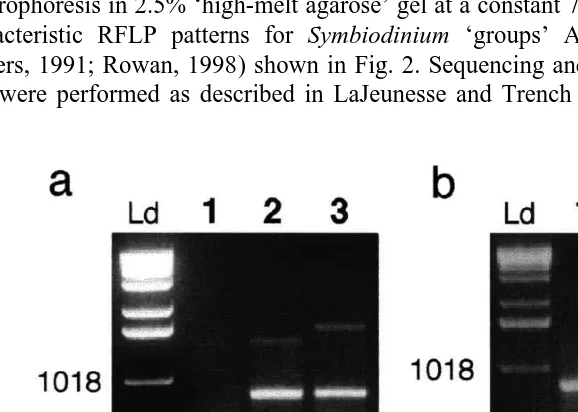
The phylotype designations used here were determined by restriction fragment length polymorphism (RFLP) analysis and partial sequencing of the small subunit ribosomal RNA gene (SSUrDNA) as described by Rowan and Powers (1991). DNA extractions were conducted on approximately 25 mg of algal material using a Proteinase-K, SDS–b-mercaptoethanol digestion, phenol–chloroform extraction, and ethanol precipi-tation protocol. SSUrDNA was amplified from resuspended genomic DNA using the primers described by Rowan and Powers (1991) and a Perkin-Elmer 2400 Thermal Cycler. The amplification conditions were: an initial denaturation step of 3 min at 928C followed by 35 cycles of 30 s at 928C, 40 s at 528C and 30 s at 728C. Restriction digests were performed with the endonucleases TaqI and DpnII. Fragments were separated by electrophoresis in 2.5% ‘high-melt agarose’ gel at a constant 70 V for 3 h, producing the characteristic RFLP patterns for Symbiodinium ‘groups’ A, B and C (Rowan and Powers, 1991; Rowan, 1998) shown in Fig. 2. Sequencing and phylogenetic reconstruc-tion were performed as described in LaJeunesse and Trench (2000).
3. Results and discussion
Rowan and Powers (1991) established the relation between RFLP typing and the phylogenetic affiliation of symbiotic dinoflagellates in the genus Symbiodinium. Using the same RFLP methods (Fig. 2), we find the same relation (sequence data not shown). It should be noted that the phylotype designations are not species (Rowan and Powers, 1991), as there are several distinguishable species that occur in each of the phylotypes A, B and C (Rowan, 1998). From our analyses, the alga Symbiodinium californium ([383) from Anthopleura elegantissima that we have cultured, does not conform to
phylotype B, and is in a group unto itself, and is most closely related to the non-symbiotic Gymnodinium varians (LaJeunesse and Trench, 2000). The SSUrDNA sequence indicates that this is not the same alga as that found to be the phylotype B in Rowan and Powers (1991), and this is further supported by analyses of the ITS regions (LaJeunesse and Trench, 2000). The symbiont that we have cultured from Pocillopora
damicornis from Hawaii conforms to phylotype B, and is not the same as the phylotype
C alga reported in this coral species by Rowan and Powers (1991). We have also cultured two different isolates from the coral Meandrina meandrites; isolate [130
conforms to phylotype A (cf. sequence in McNally et al., 1994), while isolate [133
conforms to phylotype C. Using RFLP analyses of the large subunit rDNA (LSUrDNA), Baker and Rowan (1997) found this same species of coral to harbour a phylotype B
Symbiodinium, while in Bermuda, Z. Billinghurst (pers. commun.), using RFLP of the
SSUrDNA, found this coral species harbouring phylotypes A or B. Thus, it appears that
P. damicornis may harbour two taxa, and M. meandrites may harbour three taxa of Symbiodinium. With the exception of the algae from Hippopus hippopus from Belau, Symbiodinium sp. ([203), which conformed to phylotype C, all of the isolates obtained
from tridacnids conformed to phylotype A.
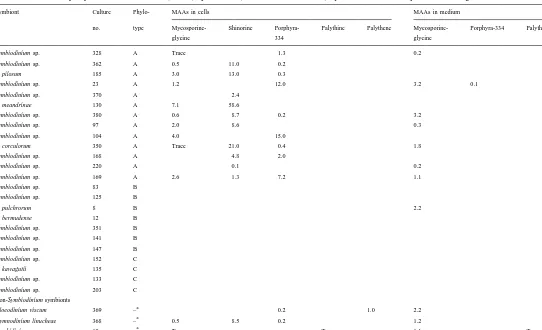
Table 2 shows that the three most frequently detected MAAs were porphyra-334 (34%), mycosporine-glycine (34%) and shinorine (29%); palythine and palythene were synthesized only by Amphidinium sp. and G. viscum, respectively. Two of the cultures produced only one MAA (shinorine in both cases), eight produced two MAAs, and six produced three. In S. pulchrorum, no MAAs were detected in the cell extracts, but mycosporine-glycine was detected in the culture medium. In other instances, either no mycosporine-glycine was detected, or detected only in low concentrations (e.g.,
Symbiodinium sp. ([220) from Tridacnacrocea, Symbiodinium sp. ([328) from
Mastigias sp., S. corculorum, and Amphidinium sp. ([19)) in the cell extracts, while the
226
Characterization of mycosporine-like amino acids (MAAs) synthesized (in nmol ml of extract) by various isolates of symbiotic dinoflagellates in culture
Symbiont Culture Phylo- MAAs in cells MAAs in medium
no. type Mycosporine- Shinorine Porphyra- Palythine Palythene Mycosporine- Porphyra-334 Palythine
glycine 334 glycine
Symbiodinium sp. 328 A Trace 1.3 0.2
Symbiodinium sp. 362 A 0.5 11.0 0.2 S. pilosum 185 A 3.0 13.0 0.3
Symbiodinium sp. 23 A 1.2 12.0 3.2 0.1
Symbiodinium sp. 370 A 2.4 S. meandrinae 130 A 7.1 58.6
Symbiodinium sp. 380 A 0.6 8.7 0.2 3.2
Symbiodinium sp. 97 A 2.0 8.6 0.3
Symbiodinium sp. 104 A 4.0 15.0
S. corculorum 350 A Trace 21.0 0.4 1.8
Symbiodinium sp. 168 A 4.8 2.0
Symbiodinium sp. 220 A 0.1 0.2
Symbiodinium sp. 169 A 2.6 1.3 7.2 1.1
Symbiodinium sp. 83 B
Gloeodinium viscum 369 – 0.2 1.0 2.2
a
Gymnodinium linucheae 368 – 0.5 8.5 0.2 1.2
a
Amphidinium sp. 19 – Trace Trace 1.1 Trace
a
bacteria themselves synthesized the mycosporine-glycine, or the algae released some MAA (not detectable in the algae themselves) which was converted to mycosporine-glycine by the bacteria. Dunlap and Shick (1998) suggested that Vibrio harveyi can hydrolyze the hydroxyamino acid substituents of shinorine and porphyra-334, to yield mycosporine-glycine. This could be consistent with our finding that in eight cultures (G.
linucheae, G. viscum, S. corculorum and Symbiodinium spp.[97, [169, [220, [328
and[380), extracts of the cells indicated the presence of shinorine and / or porphyra-334
in low concentrations, but the media contained mycosporine-glycine, sometimes in relatively high concentrations. We have tested the media from the cultures containing extracellular MAAs for bacterial contamination by plating cultures on LB bacto-agar, and we found evidence of bacterial contamination in cultures of algae from
Pseudo-pterogorgia homomalla ([380), and S. pulchrorum, but not in algae from Bartholomea
annulata ([23) where the culture medium had the same two MAAs as the cell extracts.
Our observations would therefore not be consistent with bacterial involvement in the synthesis of mycosporine-glycine. In other instances, either no MAAs were found in the media, or the same MAAs were found in cell extracts and media.
With the exception of Banaszak and Trench (1995b) and Vernet and Whitehead (1996), reported studies of MAA synthesis by microalgae do not include analyses of the culture media, and algae are regarded as synthesizers of MAAs, or not, based on whether the MAAs are detected in cell extracts. To be consistent with these other studies, in instances where no MAAs were found in cell extracts, we have scored these as ‘non-synthesizers’, even if MAAs were found in the media.
All cultured phylotype A Symbiodinium spp. synthesized from one to three identified MAAs (Table 2), including those from the tridacnids and Corculum (cf., Ishikura et al., 1997). The two isolates from T. crocea, [168 and [220, from Australia and Belau,
respectively, had identical SSUrDNA and ITS sequences (data not shown), but the former synthesized two MAAs while the latter synthesized only one, and in low concentration. Among the phylotype A algae, the three MAAs, shinorine, mycosporine-glycine and porphyra-334 are approximately equally represented, but the relative concentrations in which they were detected varied. For example, Symbiodinium
meandrinae had very high concentrations of shinorine, while the Symbiodinium sp. from T. gigas had low amounts of this MAA.
None of our cultured phylotype B Symbiodinium synthesized MAAs (Table 2). However, Lesser (1996) found two MAAs, mycosporine-glycine and shinorine, in cultured Symbiodinium sp. from Aiptasia pallida. Although Lesser (1996) did not determine the phylotype of his isolate, Rowan and Powers (1991), and our analyses characterized isolates from this host species as phylotype B. Based on the conclusion of Cairns et al. (1986) that A. pallida and A. tagetes were taxonomic synonyms, Trench (1993) suggested that the algae in the two anemones were probably the same. As a result, the binomial Symbiodinium bermudense, originally proposed for the algae in A.
tagetes (Banaszak et al., 1993), has been used in the literature to refer to the algae from
both anemone species (e.g., Lesser, 1996; Stochaj and Grossman, 1997). It would appear that this assessment may not be warranted as the algae from A. tagetes do not synthesize MAAs.
228 A. T. Banaszak, et al. / J. Exp. Mar. Biol. Ecol. 249 (2000) 219 –233
few have been cultured), and from the literature (Lesser et al., 1990) the freshly isolated algae from Palythoa caribaeorum, samples of which yielded phylotype C in our analyses, did not synthesize MAAs.
All the remaining cultures, Gloeodinium viscum, Gymnodinium linucheae and
Amphidinium sp. synthesized two or three identified MAAs.
Despite the bias in our sampling towards phylotype A Symbiodinium, the results we have presented, combined with those available in the literature are consistent with the interpretation that phylotype A Symbiodinium tend to synthesize MAAs while phylotypes B and C tend to not do so (the exception being the algae from A. pallida). This observation suggests a phylogenetic relation for MAA biosynthesis. Rowan and Powers’ (1991) and Wilcox’s (1998) phylogenetic analyses suggest that phylotype B and C algae are more closely linked to each other than either one is to phylotype A algae. These analyses also imply that phylotype A Symbiodinium are ancestral, while B’s and C’s are more derived, which would argue that phylotype B and C algae have lost the capacity to synthesize MAAs. However, other analyses (McNally et al., 1994; Saunders et al., 1997) and Rowan’s (1998) interpretation of the low sequence heterogeneity in phylotype A and high sequence heterogeneity in phylotypes B and C Symbiodinium, suggest the reverse. As stated in McNally et al. (1994), the phylogeny of symbiotic dinoflagellates, and of dinoflagellates in general, is too ill-defined to permit such evaluations, but evolutionarily, it is easier to conceive of losing a phenotypic character than gaining one.
That all the algae in our study were cultured and exposed to UVR under the same conditions would suggest that the differences we have observed are indicative of genotypic variation, but the phenotypic absence of MAA synthesis does not indicate a loss of the shikimate pathway in algae that do not synthesize MAAs. This pathway is the source of essential aromatic amino acids, and all these algae in culture are autotrophic. It is possible that those algae that we studied in culture that did not synthesize MAAs would do so when inside their hosts, the hosts providing some stimulus or substrate for MAA synthesis by the algae. Currently, this possibility has no empirical support. In those instances where the hosts contain abundant MAAs but the algae do not synthesize any (e.g., Anthopleura elegantissima and Palythoa caribaeorum), it is likely, as suggested by Banaszak and Trench (1995b), that the hosts acquire the MAAs from their diet. Animals may acquire MAAs independently of algal symbionts, as has been shown for aposymbiotic A. elegantissima (Banaszak and Trench, 1995b), the non-symbiotic A.
artemesia (J. Malcolm Shick, pers. commun.) and Actinia spp. (Lesser et al., 1990) and
the non-symbiotic coral Tubastraea coccinea (Banaszak et al., 1998). Assuming that MAAs do in fact serve a UV photo-protective function, it is quite possible that some hosts acquire some MAAs from their symbionts, and may obtain more, or other MAAs independently of their symbionts, thereby protecting the symbionts from UV damage. Carroll and Shick (1996) have demonstrated the dietary origins of MAAs in the urchin
Strongylocentrotos droebachiensis, but this phenomenon has not yet been demonstrated
in a symbiotic system.
the symbionts in the clams did not synthesize MAAs. By contrast, we found MAA synthesis in the cultured algae from T. crocea, but not in those from H. hippopus. They also found one MAA, porphyra-334, in the tissues of Corculum cardissa, but we found that S. corculorum synthesized three MAAs, including porphyra-334. Shick et al. (1999) found 10 MAAs in the tissues of the coral Stylophora pistillata, while the alga that we isolated from this coral species synthesized only one MAA. Banaszak and Trench (1995b) found six MAAs in the tissues of A. elegantissima, but the algae synthesized none of them. By contrast, they found the same three MAAs in S. microadriaticum and the tissue of Cassiopeia xamachana. Therefore, it is not possible to predict the MAA composition of hosts from the composition of the algal symbionts, or visa versa.
Symbiotic dinoflagellates isolated from hosts that may occupy shallow-water, high-UV environments (e.g., Linuche unguiculata, Corculum cardissa, Heliopora sp.,
Zoanthus sociatus, Gorgonia ventallina, Millepora dichotoma, Pseudopterogorgia bipinnata, Bartholomea annulata, Aiptasia pallida and Stylophora pistillata) synthesized
MAAs, while others (e.g., Meandrina meandrites ([133), Palythoa caribaeorum,
Aiptasia tagetes and A. pulchella and Anthopleura elegantissima) did not. Extracts of S. pulchrorum exposed to UV-B /A revealed no MAAs. Analyses of algae freshly isolated
from A. pulchella, and of the host’s tissues, also revealed no MAAs (Banaszak, unpublished; Hoegh-Guldberg, O., Shick, J.M., pers. commun.). This anemone can be found exposed in very shallow water (,0.5 m) on the reef flats in Kaneohe Bay, Oahu, Hawaii, and presumably uses some mechanism other than MAAs to protect itself from UVR. For example, it is possible that this species has active and efficient repair mechanisms to counteract UV damage. A possible mechanism of UV tolerance in corals and their symbiotic algae which has not been studied in depth is the ability to repair UV damage. Differences in rates of repair or variations in the pathways used for repair of UV damage may account for some of the differences observed at the species level. Like
A. elegantissima (Banaszak and Trench, 1995b), animal tissues of the shallow-water
Caribbean zoanthid Palythoa caribaeorum (Lesser et al., 1990) and the corals
Pocillo-pora damicornis and MontiPocillo-pora verrucosa (Teai et al., 1997), contained MAAs, but our
cultured isolates from these coral species did not synthesize MAAs, and Lesser et al. (1990) reported no MAAs in the algae from P. caribaeorum. By contrast, Amphidinium sp. synthesized two MAAs (and released both to the medium). The consortium,
Amphiscolops australis (?) occurs in rather low light environments in murky ‘marine
lakes’ in Belau (Hamner and Hauri, 1981; Trench and Winsor, 1987) where UVR is probably not significant. Therefore, there appears to be no direct relation between the habitat occupied by the consortium and synthesis of MAAs by the symbiotic algae (cf., Banaszak et al., 1998). Some dinoflagellates ((S. microadriaticum, Symbiodinium sp. from A. pallida and Gymnodinium sanguineum (Banaszak and Trench, 1995b; Lesser, 1996; Neale et al., 1998)) synthesize MAAs without the stimulus of UVR, suggesting that MAAs may serve a photo-protective function independent of UV, or that the pathway is stimulated by high PAR and enhanced by UVR (Jokiel et al., 1997).
230 A. T. Banaszak, et al. / J. Exp. Mar. Biol. Ecol. 249 (2000) 219 –233
Trench, 1994, 1997a,b). Like the low-light adapted S. kawagutii, the isolate ([203) from
H. hippopus is also low-light adapted, being very sensitive to PAR, and showing
22 21
photo-destruction of photosynthetic pigments at 125 mmol quanta m s PAR
(LaJeunesse and Trench, unpublished). But such is not the case in the phylotype C isolates from Discosoma sancti-thomae ([152) and Meandrina meandrites ([133).
Therefore, all phylotype C Symbiodinium are not low-light adapted.
The observations that we have reported on MAA synthesis, taken in conjunction with what is available in the literature on photo-acclimation to PAR (Iglesias-Prieto and Trench, 1994, 1997a), show that phylotype A Symbiodinium are adapted to high light conditions. However, empirical observations of temperature and light tolerance (e.g., Fitt and Warner, 1995; Warner et al., 1996), of Symbiodinim (as a group) suggest a more complex explanation than a correlation between genetic type, ecological distribution and function (Rowan et al., 1997).
We propose that the interplay between acclimation to PAR, MAA synthesis in response to UVR or high PAR, and temperature, in Symbiodinium spp. is rather complex, and is not only a function of their phylogenetic affiliation. For example, S.
mi-croadriaticum (phylotype A) readily acclimates to high and low PAR (Iglesias-Prieto
and Trench, 1994, 1997a) and synthesizes MAAs, even without a UV stimulus (Banaszak and Trench, 1995b), but has a low temperature tolerance (Iglesias-Prieto et al., 1992). S. pilosum (phylotype A) is exclusively high-light adapted (i.e., it responds poorly to low PAR), and synthesizes MAAs, but tolerates high temperatures (R. Iglesias-Prieto, unpublished). Symbiodinium sp. from Pocillopora damicornis (phylotype B) tolerates high PAR, does not synthesize MAAs, and has a high temperature tolerance, while S. pulchrorum (phylotype B) tolerates high PAR (Iglesias-Prieto and Trench, 1994, 1997a), does not synthesize MAAs, but has a lower temperature tolerance (Nii and Muscatine, 1997; R. Iglesias-Prieto, unpublished). By contrast, S. kawagutii (phylotype C) is low-light adapted (responds poorly to high PAR), (Iglesias-Prieto and Trench, 1994, 1997a), does not synthesize MAAs, but has a higher temperature tolerance than S.
microadriaticum (Iglesias-Prieto, 1997), but lower than S. pilosum. These observations
lead us to recall Hutchinson’s (1959) concept of the ‘n-dimensional hypervolume’ which separates niches, and ultimately defines functional species. Given that animal tissues may also attenuate, reflect, or otherwise dissipate energy from photons (PAR and UVR), a combination of the individual species characteristics of the algae, combined with those of their hosts (including the independent acquisition of MAAs), may allow low-light adapted algae to inhabit shallow-water hosts and resist environmental stress, if their other attributes are appropriate.
4. Notation
HPLC, high-performance liquid chromatography LSUrDNA, large subunit ribosomal DNA MAAs, mycosporine-like amino acids
SSUrDNA, small subunit ribosomal DNA UVR, ultraviolet radiation (280–400 nm) UV-B, (280–320 nm)
UV-A, (320–400 nm)
Acknowledgements
ATB acknowledges support from a Smithsonian Institution Postdoctoral Fellowship. TCL and RKT thank Alex Pappas for assistance with the gene typing analyses. Professor J. Malcolm Shick and Dr. Roberto Iglesias-Prieto read earlier drafts of this manuscript and provided comments that significantly improved its quality. [SS]
References
Baker, A.C., Rowan, R., 1997. Diversity of symbiotic dinoflagellates (zooxanthellae) in scleractinian corals of the Caribbean and Eastern Pacific. Proc. 8th Int. Coral Reef Symp. Vol. 2, pp. 1301–1306.
Baker, K.S., Smith, R.C., Green, A.E.S., 1980. Middle ultra-violet radiation reaching the ocean surface. Photochem. Photobiol. 32, 367–374.
Banaszak, A.T., Iglesias-Prieto, R., Trench, R.K., 1993. Scrippsiella velellae sp. nov. (Peridiniales) and Gloeodinium viscum sp. nov. (Phytodiniales), dinoflagellate symbionts of two hydrozoans (Cnidaria). J. Phycol. Vol. 29, pp. 517-528.
Banaszak, A.T., Trench, R.K., 1995a. Effects of ultraviolet (UV) radiation on microalgal-invertebrate symbiosis. I. Response of the algal symbionts in culture and in hospite. J. Exp. Mar. Biol. Ecol. 194, 213–232.
Banaszak, A.T., Trench, R.K., 1995b. Effects of ultraviolet (UV) radiation on microalgal-invertebrate symbiosis. II. The synthesis of mycosporine-like amino acids in response to UV in Anthopleura elegantissima and Cassiopeia xamachana. J. Exp. Mar. Biol. Ecol. 194, 233–250.
Banaszak, A.T., Lesser, M.P., Kuffner, I.B., Ondrusek, M., 1998. Relationship between ultraviolet (UV) radiation and mycosporine-like amino acids (MAAs) in marine organisms. Bull. Mar. Sci. Vol. 63, pp. 617–628.
Bandaranayake, W.M., 1998. Mycosporines: are they nature’s sunscreens? Nat. Prod. Rep. 15, 159–172. Bentley, R., 1990. The shikimate pathway—a metabolic tree with many branches. Crit. Rev. Biochem. 25,
307–384.
Blank, R.J., 1987. Cell architecture of the dinoflagellate Symbiodinium sp. inhabiting the Hawaiian coral Montipora verrucosa. Mar. Biol. 94, 143–155.
¨
Cairns, S., den Hartog, J.C., Arnson, C., Rutzler, K., 1986. Anthozoa. In: Sterrer, W. (Ed.), Marine Fauna and Flora of Bermuda, Wiley, New York, pp. 159–194.
Carreto, J.I., Carignan, M.O., Daleo, D., DeMarco, S.G., 1990a. Occurrence of mycosporine-like amino acids in the red-tide dinoflagellate Alexandrium excavatum: UV-photoprotective compounds. J. Plankton. Res. 12, 909–921.
Carreto, J.I., Lutz, V.A., DeMarco, S.G., Carignan, M.O., 1990b. Fluence and wavelength dependence of ´
mycosporine-like amino acid synthesis in the dinoflagellate Alexandrium excavatum. In: Graneli, E., Edler, ¨
L., Sundstrom, B., Anderson, D.M. (Eds.), Toxic Marine Phytoplankton, Elsevier, Amsterdam, pp. 275–279.
Carroll, A.K., Shick, J.M., 1996. Dietary accumulation of mycosporine-like amino acids (MAAs) by the green sea urchin (Strongylocentrotus droebachiensis). Mar. Biol. 124, 561–569.
232 A. T. Banaszak, et al. / J. Exp. Mar. Biol. Ecol. 249 (2000) 219 –233
Dunlap, W.C., Shick, J.M., 1998. Ultraviolet radiation-absorbing mycosporine-like amino acids in coral reef organisms: a biochemical and environmental perspective. J. Phycol. 34, 418–430.
Dunlap, W.C., Chalker, B.E., Oliver, J.K., 1986. Bathymetric adaptations of reef-building corals at Davies Reef, Great Barrier Reef, Australia. III. UV-B absorbing compounds. J. Exp. Mar. Biol. Ecol. 104, 239–248.
Dunne, R.P., 1994. Radiation and coral bleaching. Nature 368, 697.
Favre-Bonvin, J., Bernillon, J., Salin, N., Arpin, N., 1987. Biosynthesis of mycosporine: mycosporine glutaminol in Trichothecium roseum. Phytochemistry 26, 2509–2514.
Fitt, W.K., Warner, M.E., 1995. Bleaching patterns of four species of Caribbean reef corals. Biol. Bull. 189, 298–307.
Gleason, D.F., Wellington, G.M., 1993. Ultraviolet radiation and coral bleaching. Nature 365, 836–838. Gleason, D.F., Wellington, G.M., 1995. Variation in UVB sensitivity of planula larvae of the coral Agaricia
agaricites along a depth gradient. Mar. Biol. 123, 693–703.
Hamner, W.M., Hauri, I.R., 1981. Long distance horizontal migrations of zooplankton (Scyphomedusae: Mastigias). Limnol. Oceanogr. 26, 414–423.
Hutchinson, G.E., 1959. Homage to Santa Rosalia, or why are there so many kinds of animals. Am. Nat. 94, 145–159.
Iglesias-Prieto, R., 1997. Temperature-dependent inactivation of photosystem II in symbiotic dinoflagellates. Proc. 8th Int. Coral Reef Symp. Vol. 2. pp. 1313–1318.
Iglesias-Prieto, R., Trench, R.K., 1994. Acclimation and adaptation to irradiance in symbiotic dinoflagellates. I. Responses of the photosynthetic unit to changes in photon flux density. Mar. Ecol. Prog. Ser. 113, 163–175. Iglesias-Prieto, R., Trench, R.K., 1997a. Acclimation and adaptation to irradiance in symbiotic dinoflagellates. II. Responses of chlorophyll-protein complexes to different photon-flux densities. Mar. Biol. 130, 23–33. Iglesias-Prieto, R., Trench, R.K., 1997b. Photoadaptation, photoacclimation and niche diversification in
invertebrate-dinoflagellate symbioses. Proc. 8th Int. Coral Reef Symp. Vol. 2, pp. 1319–1324.
Iglesias-Prieto, R., Matta, J.L., Robins, W.A., Trench, R.K., 1992. Photosynthetic response to elevated temperature in the symbiotic dinoflagellate Symbiodinium microadriaticum in culture. Proc. Natl. Acad. Sci. USA 89, 10302–10305.
Ishikura, M., Kato, C., Maruyama, T., 1997. UV-absorbing substances in zooxanthellate and azooxanthellate clams. Mar. Biol. 128, 649–655.
Jokiel, P.L., 1980. Solar radiation and coral reef epifauna. Science 207, 1069–1071.
Jokiel, P.L., York, Jr. R.H., 1982. Solar ultraviolet photobiology of the reef coral Pocillopora damicornis and symbiotic zooxanthellae. Bull. Mar. Sci. 32, 301–315.
Jokiel, P.L., Lesser, M.P., Ondrusek, M.E., 1997. UV-absorbing compounds in the coral Pocillopora damicronis: interactive effects of UV radiation, photosynthetically active radiation, and water flow. Limnol. Oceanogr. 42, 1468–1473.
Kuffner, I.B., Ondrusek, M.E., Lesser, M.P., 1995. Distribution of mycosporine-like amino acids in the tissues of Hawaiian Scleractinia: a depth profile. In: Gulko, D., Jokiel, P.L. (Eds.), Ultraviolet Radiation and Coral Reefs, Hawaii Institute of Marine Biology Tech. Report 41, University of Hawaii, Honolulu, pp. 77–85. LaJeunesse, T.C., Trench, R.K., 2000. The biogeography of two species of Symbiodinium (Freudenthal)
inhabiting the intertidal sea anemone, Anthopleura elegantissima (Brandt). Biol. Bull. (in press). Lesser, M.P., 1996. Elevated temperatures and ultraviolet radiation cause oxidative stress and inhibit
photosynthesis in symbiotic dinoflagellates. Limnol. Oceanogr. 41, 271–283.
Lesser, M.P., Stochaj, W.R., Tapley, D.W., Shick, J.M., 1990. Bleaching in coral reef anthozoans: effects of irradiance, ultraviolet radiation and temperature on the activities of protective enzymes against active oxygen. Coral Reefs 8, 225–232.
Maragos, J.E., 1972. A study of the ecology of Hawaiian reef corals. Ph.D. dissertation, Univ. of Hawaii, Honolulu, 290 pp.
´
McNally, K.L., Govind, N.S., Thome, P.E., Trench, R.K., 1994. Small-subunit ribosomal DNA sequence analysis and a reconstruction of the inferred phylogeny among symbiotic dinoflagellates (Pyrrophyta). J. Phycol. 30, 316–329.
Nii, C.M., Muscatine, L., 1997. Oxidative stress in the symbiotic sea anemone Aiptasia pulchella (Carlgren, 1943): contribution of the animal to superoxide ion production at elevated temperature. Biol. Bull. 192, 444–456.
Rowan, R., 1998. Diversity and ecology of zooxanthellae on coral reefs. J. Phycol. 34, 407–417.
Rowan, R., Powers, D.A., 1991. A molecular genetic classification of zooxanthellae and the evolution of animal–algal symbioses. Science 251, 1348–1351.
Rowan, R., Knowlton, N., Baker, A., Jara, J., 1997. Landscape ecology of algal symbionts creates variation in episodes of coral bleaching. Nature 388, 265–269.
Sadler, L.A., L McNally, K., Govind, N.S., Brunk, C.F., Trench, R.K., 1992. The nucleotide sequence of the small subunit ribosomal RNA gene from Symbiodinium pilosum, a symbiotic dinoflagellate. Curr. Genet. 21, 409–416.
Saunders, G.W., Hill, D.R.A., Sexton, J.P., Andersen, R.A., 1997. Small-subunit ribosomal RNA sequences from selected dinoflagellates: testing classical evolutionary hypotheses with molecular systematic methods. Plant Syst. Evol. (Suppl.) 11, 237–259.
Shibata, K., 1969. Pigments and a UV-absorbing substance in corals and a blue-green alga living on the Great Barrier Reef. Plant Cell Physiol. 10, 325–335.
Shick, J.M., Lesser, M.P., Dunlap, W.C., Stochaj, W.R., Chalker, B.E., Wu Won, J., 1995. Depth-dependent responses to solar ultraviolet radiation and oxidative stress in the zooxanthellate coral Acropora microphthalma. Mar. Biol. 122, 41–51.
`
Shick, J.M., Romaine-Lioud, S., Ferrier-Pages, C., Gattuso, J.-P., 1999. Ultraviolet-B radiation stimulates shikimate pathway-dependent accumulation of mycosporine-like amino acids in the coral Stylophora pistillata despite decreases in its population of symbiotic dinoflagellates. Linol. Oceanogr. 44, 1667–1682. Smith, R.C., Baker, K.S., 1979. Penetration of UV-B and biologically effective dose-rates in natural waters.
Photochem. Photobiol. 29, 311–323.
Stochaj, W.R., Grossman, A.R., 1997. Differences in the protein profile of cultured and endosymbiotic Symbiodinium sp. (Pyrrophyta) from the anemone Aiptasia pallida (Anthozoa). J. Phycol. 33, 44–53. Stochaj, W.R., Dunlap, W.C., Shick, J.M., 1994. Two new UV-absorbing mycosporine-like amino acids from
the sea anemone Anthopleura elegantissima and the effects of zooxanthellae and spectral irradiance on chemical composition and content. Mar. Biol. 118, 149–156.
Teai, T., Drollet, J.H., Bianchini, J.-P., Cambon, A., Martin, P.M.V., 1997. Widespread occurrence of mycosporine-like amino acid compounds in scleractinians from French Polynesia. Coral Reefs 16, 169–176.
Trench, R.K., 1993. Microalgal-invertebrate symbioses: a review. Endocytobiosis Cell Res. 9, 135–175. Trench, R.K., Winsor, H., 1987. Symbiosis with dinoflagellates in two pelagic flatworms, Aphiscolops sp. and
Haplodiscus sp. Symbiosis 3, 1–21.
Vernet, M., Whitehead, K., 1996. Release of ultraviolet-absorbing compounds by the red-tide dinoflagellate Lingulodinium polyedra. Mar. Biol. 127, 35–44.
Warner, M.E., Fitt, W.K., Schmidt, G.W., 1996. The effects of elevated temperature on the photosynthetic efficiency of zooxanthellae in hospite from four different species of reef coral: a novel approach. Plant Cell Environ. 19, 291–299.
Warner, M.E., Fitt, W.K., Schmidt, G.W., 1999. Damage to Photosystem II in symbiotic dinoflagellates: a determinant of coral bleaching. Proc. Natl. Acad. Sci. USA 96, 8007–80012.