www.elsevier.com/locate/ibmb
Differential glycosylation produces heterogeneity in elevated
esterases associated with insecticide resistance in the brown
planthopper Nilaparvata lugens Sta˚l
Graham J. Small
*, Janet Hemingway
Cardiff School of Biosciences, University of Wales Cardiff, P.O. Box 915, Cardiff CF1 3TL, UK
Received 9 April 1999; received in revised form 29 December 1999; accepted 4 January 2000
Abstract
The major insecticide resistance mechanism in the brown planthopper Nilaparvata lugens involves overproduction of esterases. Esterases purified from a resistant strain appeared as a ladder of bands on isoelectric focussing (IEF) gels from pI 4.7 to 5.0. Two-dimensional electrophoresis showed that isozymes ranged in size from 66 to 68 kDa with those of lower pI being apparently smaller. All isozymes detected by two-dimensional electrophoresis were glycosylated. N-glycosidase A reduced the number of isozymes on IEF to two, with increased pI and an increased molecular weight of 69 kDa. No O-linked glycans were detected. Deglycosylation had no effect on esterase activity, hence glycosylation is not involved in active site conformation. As N-glycosidase F completely deglycosylated the esterases, none of the glycans has anα1,3-bound core fucose. Reactivity with the lectins GNA, MAA and DSA, combined with differential cleavage of N-linked glycans with endoglycosidases F1 and F2, indicated that terminally linked mannose is present in high mannose and/or hybrid type glycans and that terminally linked sialic acid and galactose-β (1-4)-N-acetylglucosam-ine are present in biantennary complexes. Neuraminidase treatment had the same effect on pI of isozymes as complete deglycosyl-ation. Therefore, the majority of the heterogeneity of elevated esterases on IEF is due to differential attachment of sialic acid to glycans of the two proteins.2000 Elsevier Science Ltd. All rights reserved.
Keywords: Nilaparvata lugens; Carboxylesterase; Insecticide; Resistance; Glycoprotein; Glycosylation
1. Introduction
Nilaparvata lugens, the brown planthopper (BPH), is
a major pest of rice in many parts of Asia, with severe infestations causing hopperburn and a reduction in rice yields. As a consequence of intensive chemical control measures, resistance to organophosphorus (OP) and car-bamate insecticides has been selected in populations from Japan, the Philippines, the Solomon Islands, Sri Lanka, Indonesia and Japan (Hemingway et al., 1999; Hasui and Ozaki, 1984; Tranter, 1983). The underlying mechanism of resistance to these insecticides, and poss-ibly the pyrethoid permethrin, is an elevation of car-boxylesterases (Karunaratne et al., 1999; Chen and Sun, 1994; Chung and Sun, 1983). Chen and Sun (1994) pur-ified and characterised several of the BPH esterase
iso-* Corresponding author for correspondence. Fax:+44-1222-876807.
E-mail address: [email protected] (G.J. Small).
0965-1748/00/$ - see front matter2000 Elsevier Science Ltd. All rights reserved. PII: S 0 9 6 5 - 1 7 4 8 ( 0 0 ) 0 0 0 0 7 - 2
zymes from Japanese strains. They reported that the esterases had a dual role, hydrolyzing malathion and
trans-permethrin and sequestering the oxon analogs of
the OPs, the oxons acting as poor substrates for the ester-ases which bind them rapidly but then hydrolyse them slowly. However, a recent study of partially purified BPH esterases from a Sri Lankan strain, whilst con-firming their role in sequestering the oxons of OPs, did not detect any interaction with malathion or permethrin (Karunaratne et al., 1999).
their interaction with the oxon analogs of organophosph-orus insecticides (Chen and Sun, 1994). A decrease in pI of the various isozymes was correlated with a slight increase in molecular weight (Chen and Sun, 1994). From these results it was suggested that all BPH esterase isozymes may represent the products of different post-translational modifications of the nascent protein (Chen and Sun, 1994). In the small brown planthopper
Laodel-phax striatellus Falle´n, where a similar complex of
ester-ase isozmes of differing pI is found and where the corre-lation with molecular weight also holds true, a study of the glycosylation of these isozymes suggested that these polymorphic characteristics were due to differential attachment of N-linked glycans (Sakata and Miyata, 1994). The purpose of this study was to investigate whether the BPH esterases are glycosylated and, if so, whether the nature of the attached glycans could account for the presence of the complex of BPH esterase iso-zymes and for the observed variation in their physical characteristics.
2. Materials and methods
2.1. Chemicals
Q-Sepharose, phenyl Sepharose, Superdex 75 prep
grade, PD10 columns, PhastGels (IEF 4–6.5, Gradient 4–15 and Gradient 10–15) and PhastGel buffer strips (Native and SDS) were purchased from Pharmacia LKB (Uppsala, Sweden). Hydroxylapatite, protein assay reagent and broad range markers for SDS–PAGE were
purchased from Bio-Rad Laboratories (Hemel
Hempsted, UK). The DIG glycan detection kit; DIG gly-can differentiation kit; peptide-N4-(N-acetyl-β -glucosaminyl) asparigine amidase (N-glycosidase A)
from sweet almonds; peptide-N4-(acetyl-β
-glucosaminyl) asparigine amidase (N-glycosidase F) cloned from Flavobacterium meningosepticum and expressed in E. coli; endo-β-N-acetylglucosaminidase F1 (endoglycosidase F1) cloned from F.
meningosep-ticum and expressed in E. coli; endo-β -N-acetylglucosa-minidase F2 (endoglycosidase F2) cloned from F.
men-ingosepticum and expressed in E. coli and neuraminidase
(neuraminidase) from Arthobacter ureafaciens were
pur-chased from Boehringer Mannheim (Mannheim,
Germany). Hybond ECL membrane was purchased from Amersham Life Science (Little Chalfont, UK). Amicon Centriprep 10 and Centricon 10 protein concentrator units were purchased from Amicon (Stonehouse, UK). All other reagents were purchased from Sigma Chemical Co. (Poole, UK).
2.2. Insects
Two strains of N. lugens were used, both of which were derived from a heterogeneous population sampled
from rice plants in Batalagoda, Sri Lanka in 1980. Adult malathion selection at the LT80level for five generations resulted in Sri Lanka-R, a strain homogeneous for an elevated esterase-based organophosphorus insecticide resistance mechanism. A strain homozygous for low esterase activity, Sri Lanka-S, was derived by single family selection of the parental strain for five gener-ations. Sri Lanka-R was 8.5-fold more resistant to mala-thion than Sri Lanka-S at the LT50 level.
2.3. Purification of elevated esterases
The BPH elevated esterases were purified from the Sri Lanka-R strain by a further development of the method of Karunaratne et al. (1999). 4–5 g of adult Sri Lanka-R BPH was used as the starting material for purifications. During purification esterase activity was followed by incubating 10 µl aliqouts of fractions with 200 µl of 1 mM p-nitrophenyl acetate (p-NPA) in 50 mM sodium phosphate buffer (pH 7.4) in a microtitre plate well. The increase in absorbance at 405 nm was measured continu-ously for 2 min in a UVmax microtitire plate reader (Molecular Devices, USA) at 22°C. An extinction coef-ficient of 6.35 mM21
(taking into account the pH of 7.4 and the path length of 0.6 cm) was used to convert absorbance to millimoles of product in the volume of the reaction. Protein concentrations of the fractions were determined by the method of Bradford (1976) using Bio-Rad protein assay reagent with bovine serum albumin as the standard protein. 10 µl aliquots of fractions were mixed with 300µl of working solution (prepared accord-ing to manufacturer’s instructions) and the absorbance measured at 570 nm after incubation for 5 min at 22°C. After Q-Sepharose, phenyl Sepharose and hydroxylapa-tite column chromatography, fractions with esterase activity were pooled and concentrated in an Amicon Centriprep 10 unit to a volume of 2.5 ml. Buffer was exchanged with 25 mM Bistris propane, 0.15 M NaCl pH 7.4 with 10 mM dithiothreitol using a PD10 column according to manufacturer’s instructions and the sample further concentrated to a volume of 0.5–1 ml using an Amicon Centricon 10 unit. This was applied to a 2.5×50 cm Superdex 75 column at 0.5 ml/min equilibrated with the BisTris propane buffer. Elution was at 1 ml/min and the eluate was collected in 3 ml fractions. Active frac-tions were concentrated using Centriprep 10 and Cen-tricon 10 units. An equal volume of glycerol was added, the concentration of dithiothreitol made up to 25 mM and the sample stored at220°C. Under these conditions, BPH esterases were stable for several months.
2.4. Electrophoresis
To this diluted sample was added SDS to 2.5% and β -mercaptoethanol to 5%. The sample was heated at 100°C for 5 min and bromophenol blue added to 0.01%. After a brief centrifugation to remove any insoluble material, 3µg of protein was loaded onto a PhastGel gradient 10– 15 together with SDS–PAGE standard markers. Follow-ing electrophoresis, proteins were visualised usFollow-ing Coomassie Blue R250.
For isoelectric focussing, PhastGel IEF 4–6.5 gels were used. Aliquots of BPH esterase were diluted in dis-tilled water and applied to gels together with IEF stan-dard protein markers. To ensure that none of the bands seen in the purified BPH esterases were artifacts intro-duced during purification and to study the banding pat-tern of non-elevated esterases, aliquots of crude homo-genates of individual Sri Lanka-R and Sri Lanka-S adults were also applied to IEF 4–6.5 gels. Activities of the purified BPH esterases and of the Sri Lanka-R crude homogenate were adjusted to that of the Sri Lanka-S crude homogenate to achieve similar rates of staining of all samples. Following isoelectric focussing, gels were cut so that the BPH esterases and IEF standards could be stained separately, the former with an esterase activity stain and the latter with a Coomassie Blue R 250 protein stain. The esterase activity stain was 0.04% (w/v)α- and
β-naphthyl acetate, 0.1% (w/v) Fast Blue B in 100 mM sodium phosphate buffer, pH 7.4.
Native PAGE was carried out on PhastGel 4–15 gels. Samples of BPH were prepared as for isoelectric focussing. After electrophoresis, esterases were visual-ised by activity staining as for IEF.
For two-dimensional (2-D) electrophoresis, samples were prepared as for isoelectric focussing and applied to a PhastGel IEF 4–6.5. The PhastSystem was pro-grammed with a sample application step (200 V, 2.5 mA, 3.5 W, 15°C, 30 Vh) (2-D electrophoresis was found to work best without a pre-run step) and a separation step (2000 V, 2.5 mA, 3.5 W, 15°C, 770 Vh). After separ-ation of esterases on the IEF gel, the parts of the gel that the electrodes rested upon were removed and a strip of gel cut corresponding to the lane to which the sample had been applied. This strip was equilibrated for 2 min in 0.112 M Tris/HAc, 1% dithiothreitol and 2.5% SDS followed by 2 min in 0.112 M Tris/HAc, 2.5% SDS, 0.26 M iodoacetamide and 0.001% bromophenol blue adjusted to pH 6.4. The equilibrated strip was placed, gel side down, onto the stacking gel zone of a PhastGel gradient 10–15 gel that had been pre-run (250 V, 2.5 mA, 3.0 W, 15°C, 5 Vh). Standard proteins for SDS– PAGE and a sample of BPH esterases (prepared as for SDS–PAGE) were also applied. Following a sample application step (250 V, 5 mA, 3.0 W, 15°C, 5 Vh), the gel strip was removed and the proteins separated (250 V, 10 mA, 3.0 W, 15°C, 60 Vh). Proteins were then either visualised with Coomassie Blue R250 or were
electrophoretically transferred to Hybond ECL mem-brane.
Semi-dry electrophoretic transfer of proteins was achi-eved by use of a Bio-Rad Trans-blot SD Semi-Dry Transfer Cell. Ten pieces of Whatman 3 MM filter paper and a Hybond ECL membrane were equilibrated in transfer buffer (25 mM Tris, 192 mM glycine, 20% methanol, pH 8.3). After separating the 2-D PAGE gel from its plastic backing, the gel was equilibrated in transfer buffer and then placed into a blotting sandwich consisting of five pieces of filter paper, the Hybond ECL membrane, the gel and another five pieces of filter paper. Proteins were transferred for 1 h at 20 V. The mem-brane-immobilised proteins were then ready for the DIG glycan detection procedure.
2.5. Glycan detection using digoxigenin labelling
After washing the membrane in 50 ml phosphate buff-ered saline (PBS, 50 mM potassium phosphate, 150 mM NaCl, pH 6.5), glycans were visualised using the Boehr-inger Mannheim DIG Glycan Detection Kit and follow-ing manufacturer’s instructions. Incubation of the mem-brane in blocking solution was extended to 2 h as this was found to increase blocking efficiency.
2.6. Glycan differentiation using digoxigenin-labelled lectins
Glycans were differentiated using the Boehringer Mannheim DIG Glycan Differentiation Kit. 1µg of BPH esterases was dotted onto Hybond ECL membrane together with 1µg of each of four control glycoproteins: carboxypeptidase Y, having a N-linked ‘high mannose’ glycan; transferrin, having sialic acid terminally linked
α(2-6) to galactose; fetuin, having sialic acid terminally linked α(2-6) andα(2-3) to galactose; and an N-linked galactose-β(1-4)-N-acetylglucosamine; and asialofetuin, having an N-linked glycan with galactose-β (1-4)-N-ace-tylglucosamine and an O-linked glycan with
galactose-β(1-3)-N-acetylgalactosamine. After immobilisation of these proteins on the membrane it was incubated for 2 h at room temperature in blocking solution. After two 10 min washes in 50 ml TBS the membrane was equilib-rated in lectin incubation buffer (TBS; 1 mM MgCl2; 1 mM MnCl2; 1 mM CaCl2; pH 7.5). Five digoxigenin-labelled lectins were used for glycan differentiation:
Galanthus nivalis agglutinin (GNA), which recognises a
terminal mannose α(1-3), α(1-6), and α(1-2) linked to mannose in N- and O-linked glycans; Sambucus nigra agglutinin (SNA), which recognises sialic acid linked
α(2-6) to galactose in N- and O-linked glycans; Maackia
amurensis agglutinin (MAA), which recognises sialic
disaccharide galactose-β(1-3)-N-acetylgalactosamine; and Datura stramonium agglutinin (DSA), which recog-nises galactose-β(1-4)-N-acetylglucosamine in complex and hybrid N-linked glycans and N-acetylglucosamine in O-linked glycans. Each lectin was supplied as a 1 mg/ml stock in 50 mM Tris/HCl pH 7.0 containing 0.05% sodium azide (w/v). The required amount of lec-tin stock (for GNA, SNA and DSA 10µl each, for MAA 50 µl and for PNA 100µl) was added to 10 ml of the lectin incubation buffer and the membrane incubated in this solution for 1 h. Thereafter, the incubation with anti-digoxigenin-AP and staining of the membrane was as described in the manufacturer’s instructions.
2.7. Enzymatic cleavage of N-linked glycans and of sialic acids
The different specificities of N-glycosidase A, N-gly-cosidase F, endoglyN-gly-cosidase F1, endoglyN-gly-cosidase F2 and neuraminidase were exploited in conjunction with gly-can detection, glygly-can differentiation and electrophoresis to characterise the N-linked glycans of BPH esterases. Whilst N-glycosidase A has the ability to cleave all types of N-linked glycans from proteins, N-glycosidase F can only cleave those N-linked glycans lacking α1,3-bound core fucose residues (Tretter et al., 1991). Endoglycosid-ase F1 cleaves high mannose type and biantennary hybrid type glycans but not biantennary complex type glycans. N-glycosidase F2 cleaves only biantennary and to some extent high mannose type and biantennary hybrid type glycans (Trimble and Tarentino, 1991). Neu-raminidase cleaves terminal N- (or O-) sialic acids which are α2-3, α2-6 or α2-8 linked to glycans (Uchida et al., 1979).
N-glycosidase A was used to cleave N-linked glycans from BPH esterases in both their native form and denatured form. For cleavage of N-linked glycans from the native esterases, 12µl of esterases (1µg/µl) in incu-bation buffer (100 mM citrate/phosphate buffer, pH 5.0) was mixed with 24 µl (0.6 mU) N-glycosidase A (in incubation buffer, glycerol 50% w/v) and 8µl incubation buffer at 37°C. As a control, the same amount of ester-ases was incubated in the same buffer minus the glycosi-dase. Aliquots were withdrawn at 0, 12, 24, 48 and 120 h and frozen at 220°C prior to electrophoresis. 1 µl of each aliquot was run on a PhastGel IEF 4–6.5 and stained for esterase activity as previously described. To detect the presence of glycans attached to the glycosid-ase-treated BPH esterases, 1µl of the aliquots withdrawn at 0 and 120 h of glycosidase cleavage and 1 µl of the control incubation (120 h) were dotted onto Hybond ECL membrane and glycan detection performed as pre-viously described. The effect of deglycosylation on esterase activity was investigated by assaying the ali-quots from each of the time points of the
glycosidase-treated and control esterases for activity as previously described.
The effect of deglycosylation on BPH esterase mol-ecular weights was studied by cleaving N-linked glycans with N-glycosidase A under denaturing conditions. 0.5
µl (0.5 µg) of purified esterases was incubated with 1
µl (50 µU) of N-glycosidase A in 8.5 µl of denaturing incubation buffer (10 mM sodium actetate buffer, 0.5 M sodium thiocyanate, 0.1 M β-mercaptoethanol, pH 5.1) for 24 h at 37°C. As a control, the same incubation was set-up without glycosidase. 200 µg of both deglycosyl-ated and control incubations of BPH esterases were elec-trophoresed on two SDS–PAGE gels as previously described. One gel was Coomassie stained for proteins and the other transferred to Hybond ECL membrane and glycan detection performed as previously described.
To determine if N-linked glycans had an α1,3-bound core fucose residue, 0.5µl (0.5µg) of purified esterases was boiled for 5 min in 8.5 µl of denaturing incubation buffer (20 mM sodium phosphate buffer, 10 mM EDTA, 0.5% (v/v) Nonidet P-40, 0.2% SDS, 1% (v/v) β -mer-captoethanol, pH 7.2), cooled to 37°C, 1 µl (0.2 U) of N-glycosidase F added and the whole incubated at 37°C for 24 h. The control had the same incubation mixture without glycosidase. Both deglycosylated and control incubations of BPH esterases were dotted onto Hybond ECL membrane and glycan detection performed as pre-viously described.
To characterise the structure of N-linked glycans of the BPH esterases, 0.5µl of a 1 µg/µl stock of purified esterases was boiled for 5 min in 8.5 µl of denaturing incubation buffer (20 mM sodium citrate phosphate buffer, 10 mM EDTA, 0.5% (v/v) Nonidet P-40, 0.2% SDS, 1% (v/v) β-mercaptoethanol, pH 4.75), cooled, 1
µl (0.2 U) of either endoglycosidase F1 or endoglycosid-ase F2 added and the whole incubated at 37°C for 24 h. Each of the posistive control proteins used in glycan differentiation were treated in the same way to confirm results obtained with the BPH esterases. All incubations were dotted onto Hybond ECL membrane and glycan differentiation performed as previously described.
The effect of removal of terminal sialic acid residues on the pI of BPH esterases was investigated by removal of these residues with neuraminidase. 4µl (4µg) of BPH esterases was incubated with 6µl (60 mU) of neuramini-dase and 10 µl 100 mM sodium acetate buffer, pH 5.0 at 37°C. Aliquots were removed at 0, 4, 8 and 24 h and frozen at 220°C. From each aliquot 0.5 µl was elec-trophoresed on PhastGel IEF 4–6.5 and activity stained as previously described.
3. Results
3.1. Purification of BPH esterases
Esterase purification was achieved using sequential
phenyl-Sepharose, hydroxylapatite and Superdex 75 prep grade.
An overloaded SDS–PAGE PhastGel 10–15 (3 µg)
showed only a broad single band of 67.6±1.0 kDa (SE;
n=4) (Fig. 1). The specific activity of the purified ester-ases with p-NPA was 11.85µmol/mg/min giving a puri-fication factor of 21.2 over the crude homogenate. Recovery of activity after purification was 25.2%. This low recovery was at least partly due to the sacrifice of esterase activity in the tails of peaks eluted from col-umns to ensure purity at the end of the purification pro-cedure.
3.2. Physical charcteristics of BPH esterases
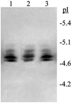
The purified esterases responsible for the elevation of activity appeared as two β-naphthyl acetate specific bands on native gradient PAGE (Fig. 2). One was intensely staining and diffuse, the other, of slightly lower mobility, less intense but tighter. On IEF gels these same esterases appeared as a ladder of five bands ranging in pI from 4.7 to 5.0 (Fig. 3). The same banding pattern on IEF gels was seen for the BPH esterases in crude homogenates of individual Sri R and Sri Lanka-S BPH. Therefore, despite the heterogeneity in the physi-cal characteristics of these esterases, they purified together throughout. On 2-D electrophoresis the purified BPH esterases ranged in size from 66 to 68 kDa [Fig. 4(a)], with the esterases having a lower pI being appar-ently smaller in size.
3.3. Glycans of BPH esterases and the effect of their removal on physical characteristics and enzymatic activity
Glycan detection of Western-blotted, 2-D separated esterases showed that all detectable isozymes were gly-cosylated [Fig. 4(b)]. Cleavage of N-linked glycans from
Fig. 1. SDS–PAGE (10–15% gradient gel) of purified brown plan-thopper esterases. SDS–PAGE standard markers were run in the adjac-ent lane. Proteins were visualised by Coomassie Blue staining.
Fig. 2. Native PAGE (4–15% gradient gel) of purified brown plan-thopper esterases. Esterases were visualised histochemically using 0.04%α- andβ-naphthyl acetate as substrates and Fast Blue B stain. The arrow indicates the direction of migration on the gel.
Fig. 4. (a) Two-dimensional electrophoresis [isoelectric focussing (pH 4–6.5) followed by SDS–PAGE (10–15% gradient gel)] of purified brown planthopper esterases, together with the same esterases applied directly to the SDS–PAGE gel (lane S). Proteins were visualised by Coomassie Blue staining. (b) Glycan detection of Western-blotted, two-dimensionally separated purified brown planthopper esterases. Glycans were detected using the Boehringer Mannheim glycan detec-tion kit following manufacturer’s instrucdetec-tions. The arrow on each fig-ure shows the orientation of the isoelectric focussing gel strip when applied to the SDS–PAGE gel.
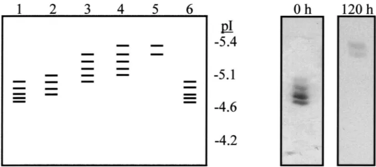
the native form of the esterases with N-glycosidase A reduced the number of esterase bands seen on IEF gels to two and increased their pIs (Fig. 5). Cleavage of N-linked glycans from the denatured form of the esterases increased the apparent molecular weight, as seen on SDS
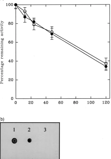
gradient PAGE to 69 kDa [Fig. 6(a)]. Western-blotting of N-glycosidase A treated esterases after separation on SDS gradient PAGE showed that they had been com-pletely deglycosylated [Fig. 6(b)]. Therefore, there are no O-linked glycans attached to the BPH esterases. The percentage remaining p-NPA activities of esterases incu-bated with and without N-glycosidase A were equivalent throughout the 120 h incubation, both glycosidase-treated and control incubations of esterases losing activity at similar rates [Fig. 7(a)]. Hence progressive deglycosylation had no effect on active site confor-mation. No glycans could be detected in the N-glycosid-ase A digested BPH esterN-glycosid-ase following incubation for 120 h [Fig. 7(b)]. Cleavage of linked glycans using N-glycosidase F again completely deglycosylated the BPH esterases (Fig. 8). Therefore, none of the N-linked gly-cans had an α1,3-bound core fucose residue.
3.4. Differentiation of BPH esterase glycans
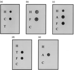
Posistive reactions were obtained with the digoxig-enin-labelled GNA, MAA and DSA [Fig. 9(a–c)] indi-cating that the N-linked glycans of BPH esterases have terminally linked mannose, sialic acid terminally linked
α(2-3) to galactose and terminally linked galactose-β (1-4)-N-acetylglucosamine. The lectins SNA and PNA both gave negative results [Fig. 9(d–e)] indicating that there are no sialic acid residuesα(2-6) linked to galactose and confirming that there are no O-linked glycans,
galactose-β(1-3)-N-acetylgalactosamine commonly forming the core unit of such glycans. Reactivity with GNA was removed on incubation of BPH esterases with endogly-cosidase F1 but not with endoglyendogly-cosidase F2 indicating that the mannose residues are terminally linked to high mannose and/or hybrid type glycans [Fig. 10(a)]. Reac-tivity with MAA and DSA was removed by incubation with endoglycosidase F2 but not with endoglycosidase F1 indicating that both the sialic acid α(2-3) linked to galactose and the galactose-β(1-4)-N-acetylglucosamine are terminally linked to biantennary complexes [Fig. 10(b,c)].
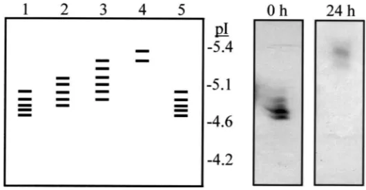
3.5. The effect of cleavage of terminal sialic acid residues on the pI of BPH esterases
Fig. 5. Diagrammatic representation of the banding pattern of brown planthopper esterases applied to an isoelectric focussing gel (pH 4–6.5) following incubation with N-glycosidase A under native conditions for 0 h (lane 1), 12 h (lane 2), 24 h (lane 3), 48 h (lane 4) and 120 h (lane 5) at 37°C. Lane 6 is the pattern of brown planthopper esterases following incubation under the same conditions without glycosidase for 120 h. The inset boxes show the actual banding pattern of these esterases on the isoelectric focussing gel at 0 h and 24 h. Esterases were visualised histochemically using 0.04%α- andβ-naphthyl acetate as substrates and Fast Blue B stain.
them away from each other. However, the change in pI of the proteins during such treatment suggests that each protein must contain three or four such glycans.
4. Discussion
Native gradient PAGE of BPH esterases in this study gave two distinct bands whereas a previous study using native PAGE found only one broad band (Karunaratne et al., 1999). This difference in banding pattern is prob-ably due to the different acrylamide gels and buffer sys-tems used. Both the molecular weights (62–64 kDa) and pIs (4.7–4.9) of the BPH esterases reported by Chen and Sun (1994) are slightly lower than those found in this study. However, in both studies the esterases of lower pI had higher apparent molecular weights. Estimated molecular weights for other insect esterases are in the same range as the BPH esterases e.g. esterases E4 and FE4 in the aphid Myzus persicae (Devonshire et al., 1986), esterase-C in the fruitfly Drosophila
melanogas-ter (Holwerda and Morton, 1983) and esmelanogas-terases E1, E2 and E3 in the brown planthopper Nilaparvata lugens (Chen and Sun, 1994) have Mrs of 62 000–66 000. The five esterase isozymes resolved by IEF in this study were fewer in number than the .10 reported by Chen and Sun (1994). This discrepancy may be due to a difference in the resolution of the esterase isozymes by IEF in the two studies as the pH 4 to 6.5 gels used in this study would not have resolved the esterase isozymes to the same extent as the pH 4.5 to 5.4 gels used in the study of Chen and Sun (1994). The pIs of the BPH esterases are all within the range of pH 4.7–6.5 typical of car-boxylesterases (Heymann, 1980).
A pattern of isoforms similar to the BPH esterases on IEF gels is seen in the esterases of the small brown
planthopper Laodelphax striatellus but in this species there is no clear relationship between pI and molecular weight (Sakata and Miyata, 1994). The change in appar-ent molecular weights of esterases on deglycosylation also differs in the two species, the L. striatellus esterases showing a marked decrease (from 70 to 66 kDa) (Sakata and Miyata, 1994) whilst the BPH esterases showed an increase. No analysis of glycosylation has been carried out for the esterases of L. striatellus, but these differ-ences suggest that the nature of glycosylation of iso-zymes is different in the two species.
Glycosylation has been noted in the E4 esterase in the peach-potato aphid Myzus persicae (Devonshire et al., 1986), EST6 in Drosophila melanogaster (Myers et al., 1996) and the juvenile hormone esterases (JHEs) from
Trichoplusia ni (Jones et al., 1993; Wozniak and Jones,
Fig. 6. (a) SDS–PAGE (10–15% gradient gel) of denatured brown planthopper esterases incubated with (lane 2) and without (lane 1) N-glycosidase A for 24 h at 37°C. Proteins were visualised by Coomassie Blue staining. (b) Glycan detection of denatured brown planthopper esterases incubated with (lane 2) and without (lane 1) N-glycosidase A for 24 h at 37°C, run on SDS–PAGE (10–15% gradient gel) and Western-blotted. Glycans were detected using the Boehringer Mannheim glycan detection kit following manufacturer’s instructions.
in the fully glycosylated enzyme (Myers et al., 1996). Glycosylation plays a role in protein folding, secretion, activity, thermostability and persistence in other serine hydrolases (Robbi et al., 1996; Sheriff et al., 1995; Mor-lockfitzpatrick and Fisher, 1995; Benzeev et al., 1994; Kronman et al., 1995; Velan et al., 1993; Abouakil et al., 1993). However, unglycosylated human acetylcholi-nesterase and human milk bile salt-stimulated lipase, like the deglycosylated BPH esterases, had the same activity as the glycosylated forms (Hernell and Blackberg, 1994; Velan et al., 1993).
In common with other insect glycoproteins, the BPH esterases had high mannose and complex glycans con-taining sialic acid attached (Altmann, 1996; Davis and Wood, 1995). However, the BPH esterases lacked the
Fig. 7. (a) Mean percentage remaining p-nitrophenyl acetate activity of brown planthopper esterases (±standard deviations) incubated with (I) and without (s) N-glycosidase A under native conditions for up
to 120 h at 37°C (n=4). 10µl aliquots of incubations were withdrawn at intervals, mixed with 200 µl of 1 mM p-nitrophenyl acetate (p-NPA) in 50 mM sodium phosphate buffer (pH 7.4) and the increase in absorbance at 405 nm was measured continuously for 2 min at 22°C. (b) Dot blot of aliquots of the same N-glycosidase A incubations with-drawn at 0 h (sample 1) and 120 h (sample 3). Sample 2 is brown planthopper esterases incubated in the same buffer without glycosidase for 120 h. Glycans were detected using the Boehringer Mannheim gly-can detection kit following manufacturer’s instructions.
Fig. 8. Dot blot of 0.5µg of brown planthopper esterases incubated with (sample 2) and without (sample 1) N-glycosidase F at 37°C for 24 h. Glycans were detected using a Boehringer Mannheim glycan detection kit following manufacturer’s instructions.
Fig. 9. Dot blots of 1µg of brown planthopper esterases probed with Galanthus nivalis agglutinin (a), Maackia amurensis agglutinin (b), Datura
stramonium agglutinin (c), Sambucus nigra agglutinin (d) and Peanut agglutinin (e). 1µg of the glycoproteins carboxypeptidase Y (C), transferrin (T), fetuin (F) and asiaolofetuin (A) were applied as positive and negative controls. Glycans were differentiated using a Boehringer Mannheim glycan differentiation kit following manufacturer’s instructions.
hormone and beta-1 intergrins from mouse melanoma cells converted the several isoforms into a single species of a higher pI indicating a single protein (Veiga et al., 1995; Isaksson and Hultberg, 1995; Canonne et al., 1995). The BPH esterase isoforms were converted into two species after neuraminidase treatment. This, together
with the presence of two species after complete deglyco-sylation, suggests there are two distinct BPH elevated esterase proteins. In the small brown planthopper, two proteins were seen after deglycosylation, but in contrast to the BPH where the esterases have the same molecular weight, these deglycosylated forms differed in molecular weight (Sakata and Miyata, 1994). As with the BPH esterases, removal of sialic acids from two porcine intes-tinal adhesion receptors caused them to migrate more slowly on SDS–PAGE. This suggests that the sialic acids are contributing to the migration of these proteins on SDS–PAGE and that their removal results in a reduced migration (higher apparent molecular weight).
A key step in the linkage of glycans to asparigine resi-dues is the transfer of the oligosacharide from the pre-cursor to the polypeptide. It is commonly found that for each potential glycosylation site transfer may or may not take place. This leads to heterogeneity in the observed physical characteristics of a protein (Shelikoff et al., 1997). Differential glycosylation has been noted in sev-eral examples of over-expressed recombinant genes in
Fig. 10. Dot blots of 0.5µg of brown planthopper esterases (H) incubated with endoglycosidase F1 (column 2), endoglycosidase F2 (column 3) or without endoglycosidases (column 1) at 37°C for 24 h and probed with Galanthus nivalis agglutinin (a), Maackia amurensis agglutinin (b),
Datura stramonium agglutinin (c). 0.5µg of the control glycoproteins carboxypeptidase Y (C), transferrin (T), fetuin (F) and asiaolofetuin (A) were incubated under the same conditions and applied to the dot blots. Glycans were differentiated using a Boehringer Mannheim glycan differentiation kit following manufacturer’s instructions.
Fig. 11. Diagrammatic representation of the banding pattern of brown planthopper esterases applied to an isoelectric focussing gel (pH 4–6.5) following incubation with neuraminidase under native conditions for 0 h (lane 1), 4 h (lane 2), 8 h (lane 3) and 24 h (lane 4) at 37°C. Lane 5 is the pattern of brown planthopper esterases following incubation under the same conditions without neuraminidase for 24 h. The inset boxes show the actual banding pattern of these esterases on the isoelectric focussing gel at 0 h and 24 h. Esterases were visualised histochemically using 0.04% α- andβ-naphthyl acetate as substrates and Fast Blue B stain.
esterases of susceptible insects. Heterogeneity may also be due to major variations in the structure of attached glycans (Rudd and Dwek, 1997). The functional signifi-cance of the individual carbohydrate variants is becom-ing ever more apparent (Rudd and Dwek, 1997). This investigation is part of an on-going project to study the nature of esterase-based insecticide resistance in the BPH. By mutational analysis of N-linked glycosylation of these esterases, the effect of differential glycosylation, and glycosylation in general, on the insecticide binding efficiency and localisation of the BPH esterases will now be investigated.
Acknowledgements
We thank J. Morgan for technical assistance. This work was funded by a BBSRC ROPA award to J.H.
References
Abouakil, N., Mas, E., Bruneau, N., Benajiba, A., Lombardo, D., 1993. Bile salt-dependent lipase biosynthesis in rat pancreatic AR2
J-cells—essential requirement of N-linked oligosaccharide for secretion and expression of a fully active enzyme. J. Biol. Chem. 268, 25755–25763.
Altmann, F., 1996. N-glycosylation in insects revisited. Trends Glyco-sci. Glyc. 8, 101–114.
Benzeev, O., Stahnke, G., Liu, G.Q., Davis, R.C., Doolittle, M.H., 1994. Lipoprotein-lipase and hepatic lipase—the role of asparigine-linked glycosylation in the expression of a functional enzyme. J. Lipid Res. 35, 1511–1523.
Bradford, M.M., 1976. A rapid and sensitive method for the quantit-ation of microgram quantities of protein utilizing the principle of protein dye binding. Anal. Biochem. 72, 248–254.
Bulet, P., Dimarcq, J.L., Hetru, C., Lagueux, M., Charlet, M., Hegy, G., Van Dorsselaer, A., Hoffmann, J.A., 1993. A novel inducible antibacterial peptide of Drosophila carries an O-glycosylated sub-stitution. J. Biol. Chem. 268, 14893–14897.
Bulet, P., Hegy, G., Lambert, J., Van Dorsselaer, A., Hoffmann, J.A., Hetru, C., 1995. Insect immunity—the inducible antibacterial pep-tide diptericin carries 2 O-glycans necessary for biological activity. Biochemistry 34, 7394–7400.
Canonne, C., Papandreou, M.J., Medri, G., Verrier, B., Ronin, C., 1995. Biological and immunochemical characterization of recombi-nant human thyrotrophin. Glycobiology 5, 473–481.
Chang, C.K., Whalon, M.E., 1987. Substrate specificities and multiple forms of esterase in the brown planthopper Nilaparvata lugens (Stal). Pest. Biochem. Physiol. 27, 30–35.
Chung, T.C., Sun, C.N., 1983. Malathion and MIPC resistance in
Nila-parvata lugens (Homoptera: Delphacidae). J. Econ. Ent. 76, 1–5.
Cociancich, S., DuPont, A., Hegy, G., Lanot, R., Holder, F., Hetru, C., Hoffmann, J.A., Bulet, P., 1994. Novel inducible antibacterial peptides from a hemipteran insect, the sap-sucking bug Pyrrhocoris
apterus. Biochem. J. 300, 567–575.
Cousin, X., Creminon, C., Grassi, J., Meflah, K., Cornu, G., Saliou, B., Bon, S., Massoulie, J., Bon, C., 1996. Acetylcholinesterase from
Bungarus venom: a monomeric species. FEBS Lett. 387, 196–200.
Davis, T.R., Wood, H.A., 1995. Intrinsic glycosylation potentials of insect cell cultures and insect larvae. In Vitro Cell. Dev. Biol. 31, 659–663.
Devonshire, A.L., Searle, L.M., Moores, G.D., 1986. Quantitative and qualitative variation in the messenger-RNA for carboxylesterases in insecticide-susceptible and resistant Myzus persicae (Sulz). Ins. Biochem. 16, 659–665.
Eldridge, R., O’Reilly, D.R., Hammock, B.D., Miller, L.K., 1992. Insecticidal properties of genetically engineered baculoviruses expressing an insect juvenile-hormone esterase gene. Appl. Envir. Micro. 58, 1583–1591.
Geisse, S., Kocher, H.P., 1999. Protein expression in mammalian and insect cell systems. Meth. Enzymol. 306, 19–42.
Hasui, H., Ozaki, K., 1984. Electrophoretic esterase patterns in the brown planthopper Nilaparvata lugens Sta˚l (Hemiptera: Delphacidae) which developed resistance to insecticides. Appl. Ent. Zool. 19, 52–58.
Hemingway, J., Karunaratne, S.H.P.P., Claridge, M.F., 1999. Insecti-cide resistance spectrum and underlying resistance mechanisms in tropical populations of the brown planthopper (Nilaparvata lugens) collected from rice and the wild grass Leersia hexandra. Int. J. Pest. Manage. 45, 215–223.
Hernell, O., Blackberg, L., 1994. Human milk bile salt-stimulated lipase: functional and molecular aspects. J. Pediatr. 125, S56–61. Heymann, E., 1980. Carboxylases and amidases. In: Jakoby, W.B.
(Ed.), Enzymatic basis of Detoxication, vol. II. Academic Press, New York, p. 291.
Holwerda, B.C., Morton, R.A., 1983. The in vitro degradation of [C14]malathion by enzymatic extracts from resistant and susceptible strains of Drosophila melanogaster. Pest. Biochem. Physiol. 20, 151–160.
Isaksson, A., Hultberg, B., 1995. Serum betahexosaminidase isoen-zymes are precursor froms. Scand. J. Clin. Lab. Invest. 55, 433– 440.
Jones, G., Manczak, M., Wozniak, M., Korrati, R., 1993. Characteriz-ation of 2 major isoforms of juvenile-hormone esterase from
Trich-oplusia ni (Lepidoptera). Biochim. Biophys. Acta 1161, 235–243.
Karunaratne, S.H.P.P., Small, G.J., Hemingway, J., 1999. Characteriz-ation of the elevated esterase-associated insecticide resistance mechanism in Nilaparvata lugens Stal and other planthopper spec-ies. Int. J. Pest. Manage. 45, 225–230.
Kronman, C., Velan, B., Marcus, D., Ordentlich, A., Reuveny, S., Shafferman, A., 1995. Involvement of oligomerization, N-glycosyl-ation and sialylN-glycosyl-ation in the clearance of cholinesterase from the circulation. Biochem. J. 311, 959–967.
Kuga, T., Hattori, S., Yoshida, M., Taniguchi, T., 1986. Expression of human T-cell leukemia virus type I envelope protein in
Saccharo-myces cerevisiae. Gene 44, 337–340.
Morlockfitzpatrick, K.R., Fisher, E.A., 1995. The effects of O-linked
and N-linked glycosylation on the secretion and bile-salt stimu-lation of pancreatic carboxyl ester lipase activity. Proc. Soc. Exp. Biol. Med. 208, 186–190.
Myers, M.A., Healy, M.J., Oakeshott, J.G., 1996. Mutational analysis of N-linked glycosylation of esterase-6 in Drosophila
melanogas-ter. Biochem. Genet. 34, 201–218.
Poly, W.J., 1997. Nongenetic variation, genetic–environmental interac-tions and altered gene expression. 3. Posttranslational modifi-cations. Comp. Biochem. Physiol. 118, 551–572.
Robbi, M., Schaftingen, E.V., Beaufay, H., 1996. Cloning and sequen-cing of rat liver carboxylesterase ES-4 (microsomal palmitoyl-CoA hydrolase). Biochem. J. 313, 821–826.
Rudd, P.M., Dwek, R.A., 1997. Glycosylation: heterogeneity and the 3D structure of proteins. Crit. Rev. Biochem. Mol. 32, 1–100. Sakata, K., Miyata, T., 1994. Biochemical characterization of
carboxy-lesterase in the small brown planthopper Laodelphax striatellus (Fallen). Pest. Biochem. Physiol. 50, 247–256.
Sato, T., Tsunasawa, S., Nikamura, Y., Emi, M., Sakiyama, F., Matsu-bara, K., 1986. Expression of the human salivary α-amylase in yeast and characterization of the secreted protein. Gene 50, 247– 257.
Shelikoff, M., Sinskey, A.J., Stephanopoulos, G., 1997. A modelling framework for the study of protein glycosylation. Biotechnol. Bioeng. 50, 73–90.
Sheriff, S., Du, H., Grabowski, G.A., 1995. Characterization of lysoso-mal acid lipase by site-directed mutagenesis and heterologous expression. J. Biol. Chem. 270, 27766–27772.
Takada, H., Murakami, Y., 1988. Esterase variation and insecticide resistance in japanese aphis-gossypii. Ent. Exp. Applic. 48, 37–41. Tranter, B.C., 1983. Elucidation of insecticide resistance mechanisms in the brown planthopper Nilaparvata lugens. PhD thesis, Univer-sity of Reading.
Tretter, V., Altmann, F., Marz, L., 1991. Peptide-N4 -(N-acetyl-b-glucosaminyl) asparigine amidase F cannot release glycans with fucose attached a-3 to the asparigine-linked N-acetylglucosamine residue. Eur. J. Biochem. 199, 647–652.
Trimble, R.B., Tarentino, A.L., 1991. Identification of distinct endog-lycosidase (endo) activities in Flavobacterium meningosepticum: Endo F1, Endo F2 and Endo F3. J. Biol. Chem. 266, 1646–1651. Uchida, Y., Tsukada, Y., Sugimori, T., 1979. Enzymatic neuraminid-ases from Arthrobacter ureafaciens. J. Biochem. 86, 1573–1585. Veiga, S.S., Chammas, R., Cella, N., Brentani, R.R., 1995.
Glycosyl-ation of beta-1 integrins in B16-F10 mouse melanoma cells as determinant of differential binding and acquisition of biological activity. Int. J. Cancer 61, 420–424.
Velan, B., Kronman, C., Ordentlich, A., Flashner, Y., Leitner, M., Cohen, E., Shafferman, A., 1993. N-glycosylation of human acetyl-cholinesterase: effects on activity, stability and biosynthesis. Biochem. J. 296, 649–656.
Wozniak, M., Jones, G., 1990. Glycosylation and isoform variation of juvenile-hormone esterase in the fat-body and hemolymph during metamorphosis of Trichoplusia ni (lepidoptera). Mol. Cell. Endoc-rin. 70, 255–262.
![Fig. 4.(a) Two-dimensional electrophoresis [isoelectric focussing(pH 4–6.5) followed by SDS–PAGE (10–15% gradient gel)] of purifiedbrown planthopper esterases, together with the same esterases applieddirectly to the SDS–PAGE gel (lane S)](https://thumb-ap.123doks.com/thumbv2/123dok/3120878.1379314/6.598.83.247.71.552/dimensional-electrophoresis-isoelectric-focussing-puriedbrown-planthopper-esterases-applieddirectly.webp)