Review
Therapeutic targeting of human immunodeficiency virus
type-1 latency: current clinical realities and future
scientific possibilities
Salvatore T. Butera *
HIV and Retro6irology Branch,Di6ision of AIDS,STD,and TB Laboratory Research,National Center for Infectious Diseases, Centers for Disease Control and Pre6ention,Atlanta,GA30333,USA
Received 4 September 2000; accepted 6 October 2000
Abstract
Factors affecting HIV-1 latency present formidable obstacles for therapeutic intervention. As these obstacles have
become a clinical reality, even with the use of potent anti-retroviral regimens, the need for novel therapeutic strategies
specifically targeting HIV-1 latency is evident. However, therapeutic targeting of HIV-1 latency requires an
understanding of the mechanisms regulating viral quiescence and activation. These mechanisms have been partially
delineated using chronically infected cell models and, clearly, HIV-1 activation from latency involves several key viral
and cellular components. Among these distinctive therapeutic targets, cellular factors involved in HIV-1 transcription
especially warrant further consideration for rational drug design. Exploring the scientific possibilities of new therapies
targeting HIV-1 latency may hold new promise of eventual HIV-1 eradication. © 2000 Published by Elsevier Science
B.V. All rights reserved.
Keywords:HIV-1 latency; Therapeutic intervention; Cell models; Viral transcription
www.elsevier.com/locate/antiviral
1. Introduction
Without question, the development and clinical
introduction of highly active anti-retroviral
ther-apy (HAART, regimens including various
combi-nations
of
nucleoside,
non-nucleoside,
and
protease inhibitors) have provided unequivocal
benefits to infected individuals by controlling the
replication of human immunodeficiency virus
type-1 (HIV-1). These clinical benefits include
dramatic and sustained reductions in the level of
circulating HIV-1 (viral load) and a halting of the
progressive CD4 T-lymphocyte loss that
accom-panies HIV-1 disease progression (Gulick et al.,
1997; Hammer et al., 1997; Gulick et al., 2000).
Furthermore, HAART implementation has
re-sulted in substantial reductions in morbidity and
* Tel.: +1-404-6391033; fax:+1-404-6391174. E-mail address:[email protected] (S.T. Butera).
mortality from various opportunistic infections
that characterize acquired immunodeficiency
syn-drome (AIDS), thus often prolonging of survival
(Detels et al., 1998; Mocroft et al., 1998; Palella et
al., 1998).
However, these clinical benefits of HAART still
hold limitations. First, the high cost and limited
availability of drugs used in HAART make the
clinical benefits largely available only to persons
in developed countries. Second, HAART
regi-mens require patients to take many tablets per
day. Furthermore, the potential side effects and
the risk of developing HIV-1 resistant to any of
the antiretroviral compounds is concerning and
makes long-term maintenance on HAART
unre-alistic. Lastly, the initial hope that prolonged and
maintained
suppression
of
HIV-1
load
by
HAART regimens alone could result in the
even-tual clearance or eradication of virus from an
individual (Perelson et al., 1997) is now clearly
improbable. It soon became apparent that issues
related to HIV-1 latency would limit the
possibil-ity of complete viral clearance and that latent
HIV-1 reservoirs would have to be addressed
therapeutically if virus eradication was to become
a reality. These subjects are the nature of this
review.
2. What has HAART taught us?
In the mid-1990s, when HAART regimens first
became available (as a result of the clinical
intro-duction of HIV-1 protease inhibitors), there was
continuing uncertainty surrounding the
mecha-nisms by which HIV-1 led to the development of
AIDS. This uncertainty arose from the
observa-tions that only a small percentage of peripheral
T-cells harbor HIV-1 in infected individuals
(Brinchmann et al., 1991) and from the inability
to define completely mechanisms by which HIV-1
infection results in progressive T-cell loss. The
introduction of effective viral suppression under
HAART regimens firmly established that
prevent-ing continued high-level viral replication will
im-pede progressive immunologic dysfunction and
reduce AIDS mortality (Gulick et al., 1997;
Ham-mer et al., 1997; Detels et al., 1998; Mocroft et al.,
1998).
The widespread use of HAART for treating
advanced HIV-1 disease among persons in
devel-oped countries has demonstrated both the
restora-tive capacity of the adult immune system as well
as its limitations. In many cohorts that have been
studied to date, HIV-1-infected persons receiving
HAART have responded with partial restoration
of immune cell populations (Bohler et al., 1999;
Hengel et al., 1999), immune functions (Autran et
al., 1997; Arno et al., 1998; Dam Nielsen et al.,
1998; Kroon et al., 1998; Lederman et al., 1998),
and homeostasis (Andersson et al., 1998;
Bous-carat et al., 1998; Evans et al., 1998; Dyrhol-Riise
et al., 1999). However, immune recovery generally
appears incomplete and variable (Connors et al.,
1997; Fleury et al., 1998; Pakker et al., 1999;
Zaunders et al., 1999). Although an extensive
detailing of immune restoration resulting from
HAART regimens is beyond the scope of this
review (reviewed by Autran et al., 1999), this
subject is relevant and discussed below because
such restoration may hold promise for eventual
viral eradication.
most likely arises due to a persistent latent
popu-lation (Chun et al., 1997a; Finzi et al., 1997;
Wong et al., 1997; reviewed in Finzi and Siliciano,
1998) and contributes to continued HIV-1
dissem-ination (Grossman et al., 1998, 1999).
Remark-ably, decay estimates now suggest that the HIV-1
latent population may persist for up to 60 years in
infected individuals receiving HAART (Finzi et
al., 1999; Ramratnam et al., 2000).
3. HIV-1 latency defined
The concept of HIV-1 latency can be
consid-ered on several levels and must be precisely
defined to avoid misunderstanding. First, HIV-1
latency can be regarded on a clinical level as the
extended and variable asymptomatic period after
acute infection but prior to the onset of
AIDS-defining immune dysfunction. But, as discussed
below, HIV-1 infection does not assume a state of
true latency, in which cessation of viral replication
accounts for the lack of disease progression
(Gar-cia-Blanco and Cullen, 1991), and to apply the
term latency in this manner is imprecise.
How-ever, as we have learned from the advent of
HAART, HIV-1 latency on a cellular level does
have clinical ramifications thus the two concepts
are inherently linked.
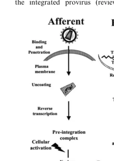
On a cellular level, HIV-1 latency can exist
during either the afferent or the efferent portion
of the viral life cycle (Fig. 1). The afferent portion
of the HIV-1 life cycle includes the necessary
events between extracellular binding to the surface
co-receptors and eventual integration of the
re-verse-transcribed viral genome into the host cell
chromosomes. However, when HIV-1 enters an
unactivated CD4 T-lymphocyte, the afferent
por-tion of the life cycle is not completed and a
temporally labile, partially reverse-transcribed
complex is created in the cytoplasm of the host
cell (Stevenson et al., 1990; Zack et al., 1990;
Bukrinsky et al., 1991). This situation is
consid-ered pre-integration latency. If cellular activation
occurs with hours to days of HIV-1 entry and
before to the degradation of the pre-integration
complex, then reverse transcription and
integra-tion ensue to complete the afferent porintegra-tion of the
life cycle. Therefore, resting CD4
+lymphocytes
can serve as a latent viral reservoir by maintaining
HIV-1 in the pre-integrated state until cellular
activation (Bukrinsky et al., 1992; Zack et al.,
1992).
The efferent portion of the HIV-1 life cycle
begins at the point of stable proviral integration
and includes subsequent viral transcription,
trans-lation, and assembly to produce infectious viral
progeny. At this point of the life cycle, HIV-1
latency is defined by the transcriptional state of
the integrated provirus (reviewed in McCune,
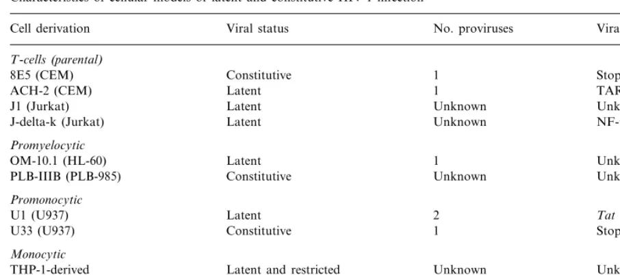
Table 1
Characteristics of cellular models of latent and constitutive HIV-1 infection
No. proviruses
Cell derivation Viral status Viral defect
T-cells(parental)
1
Constitutive Stop codon inpol
8E5 (CEM)
1
ACH-2 (CEM) Latent TAR mutations
Unknown
Latent Unknown
J1 (Jurkat)
J-delta-k (Jurkat) Latent Unknown NF-kB deletions
Promyelocytic
OM-10.1 (HL-60) Latent 1 Unknown
PLB-IIIB (PLB-985) Constitutive Unknown Unknown
Promonocytic
2
Latent Tatmutations
U1 (U937)
U33 (U937) Constitutive 1 Stop codon inen6
Monocytic
Latent and restricted
THP-1-derived Unknown Unknown
B-cells(EBV-transformed)
LL58 Latent Unknown Unknown
1995). In some instances, the integrated HIV-1
provirus is transcriptionally dormant, regardless
of the activation status of the host cell. This
condition, termed absolute latency, appears to be
a dead-end for viral replication and may result
from the integration of a transcriptionally
defec-tive provirus. However, the best characterized and
clinically most concerning state of
post-integra-tion latency exists when the HIV-1 provirus is
transcriptionally dormant until the host cell
be-comes activated by any number of first, second, or
third messenger pathways (discussed below). Viral
activation from this state, termed microbiologic
latency, results in the production of infectious
HIV-1 progeny to complete the life cycle and
further propagate infection and disease. As issues
related to viral eradication have evolved, HIV-1
microbiologic latency has come to the forefront of
clinical and therapeutic considerations regarding
HAART.
4. History of HIV-1 latency
HIV-1 latency was first demonstrated in
experi-ments examining various cellular outcomes of
acute HIV-1 infection. Working with transformed
cell lines in vitro, cells that survived the
cyto-pathic effects of acute HIV-1 replication could be
identified and cloned. Although some of these
cloned populations maintained chronic high-level
viral replication (Folks et al., 1986), others were
observed to express little or no viral proteins
unless activated by exogenous stimuli (Folks et
al., 1987, 1988, 1989; Clouse et al., 1989). This
state of HIV-1 microbiologic latency was
subse-quently established using transformed cells
repre-senting several different cellular lineages (Tozzi et
al., 1989; Mikovits et al., 1990; Butera et al., 1991;
Perez et al., 1991) (Table 1). A large body of
literature quickly became established concerning
the spectrum of soluble mediators and cytokines
that could activate HIV-1 expression from latency
and the intracellular mechanisms regulating this
process (reviewed in Butera and Folks, 1992).
Cells that harbored an integrated HIV-1 provirus
but could not be activated using the established
stimuli could also be identified. These cells may
harbor a dormant or transcriptionally defective
HIV-1 provirus or respond to extracellular stimuli
other than that tested (Mikovits et al., 1990).
infec-tion and the onset of AIDS-defining illnesses and
suggested that HIV-1 enters into a state of clinical
latency. In fact, during this early time period, a
reservoir of virally infected cells was detected in
peripheral
blood
CD4
+T-lymphocytes
(Schnittman et al., 1989). Since HIV-1 replication
was known to down-modulate cell surface CD4
expression via active expression of envelope or
other viral proteins (Butera et al., 1991), it was
considered probable that HIV-1 was
transcrip-tionally dormant in these infected lymphocytes
that retained surface CD4 expression. Shortly
fol-lowing these observations, the lymph nodes were
firmly established as a major site of HIV-1
infec-tion (Pantaleo et al., 1991) and were examined for
the extent of viral replication. Using more
sophis-ticated in situ technologies, researchers found that
lymphoid tissues from HIV-1 infected individuals
during the clinically asymptomatic phase showed
a large percentage of cells that harbored proviral
DNA without evidence of viral RNA (Embretson
et al., 1993). However, it also became quite clear
that HIV-1 expression was active at all stages of
disease
progression,
including
the
clinically
asymptomatic period, both in the lymph nodes
(Pantaleo et al., 1993) and the peripheral blood
(Michael et al., 1992; Piatak et al., 1993).
There-fore, HIV-1 did not enter a state of true clinical
latency, in which cessation of viral replication
correlated with asymptomatic infection. These
landmark findings raised an aura of doubt over
HIV-1 latency as being a possible artifact of in
vitro infections, the use of transformed and
cloned cell lines, or a virologic phenomenon
with-out any real clinical relevance.
These perceptions of HIV-1 latency
dramati-cally changed again with the advent of HAART.
Although HAART maintenance held the initial
promise of HIV-1 eradication, levels of detectable
provirus remained for extended periods of time in
individuals undergoing HAART. Furthermore,
the detectable provirus was integrated into the
host genome and could be activated in vitro to
generate infectious virus particles. These features
implied that this was not dead-end defective
provirus but rather replication-competent HIV-1
existing in a state of microbiologic latency in vivo.
It is now well accepted that HIV-1 latency is a
clinical reality and a new therapeutic challenge to
possible viral eradication.
5. Clinical complications of HIV-1 latency —
what do we know?
Recent technological advances have enabled
re-searchers to address the impact of HIV-1 latency
on a clinical level. Much of the difficulty in
detect-ing and quantitatdetect-ing this viral reservoir arose
from the large amount of patient material
re-quired and the low frequency at which these
latently infected cells exist. Techniques were first
established to demonstrate the existence of this
population even before HAART was available to
reduce the background of active HIV-1
replica-tion (Chun et al., 1995). These pioneering studies
assumed that HIV-1 microbiologic latency would
exist in unactivated T-lymphocytes that retain
surface CD4 expression and rigorously purified
resting cells from the CD4
+T-cell population by
selecting against those expressing activation
sur-face markers (HLA-DR, CD25, etc.). A culture
period of several days was also implemented to
permit the degradation of temporally labile,
HIV-1 pre-integration complexes. PCR-based
tech-niques were then applied that detected HIV-1
proviral DNA only when integrated into cellular
DNA.
cellular
phenotype
harboring
latent
HIV-1
provirus was the memory T-cell expressing the
CD45RO surface antigen (as opposed to the naı¨ve
T-cell expressing CD45RA) and that the
frequen-cies of these cells were similar in blood and lymph
nodes (Chun et al., 1997b).
When these techniques were applied to patients
receiving HAART (Chun et al., 1997a; Finzi et
al., 1997; Wong et al., 1997), a more complete
appreciation of the clinical complications arising
from HIV-1 microbiologic latency became
appar-ent. Although early predictions estimated that the
decay of infected cells occurred
3 years after
effectively halting HIV-1 replication, the decay of
the latently HIV-1 reservoir was determined to be
extremely protracted in all individuals examined.
It is now estimated that complete eradication of
the estimated less than 1 million cells harboring
replication-competent latent HIV-1 will require
up to 60 years of continued viral suppressive
therapy (Finzi et al., 1999; Ramratnam et al.,
2000). The mean half-life for this cellular reservoir
was estimated between 6 and 44 months, with the
rate of decay possibly influenced by the
effective-ness of suppressive HIV-1 therapy in a given
individual (Finzi et al., 1999; Ramratnam et al.,
2000).
Equally sobering was the finding that the
popu-lation of cells harboring HIV-1 in a state of
microbiologic latency was established very early,
during primary HIV-1 infection (Chun et al.,
1998a). In patients initiating suppressive HAART
regimens within 10 days of the onset of
HIV-re-lated acute retroviral syndrome (probably 2 – 3
weeks post-exposure but after the burst of viremia
and viral dissemination), the latent population of
cells had already been established. Therefore, even
with effective intervention at the earliest
recogni-tion of symptoms, latent HIV-1 infecrecogni-tion will
present a significant therapeutic challenge.
Fur-thermore, in vitro experiments with latently
in-fected cells from patient material indicated that
HIV-1 expression could be activated either by
direct stimulation of the T-cell receptor (via
anti-CD3 cross-linking) or by the addition of a
combi-nation of proinflammatory and T-cell activating
cytokines (Chun et al., 1998b). These conditions
are most likely to continually exist within the
normal cellular milieu and microenvironment of
lymph nodes and further emphasize the challenges
facing HIV-1 interventions.
num-ber of latently infected cells (between 10
5and 10
6)
are observed in almost all individuals examined
thus far, regardless of stage of disease (Chun et
al., 1997a; Finzi et al., 1997; Wong et al., 1997).
Ongoing viral replication during highly
sup-pressive therapy also suggests the presence of
sequestered sites unavailable or insensitive to the
effects of antiretroviral drugs (reviewed in
Hoetel-mans 1998; Schrager and D’Souza 1998; Clarke et
al., 2000). Several anatomical sites are suspected
to support ongoing viral replication during
HAART regimens; however, the lymphoid
com-partments are especially concerning (Cavert et al.,
1997; Lafeuillade et al., 1997; Rosok et al., 1997a;
Stellbrink et al., 1997; Pantaleo et al., 1998; Perrin
et al., 1998; Tenner-Racz et al., 1998) because
they contribute extensively to HIV-1 pathogenesis
(Embretson et al., 1993; Pantaleo et al., 1993;
Hufert et al., 1997). The lymphoid
microenviron-ment also provides a rich source of soluble factors
capable of perpetuating HIV-1 replication
(Rieck-mann et al., 1991; Schnittman et al., 1991).
Fur-thermore, the follicular dendritic cell network
with lymphoid compartments may serve as an
additional reservoir for HIV-1, even if these cells
are not productively infected (Cameron et al.,
1992; Spiegel et al., 1992; Heath et al., 1995).
Other anatomical HIV-1 reservoirs of concern
include the rectal mucosa (Kotler et al., 1998), the
genital tract (Byrn and Kiessling, 1998; Coombs
et al., 1998; Cu-Uvin et al., 1998; Mayer et al.,
1999), and the central nervous system (Bagasra et
al., 1996; Cinque et al., 1998), the latter being
complicated by issues of drug penetration.
Furthermore, the numerous cellular reservoirs
of latent HIV-1 infection during HAART are
important obstacles for viral eradication. The
complexity of this issue was recently made
appar-ent by the demonstration that the reemergence of
HIV-1 replication upon intermittent cessation of
HAART may not correspond in some individuals
to expression of virus sequestered in resting CD4
lymphocytes (Chun et al., 2000). These findings
imply that latent HIV-1 may persist in other
cellular compartments, including infected cells of
the monocyte lineage. Circulating monocytes and
tissue macrophages are a critical component of
HIV-1 pathogenesis and can serve as an
impor-tant source of virus (Orenstein et al., 1997; Lewin
et al., 1998; Lawn et al., 2000). These infected
cells are long-lived, and HIV-1 replication does
not induce cytopathic effects as observed with
infected T-lymphocytes — features that originally
placed infected monocytes as a primary concern
for HIV-1 eradication by HAART (Perelson et
al., 1997). Although residual HIV-1 has been
de-tected in circulating monocytes from individuals
on HAART regimens (Lambotte et al., 2000),
methods for quantitating latent virus in
mono-cytes have not been developed and refined as have
those for resting T-cells. Quantitating latent virus
in macrophages is further complicated by the
long-term persistence of the majority of these cells
as tissue macrophages, unavailable to sampling
via the peripheral blood. While it is difficult to
ascertain the magnitude of latent infection among
macrophages, HIV-1 latency in this population of
cells has been documented (Mikovits et al., 1992;
Lambotte et al., 2000) and should be considered
in therapeutic strategies for HIV-1 eradication.
Latent HIV-1 may also persist in some cellular
reservoirs that are either not generally considered
primary targets of active infection, such as
CD8
+
T-lymphocytes (Semenzato et al., 1998;
Yang et al., 1998), or not yet described.
6. Mechanisms regulating HIV-1 expression,
latency, and activation
6
.
1
.
Mechanisms controlling HIV
-
1
expression
CD4 lymphocytes and by which cellular activation
can result in renewed viral expression.
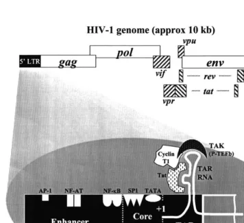
The HIV-1 core promoter, contained in the
5
%
-long terminal repeat (LTR), contains a
consen-sus TATA sequence and three binding sites for the
constitutive cellular transcription factor Sp1 (Fig.
2). This region is flanked upstream by the viral
enhancer region that contains binding regions for
several well-recognized host transcription factors.
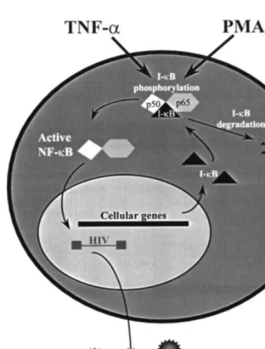
Most notable among these transcription factors is
nuclear factor kappa B (NF-
k
B), a heterodimeric
inducible enhancer that exists in the host cell
cytoplasm as an inactive complex with its inhibitor,
I-
k
B. Upon cellular activation via several first and
second messenger pathways involving cellular
ki-nases (Kinter et al., 1990; Critchfield et al., 1997),
phosphorylation of I-
k
B results in its proteolytic
degradation. As a result, active NF-
k
B is liberated
and translocates to the nucleus to stimulate
expres-sion of genes that contain NF-
k
B-specific binding
sites in the enhancer (Fig. 3). Activation of HIV-1
expression via the NF-
k
B response pathway is a
well-characterized aspect of viral regulation and
latency (Duh et al., 1989; Griffin et al., 1989;
Osborn et al., 1989). Furthermore, cooperative
interaction between NF-
k
B and other elements,
especially Sp1 (Perkins et al., 1993), may be
re-quired for full proviral expression.
Fig. 3. A schematic of nuclear factor-kB (NF-kB) activation.
The NF-kB heterodimer (p50 and p65) is present in the
cytoplasm associated with its inhibitor, I-kB. Cellular
stimula-tion by TNF-a, phorbol esters (PMA), or other agents that
activate the appropriate cellular kinase cascades results in the phosphorylation-directed dissociation and degradation of
I-kB. As a result, active NF-kB is translocated to the nucleus
where it stimulates selected gene expression by binding to a motif in the promoter of responsive genes. NF-kB-responsive
genes include its inhibitor, I-kB, and HIV-1.
1996) and with HIV-1 proviruses deleted of the
NF-
k
B binding regions (Antoni et al., 1994).
The studies cited above all suggest that cellular
factors in addition to NF-
k
B are necessary for
HIV-1 regulation and activation from latency.
Other transcription factor binding sites contained
in the enhancer region of the HIV-1 LTR include
that for activation protein-1 (AP-1) and nuclear
factors of activated T-cells (NF-AT). Some
evi-dence for the contribution of these factors in
regulating HIV-1 expression has been established
(Kinoshita et al., 1998; Navarro et al., 1998).
HIV-1 expression is also responsive to a variety of
other identified cellular factors, including the
c-myc proto-oncogene (Sun and Clark, 1999), the
tumor suppressor protein p53 (Duan et al., 1994a;
Gualberto et al., 1995), and p38
mitogen-acti-vated protein (MAP) kinase (Cohen et al., 1997;
Shapiro et al., 1998a). Furthermore, depending
upon the primary cellular stimulus, HIV-1
expres-sion may be responsive to still poorly
character-ized factors (Coudronniere and Devaux, 1998;
Briant et al., 1999) or factors that regulate viral
replication in a cell lineage-selective manner
(Hen-derson and Calame, 1997). Therefore, the
activa-tion of HIV-1 expression is intricately linked to
cellular activation, involves a variety of second
messenger kinase and third messenger nuclear
fac-tor signals, and can be regulated in a cell
type-spe-cific manner.
Through the molecular manipulation of the
LTR and transient transfection experiments using
LTR-reporter gene constructs, numerous other
binding regions for a variety of cellular factors
have been implicated in and around the LTR
(reviewed in Gaynor, 1992). Delineating all the
cellular pathways and components involved in
regulating HIV-1 expression and establishing their
importance using latently infected cells derived
from patient material ex vivo will be a necessary
undertaking. Each cellular component involved in
regulating HIV-1 expression is a potential
thera-peutic target for blocking HIV-1 activation from
latency (reviewed in Baba, 1997). Therapeutic
targeting of cellular factors has proven effective
using a variety of agents that inhibit NF-
k
B-medi-ated HIV-1 expression (Mihm et al., 1991; Li et
al., 1993; Biswas et al., 1994; Navarro et al.,
Translocation of NF-
k
B to the nucleus also
stimulates renewed expression of its inhibitor,
I-k
B, to limit the impact of the original cellular
stimulus (Sun et al., 1993); however, this
regula-tory mechanism does not appear to restrict
con-tinued HIV-1 expression (Butera et al., 1995).
Furthermore, although NF-
k
B is recognized as a
critical element for HIV-1 activation, binding of
NF-
k
B to the HIV-1 enhancer without the
availability of additional cellular and viral
compo-nents is not sufficient to activate HIV-1 expression
(Doppler et al., 1992; Butera et al., 1995).
Activa-tion of HIV-1 expression has also been observed
with stimulating agents that do not involve
NF-k
B as a third messenger component (Stanley et
1998). This conceptual approach, which deserves
renewed attention for rational drug design, has
the distinct advantage of limiting the opportunity
for the emergence of viral escape mutants.
How-ever, for targeting of cellular signaling pathways
and transcription factors to become a therapeutic
reality for inhibiting HIV-1 activation from
la-tency, a great degree of specificity for HIV-1
expression will have to be achieved. Even under
circumstances where redundant cellular pathways
exist, these cellular components are involved in
vital cellular gene expression and inhibitors that
block HIV-1 expression may also result in
unfore-seen cellular toxicities.
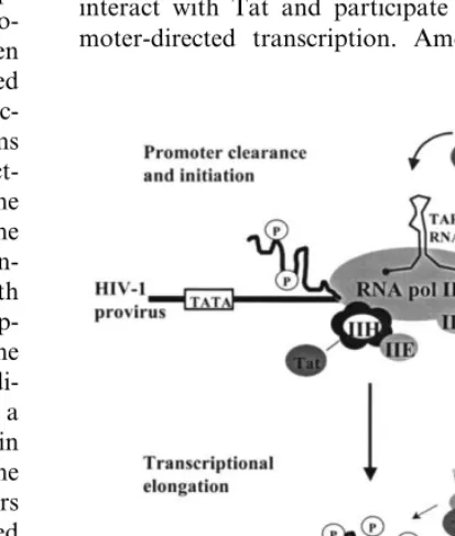
Regulation of HIV-1 transcription also
criti-cally involves the participation of a virally
en-coded transactivation protein, Tat. The primary
contribution of Tat is during the process of HIV-1
transcriptional elongation (rather than
transcrip-tional initiation, reviewed in Jones, 1997).
Pro-moter-proximal HIV-1 transcripts have been
detected in peripheral blood cells from infected
individuals, suggesting a block of the
Tat-transac-tivation axis to maintain latency in vivo (Adams
et al., 1994). Tat functions by physically
interact-ing with a hairpin loop structure, known as the
TAR (transactivation response) element, in the
proximal portion of the nascent viral RNA
tran-script (Fig. 2). Tat also physically interacts with
cellular factors necessary for HIV-1
transcrip-tional elongation, thereby recruiting them to the
HIV-1 promoter. From a multitude of
site-di-rected mutagenesis studies on the TAR element, a
consensus binding pattern was established in
which Tat binds to a bulge in the stem of the
hairpin loop and Tat-associated cellular factors
bind to a loop at the distal end (Fig. 4; reviewed
in Jones, 1997).
By all indications, the cellular components that
participate in Tat-directed HIV-1 transcriptional
elongation include a kinase that functions to
hy-perphosphorylate the carboxy-terminal domain
(CTD) of cellular RNA polymerase II (pol II) and
permit polymerase processivity. Within recent
years, the identity of such a Tat-associated kinase
(TAK) was described as the positive transcription
elongation factor-b (P-TEFb) (Mancebo et al.,
1997; Zhu et al., 1997), a finding that has been
confirmed by several additional studies (Flores et
al., 1999; Kanazawa et al., 2000). P-TEFb is a
complex comprised of multiple cellular proteins,
one of which, CDK9, is a kinase capable of CTD
hyperphosphorylation resulting in HIV-1
tran-scriptional elongation. This kinase demonstrates
cell lineage-selective regulation (Herrmann et al.,
1998) which could once again account for
differ-ences in the regulation of HIV-1 latency in
mono-cytes and T-lymphomono-cytes.
Tat-dependent HIV-1 transactivation also
in-volves several other key cellular proteins. The
interaction between Tat and P-TEFb is mediated
by an additional cellular protein, Cyclin T1, that
directly interacts with Tat and allows for
high-affinity binding of the complex to TAR RNA
(Wei et al., 1998). Furthermore, other cellular
transcription complexes and associated kinases
interact with Tat and participate in HIV-1
pro-moter-directed transcription. Among these, the
multi-subunit transcription complex, TFIIH, and
its kinase subunit, CDK7, demonstrate enhanced
CTD phosphorylation in vitro via an interaction
with Tat (Cujec et al., 1997). However, this
inter-action does not satisfy all the necessary aspects of
Tat-directed HIV-1 transcriptional elongation and
may be more critically involved in the process of
transcriptional
initiation
(reviewed
in
Jones,
1997).
As with the cellular second and third messenger
signaling components involved in HIV-1
activa-tion, understanding the full complement of host
cell factors required for Tat activity will identify
potential new targets for therapeutic intervention
against HIV-1 latency. The involvement of these
cellular components in the regulation of HIV-1
latency from material derived from infected
indi-viduals still awaits confirmation. However, in this
situation, the issue of drug specificity and cellular
toxicity may be of slightly less concern because of
the ability to target a process involving a virally
encoded
protein
with
no
known
cellular
homologue.
6
.
2
.
Mechanisms maintaining HIV
-
1
latency
The potential mechanisms regulating the state
of HIV-1 latency are numerous, diverse, and
in-volve both cellular and viral factors (reviewed in
Bednarik and Folks, 1992). Regulatory events
involving the HIV-1 LTR promoter may be of
primary importance in controlling the latent state.
Key to this process is chromatin remodeling of
the HIV-1 promoter region to permit an open and
transcriptionally active configuration. A single
nu-cleosome (termed nuc-1) positioned in the region
immediately after the HIV-1 transcription start
site was conserved in a number of different
la-tently HIV-1-infected cell lines and disrupted
upon HIV-1 transcriptional activation (Verdin et
al., 1993). Additional reconstitution studies (Pazin
et al., 1996) and studies demonstrating HIV-1
transcriptional activation following chromatin
modification (histone acetylation, Van Lint et al.,
1996) further support the importance of this
regu-latory process. HIV-1 latency may be further
reg-ulated
by
proviral
integration
into
a
transcriptionally dormant region of the host
genome (Winslow et al., 1993) or by the extent of
DNA methylation in the HIV-1 LTR (Bednarik et
al., 1991).
HIV-1 encodes several accessory proteins (Nef,
Vpu, and Vpr) that have been implicated in
con-trolling both the establishment of cellular latency
upon infection and the subsequent conversion to
productive viral expression (reviewed in Ikuta et
al., 1997). Mutations in these viral regulatory
genes are associated with deceased HIV-1
cyto-pathicity, viral persistence, and a more rapid
con-version to a nonproductive viral infection (Kishi
et al., 1995; Song et al., 1996; Kawano et al.,
1997). Furthermore, extracellular Vpr can directly
activate latent HIV-1 from several cellular models
examined and serum antibodies against this viral
protein may prevent viral activation in vivo (Levy
et al., 1994). The contribution of Nef to viral
latency may be related to its ability to permit
HIV-1 infection of resting CD4 lymphocytes
(Miller et al., 1994; Spina et al., 1994) and the
unique viral-host cell relationship that is
estab-lished under these circumstances (Spina et al.,
1995).
The regulatory proteins of HIV-1 can also be
major determinants of viral latency. As might be
anticipated, defects in the coding region for Tat
(Emiliani et al., 1998) or mutations that disrupt
binding to TAR (Emiliani et al., 1996) restrict
HIV-1 transcriptional activity, as observed in
sev-eral chronically infected cell lines (discussed
be-low). Interestingly, these chronically infected cell
lines can still undergo transcriptional activation
and respond differently to the intracellular
intro-duction of exogenous Tat (Duan et al., 1994b).
pro-duction of viral structural proteins and progeny
virions (Pomerantz et al., 1992). This mechanism,
termed blocked early-stage latency, can be
ob-served as an accumulation of multiply spliced
HIV-1 mRNA transcripts and was first
docu-mented
using
chronically
infected
cell
lines
(Pomerantz et al., 1990a). Although not a
consis-tent mechanism of latency in several additional
cell lines examined (Butera et al., 1994), evidence
for blocked early-stage latency has been
demon-strated using material from HIV-1-infected
indi-viduals (Seshamma et al., 1992).
6
.
3
.
Mechanisms of HIV
-
1
acti
6
ation from
latency
During the past decade, an enormous body of
literature has accumulated from studies conducted
in vitro and in vivo to examine the participation
of various factors and soluble products in
regulat-ing the activation of HIV-1 expression. While
truly beyond the scope of this review, several key
points concerning the mechanisms of HIV-1
acti-vation are pertinent. Most importantly, immune
activation at all levels is a driving force behind
HIV-1 expression (reviewed in Wahl and
Oren-stein, 1997) and, most likely, behind activation of
HIV-1 from latency. This concept is based on
findings from initial studies performed using
chronically infected cell models demonstrating
that the proinflammatory cytokines, in particular,
tumor necrosis factor-alpha (TNF-
a
), stimulate
HIV-1 expression from latency (Folks et al., 1987;
Clouse et al., 1989; Folks et al., 1989; Poli et al.,
1990a; Butera et al., 1993). These findings were
subsequently extended to other proinflammatory
cytokines (Poli et al., 1990b, 1994; Granowitz et
al., 1995; Marshall et al., 1999) and a host of
other cytokines and monokines (Broor et al.,
1994; Smithgall et al., 1996; Shapiro et al., 1998b;
Klein et al., 2000; reviewed in Butera, 1993).
Mechanistically, the proinflammatory cytokines
activate HIV-1 expression from latency via
recep-tor-mediated second messenger signal
transduc-tion that ultimately results in NF-
k
B activation
(Duh et al., 1989; Osborn et al., 1989; Beg et al.,
1993). Production of endogenous TNF-
a
during
acute HIV-1 infection in vitro may also affect the
establishment of the latent state (Fujinaga et al.,
1998).
The positive impact of TNF-
a
upon HIV-1
replication has also been documented in primary
human
peripheral
blood
mononuclear
cells
(Vyakarnam et al., 1990; Munoz-Fernandez et al.,
1997), although TNF-
a
may also directly
stimu-late cell death in these populations (Klein et al.,
1996; Lazdins et al., 1997). TNF-
a
was among a
small group of cytokines, that also included the
T-cell activating cytokine interleukin-2 (IL-2),
which is capable of stimulating ex vivo HIV-1
expression from latently infected resting CD4 cells
derived from infected individuals (Chun et al.,
1998b). Importantly, elevated systemic TNF-
a
levels also correlate with treatment failure in
indi-viduals receiving HAART (Aukrust et al., 1999).
It is necessary, however, to consider the impact
of proinflammatory mediators upon HIV-1
repli-cation in the context of the complex cytokine
network (Butera, 1994; Kinter et al., 1995a, 1996;
Goletti et al., 1998). Synergistic activity among
cytokines for HIV-1 activation has been
docu-mented (Poli et al., 1990b; Finnegan et al., 1996;
Rabbi et al., 1998). Furthermore, inhibitory
cy-tokines (Poli et al., 1991; Truong et al., 1999) and
antagonistic cytokine relationships (Weissman et
al., 1994; Kubo et al., 1997; Moriuchi et al., 1998)
have been described. Systemic immune activation
by a variety of means will result in the liberation
of a spectrum of soluble mediators, the balance of
which will ultimately determine HIV-1 activation
(Rieckmann et al., 1991; Bollinger et al., 1993;
Staprans et al., 1995; Moriuchi et al., 1999).
Furthermore, soluble bacterial products may
di-rectly activate HIV-1 expression from latently
in-fected macrophages (Pomerantz et al., 1990b;
Toossi et al., 1997; Goletti et al., 1998) via signal
transduction through the CD14 cell surface
molecule (Bagasra et al., 1992).
Other non-cytokine agents have been described
that directly activate latent HIV-1 in vitro. In
fact, one of the first observations of HIV-1
activa-tion from latency involved the use of chemical
agents belonging to the phorbol ester family
(Folks et al., 1988) and mediated via activation of
NF-
k
B (Griffin et al., 1989). As mentioned
previ-ously, other non-NF-
k
B-related chemical agents
(Vlach and Pitha, 1993; Antoni et al., 1994;
Laughlin et al., 1995) and cellular pathways
(Stanley et al., 1990; Hashimoto et al., 1996;
Nagai et al., 1997; Cole et al., 1998) have since
been described. While the use of these agents have
helped dissect discrete intracellular pathways
in-volved in signal transduction and resultant HIV-1
activation (Kinter et al., 1990; Critchfield et al.,
1997; Cole et al., 1998), their application to
HIV-1 latency on a clinical level remains to be
established.
7. Cell models representing latent HIV-1 infection
To decipher the cellular and molecular events
that regulate HIV-1 latency and transcriptional
activation, cell models representing several
differ-ent lineages have been developed. As previously
mentioned and detailed in Table 1, most of these
cell models were obtained after an acute infection
of a transformed cell line and the surviving cells
were expanded and cloned. Generally, among the
clonal populations derived in this manner are
some that maintain HIV-1 expression at various
levels, from a high constitutive expression to
ab-solute latency. These cell models provide
conve-nient and coordinated systems in which to study
the events of HIV-1 activation and therapeutic
interventions (reviewed in Butera and Folks,
1992).
Among the first models to be characterized
were the constitutively expressing 8.E5 line (Folks
et al., 1986) and the latently infected ACH-2 line
(Clouse et al., 1989), both developed after an
acute HIV-1 infection of a derivative of the
trans-formed T-cell line, CEM. Concurrently, a latently
infected promonocytic line, U1 (from the parental
line U937), was developed and further
character-ized (Folks et al., 1987, 1988). These lines are
noteworthy because much of the early
informa-tion concerning cytokine regulainforma-tion of HIV-1
ex-pression and the intracellular signaling pathways
and the molecular events resulting in virus
expres-sion were generated by studying coordinated
acti-vation in these systems.
Other cell systems of HIV-1 latency with
vari-ous features have since been characterized and
allow for important comparative studies. A
promyelocytic
cell
model,
OM-10.1
(HL-60
derived) that maintains surface CD4 expression
until HIV-1 activation was described (Butera et
al., 1991) and found to be convenient and
infor-mative in regard to studying therapeutic
interven-tions (Feorino et al., 1993; Butera, 1998). Two cell
lines with unique features were developed using
the Jurkat transformed T-cell line, J1 and
J-delta-K. J1 (Perez et al., 1991) is a latent cell line that
maintains T-cell receptor surface expression and
can be used to study signaling and viral-induced
defects involving this receptor. J-delta-K (Antoni
et al., 1994) harbors an HIV-1 provirus from
which the NF-
k
B binding sites of the LTR have
been deleted and can be used to study third
messenger requirements for HIV-1 activation via
selected stimuli.
Shattock et al., 1996). Promyelocytic OM-10.1
cells may also be compared with constitutively
expressing
promyelocytes,
PLB-IIIB
(derived
from transformed PLB-985 cells; Roulston et al.,
1992), just as a comparison of ACH-2 and 8E.5
T-cell lines would be informative.
However, while the transformed cell models of
HIV-1 latency are convenient due to coordinated
expression, how they relate to the mechanisms
controlling latency in resting CD4 T-lymphocytes
and latently infected macrophages in vivo is not
at all certain. In fact, some of the cell models
harbor defective proviruses that confound their
use as representatives of latently infected cells in
vivo (Duan et al., 1994b; Emiliani et al., 1996,
1998). Other groups have attempted to develop in
vitro cell systems of dormant post-integrated
HIV-1 infection using highly purified resting
T-lymphocytes (Spina et al., 1995; Tang et al.,
1995). This method, however, has not always been
successful (Chou et al., 1997) and mild cytokine
activation of the cells (Unutmaz et al., 1999) may
be required to complete the afferent portion of the
HIV-1 life cycle. However, with resting CD4 cells
also capable of supporting viral replication in vivo
(Zhang et al., 1999b), infection of these cells in
vitro may not result in a viable model of in vivo
post-integration HIV-1 latency. Development of
such an in vitro cell system that accurately reflects
the state of HIV-1 latency in vivo would be an
important advance in understanding the
mecha-nisms controlling HIV-1 expression in these cells
and testing putative therapeutics for activity. It
may be possible to use novel infection systems
involving dendritic-T-cell interactions (Weissman,
et al., 1996), discrete populations of T-cells with
unique functional properties (Marodon et al.,
1999; Wallace et al., 2000), or inhibitors during
the acute infection process (Korin and Zack,
1999) to better establish an in vitro system of
HIV-1 latency in primary cells for use in
evaluat-ing therapeutic interventions.
What is unique about latently infected cells in
vivo that permits the establishment of this state
and their longevity? Currently, only speculation is
available. It has been proposed that these cells
become activated during the process of HIV-1
infection to complete the afferent viral life cycle
and then revert to a resting state to establish viral
latency, only to express virus upon reactivation
(Finzi and Siliciano, 1998). If HIV-1 expression
does occur during the first activation period, these
cells would have to survive the cytopathic effects
of viral replication and immune surveillance
mechanisms. These cells may be unique in their
differential susceptibility to viral cytopathic
ef-fects (Chun et al., 1997c) or may benefit from a
viral-induced change in cellular processes
regulat-ing cell death and survival (Aillet et al., 1998). It
is also curious that the frequency of these cells is
roughly similar in most individuals and does not
increase in individuals along with increasing time
of infection. Does this suggest that these cells are
indeed unique in their survival capacity or,
possi-bly, their antigen specificity rather than being
simply slow in their kinetics of turn-over and
replacement? Is it possible that latent cells develop
during the process of antigen activation in naı¨ve
T-cells, known to be unique in their requirements
for HIV-1 infection (Roederer et al., 1997; Spina
et al., 1997; Woods et al., 1997; Ostrowski et al.,
1999), that results in a latently infected memory
cell of predetermined antigen specificity? Until
experimental data are available to address some
of these issues, determining whether any of the
current in vitro models accurately reflect the
mechanisms controlling in vivo latency will be
difficult.
8. Current attempts at clearing latently HIV-1
infected cells
8
.
1
.
HAART
+
IL
-
2
regimens
parameters of immune function were further
en-hanced by this combined antiviral and
immune-based treatment; however, the hope was that IL-2
immunotherapy would stimulate latent HIV-1 and
accelerate the clearance of this population of cells.
In the studies that examined the impact upon the
latent pool, these cells were observed to slowly
decline with therapy (Stellbrink et al., 1998) and
in several individuals receiving HAART plus
IL-2, no recoverable latent virus could be found
under extreme culture conditions (Chun et al.,
1999a).
While
these
results
seemed
initially
promising, the clinical reality was that when the
individuals showing clearance of latent virus went
off antiretroviral therapy, reemergence of HIV-1
in the blood was quickly observed (Chun et al.,
1999b). Other attempts at immune-based therapy
to eradicate HIV-1 during HAART have been
implemented (Prins et al., 1999) or proposed
(Pantaleo, 1997) based on the accumulative effects
provided by additional T-cell activating cytokines
(Bayard-McNeeley et al., 1996; Al-Harthi et al.,
1998). These approaches may be more effective
when combined with immunotoxin-based agents
that specifically target HIV-1 activated cells
(Goldstein et al., 2000) or nonselectively target
latently infected cells (McCoig et al., 1999) and
induce cytolysis.
8
.
2
.
Structured therapy interruptions
Another means for enhancing the elimination
of latently infected cells in vivo currently under
evaluation is through structured antiretroviral
therapy interruptions (STIs). Although the safety,
efficacy, and limits of applicability have not been
firmly established in clinical trials, this is a rapidly
emerging therapeutic approach that aims to
en-hance HIV-specific immunity in individuals by
periodically allowing viral rebound and immune
recognition (reviewed in Garcia et al., 2000). The
use of STIs in this manner would essentially be a
form of immune-based therapy or natural
thera-peutic vaccination. While it is quite clear that
nearly all individuals will experience a rebound of
virus upon stopping therapy (Davey et al., 1999;
Garcia et al., 1999; Harrigan et al., 1999) and that
this rebound has little or no deleterious effects if
monitored and kept in check (Neumann et al.,
1999), it is not clear if periodic exposure to viral
antigens will bolster immune clearance of HIV-1.
Further complicating this issue, HIV-1-specific
and -nonspecific immune responses decline during
prolonged suppressive therapy (Kalams et al.,
1999; Pitcher et al., 1999; Wilkinson et al., 1999)
and may not be adequate to restrict HIV-1
repli-cation when therapy is interrupted. Initiation of
HAART early in infection has attempted to
pre-serve
HIV-1-specific
T-helper
cell
responses
(Rosenberg et al., 1997) but may actually restrict
development of this response (Plana et al., 1998)
or the diversity of T-cell clones responding to
infection (Soudeyns et al., 2000). More
informa-tion on STIs and the impact of this approach on
the latent HIV-1 pool is certain to emerge in the
near future.
8
.
3
.
Other attempts at immune
acti
6
ation
-
nonspecific
6
ersus specific
(therapeutic
6
accination)
Various forms of HIV-1-specific therapeutic
vac-cinations have been proposed (Gotch et al.,
1999) or implemented (Calarota et al., 1999) to
further enhance immune clearance of latently
in-fected cells upon reactivation. While this
ap-proach seems reasonable and promising, the
proof-of-concept experiments for many of these
therapeutic vaccine candidates await a more
complete evaluation.
8
.
4
.
Natural suppressi
6
e agents
Since its description more than a decade ago, a
soluble factor produced by CD8 T-lymphocytes
and capable of suppressing HIV-1 replication
(Walker and Levy, 1989; Brinchmann et al.,
1990) has remained somewhat of a mystery.
While very little biochemical information on this
novel suppressive agent, including its
identifica-tion, has become available, several groups have
confirmed production of this activity (Moriuchi
et al., 1996; Rosok et al., 1997b; Leith et al.,
1999; Wilkinson et al., 1999). It is clear that this
CD8-cell-derived soluble factor does not function
via a cytotoxic mechanism (Walker et al., 1991;
Leith et al., 1999) and is distinct from
CD8-cell-derived chemokines that function to inhibit
HIV-1 entry into cells (Moriuchi et al., HIV-1996; Barker
et al., 1998). Available data further suggests that
CD8-derived suppressive activity functions to
re-strict HIV-1 transcription (Leith et al., 1999;
Mackewicz et al., 2000) making this factor a
putative natural therapy to block activation of
latent HIV-1 expression. With the development
and characterization of transformed CD8 T-cell
lines that produce such an activity (Mackewicz et
al., 1997; Moriuchi et al., 1998), possible
thera-peutic application of such activity remains
feasi-ble.
Other approaches to modulating the activity of
natural HIV-1 suppressive agents have been
pro-posed or attempted. Data from several groups
indicate that a newly characterized interleukin,
IL-16, holds promise as a natural suppressive
agent against HIV-1 replication (Viglianti et al.,
1997; Idziorek et al., 1998; Truong et al., 1999;
Zhou et al., 1999), possibly acting at the level of
viral transcription or mRNA stability (Zhou et
al., 1997). Furthermore, interferons as a family
of soluble mediators show antiviral activity, and
type I interferon has been proven to be
particu-larly effective against HIV-1 replication (Lapenta
et al., 1999). Chemical agents (Navarro et al.,
1996; Dezube et al., 1997; Biswas et al., 1998;
Moulton et al., 1998; Traber et al., 1999) and
receptor antagonists (Poli et al., 1994; Granowitz
et al., 1995) designed to limit the activity of
HIV-1 activating cytokines in vivo may also
prove to be useful adjuncts to restrict viral
acti-vation from latency. Identifying additional agents
that function in this manner should be a goal of
further therapeutic development, especially if
spe-cific response pathways can be targeted.
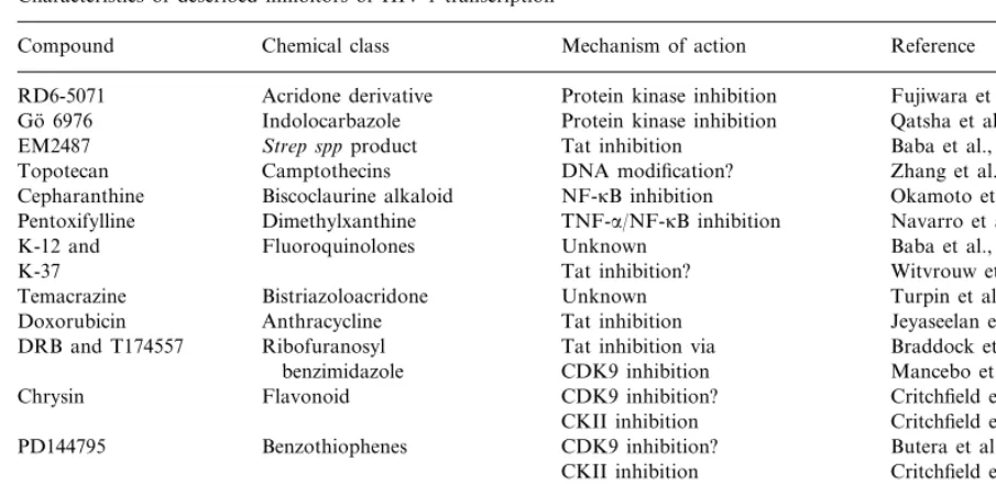
9. Experimental approaches targeting HIV-1
latency
Table 2
Characteristics of described inhibitors of HIV-1 transcription
Mechanism of action
Compound Chemical class Reference
Acridone derivative
RD6-5071 Protein kinase inhibition Fujiwara et al., 1999
Protein kinase inhibition
Indolocarbazole Qatsha et al., 1993
Go¨ 6976
Tat inhibition
EM2487 Strep sppproduct Baba et al., 1999
DNA modification?
Camptothecins Zhang et al., 1997
Topotecan
NF-kB inhibition Okamoto et al., 1998
Cepharanthine Biscoclaurine alkaloid
TNF-a/NF-kB inhibition
Dimethylxanthine Navarro et al., 1996
Pentoxifylline
Fluoroquinolones
K-12 and Unknown Baba et al., 1997
K-37 Tat inhibition? Witvrouw et al., 1998
Unknown
Bistriazoloacridone Turpin et al., 1998
Temacrazine
Anthracycline
Doxorubicin Tat inhibition Jeyaseelan et al., 1996
Tat inhibition via
Ribofuranosyl Braddock et al., 1991
DRB and T174557
CDK9 inhibition Mancebo et al., 1997
benzimidazole
CDK9 inhibition?
Flavonoid Critchfield et al., 1996
Chrysin
CKII inhibition Critchfield et al., 1997 CDK9 inhibition?
Benzothiophenes Butera et al., 1995
PD144795
CKII inhibition Critchfield et al., 1997 Isoquinoline
H-7 and HA100 CDK9 inhibition Mancebo et al., 1997
Transcript. elongation
sulfonamide Critchfield et al., 1999
T172298 Benzimidazole CDK9 inhibition Mancebo et al., 1997