© 2013 Ida Musfiroh et al. This is an open access article distributed under the terms of the Creative Commons Attribution License -NonCommercial-ShareAlike Unported License (http://creativecommons.org/licenses/by-nc-sa/3.0/).
Journal of Applied Pharmaceutical Science Vol. 3 (06), pp. 007-015, June, 2013 Available online at http://www.japsonline.com
DOI: 10.7324/JAPS.2013.3602 ISSN 2231-3354
Cytotoxicity Studies of Xanthorrhizol and Its Mechanism Using
Molecular Docking Simulation and Pharmacophore Modelling
Ida Musfiroh
1*, Muchtaridi Muchtaridi
1**, Ahmad Muhtadi
1, Ajeng Diantini
1, Aliya Nur Hasanah
1, Linar Zalinar Udin
2,
Yasmiwar Susilawati
1, Resmi Mustarichie
1, Rahmana E Kartasasmita
3, Slamet Ibrahim
31
Faculty of Pharmacy Universitas Padjadjaran, Bandung, Indonesia.
2
Laboratorium Bio-Assay Antikanker, Lembaga Ilmu Pengetahuan Indonesia, Bandung.
3
School of Pharmacy, Institut Teknologi Bandung (ITB).
ARTICLE INFO ABSTRACT
Article history:
Received on: 12/05/2013 Revised on: 28/05/2013 Accepted on: 12/06/2013 Available online: 27/06/2013
Xanthorrhizol (XNT) is one of major compounds from temulawak`s rhizome and its activity in several cancer cells is known. The aim of this study was to identify mechanism of xanthorrhizol from temulawak`s rhizome as an hERα inhibitor against breast cancer human cell lines. The cytotoxicity of XNT from temulawak`s rhizome on T47D human breast cancer cells lined by sulforhodamine B (SRB) method has been carried out, while molecular docking simulation and pharmacophore modelling methods were employed to predict mechanism of xanthorrhizol as hER inhibitor. Cytotoxicity studies showed that XNT of the isolated and standard had an IC50
100 and 55.50 µg/mL in T47D cells, respectively. Subsequently, molecular docking interaction showed that XNT
might be able to compete with estradiol (E2) as a potential ERα inhibitor with the calculated binding free-energy of -8.2 kcal/mol, even the compound superimposed with tamoxifen (4-OHT). XNT formed hydrogen bonds with Arg394 and Glu353 as mention E2 and tamoxifen also formed same interaction with same residue and interacted hydrophobic bonds similar to 4-OHT with: Leu387, Leu384, Leu391, Phe404, L349, Leu346, Met388, and Leu525 of estrogen alpha Ligan Binding Domain (LBD), although 4-OHT indicated stronger hydrophobic when the tail of tamoxifen interacted with Tyr347, Asp351, Trp383 and Leu428. XNT missed two chemical features into HipHop models pharmacophore thus may result in reduced inhibitory activity against T47D compared than 4-OHT. The xanthorrhizol mechanism as a hER inhibitor is postulated as partial estrogen antagonist, is justifiable based on its competitive characteristic versus tamoxifen (OHT-200) which was located on the active side of HER-α.
Key words:
Curcuma xa nthorrhiza Roxb., antiproliferative,
Xanthorrhizol, T47D cell, molecular docking simulation, HipHop models phamacophore
INTRODUCTION
Temulawak (Curcuma xanthorrhiza Roxb.) is one of the kinds of herbs that have been used for thousands of years as traditional medicinal herbs by some Asian countries. The
identification of essential oil of temulawak’s rhizome showed that
it consisted of several sesquiterpene derivative components, such as: artumeron, isofuranogermacene, kurlon, p-tolilmethylcarbinol and Xanthorrhizol (XNT). Essential oil fraction of temulawak was reached 3-12%, in that fraction the percentage of XNT was reached 11.6% (Sidik and Wardoyo, 1992; Hwang et al., 2000). XNT and alicyclic or tumeron aromatic (ar) will shows higher Rf value than the timol standard (Wagner et al., 1984). Based on the previous research, it is known that XNT restrained the growth of skin cancer by inhibiting the expression of ornithine
.
* Corresponding Author
Ema ils: * ida [email protected] c.id; ** muchta [email protected] c.id
decarboxylase, cyclooxygenase-2 and by inducting nitric oxide synthase through the activation of mitogen protein kinase and nuclear factor-KB (Chung et al., 2007).
XNT has been known to curb the proliferation of liver cancer by inducing apoptosis through bax control and p53 in a cell (Ismail et al., 2005). Furthermore, XNT has also been known to possess antiproliferative characteristics against breast cancer MCF-7 (Cheah et al., 2006).
Furthermore, human estrogen receptor-α is a part of a bigger core receptor family that shares structural similarity with human estrogen receptor-β, and other core receptors (Lupien et al., 2007). These two receptors are known to act as a medium for the estrogen signals. The function of this estrogen is to be a transcription factor that depends on ligands that interact with it (Lin et al., 2004).
Only recently has α estrogen inhibitor been developed as
a new type of medicine in preventing breast cancer. The slowing mechanism of this drug against estrogen has been researched and classified as: (a) estrogen partial antagonists such as tamoxifen; and as: (b) estrogen pure antagonists such as fulseverant (Jordan, 1994).
In this research, the mechanism possibility of
temulawak’s XNT as an inhibitor to human receptor estrogen
alpha (hER) was scrutinized by combining both the in vitro test or cytotoxic test and then in silico test using the molecular docking simulation and pharmacophore modelling The cytotoxicity test was done by using the Sulforhodamine B (SRB) method.
The cytotoxicity test was done into essential oils and isolate of xanthorrhizol which isolated from Curcuma xanthorrhiza. Subsequently, XNT was docked into estrogen alpha receptor (hER) with PDB code: 3ERT (Shiau et al., 1998) and compared to tamoxifen (OHT) and also E2 to clarify the mechanism of XNT. The aim of this study was to analyze the mechanism of xanthorrhizol from temulawak`s rhizome as an
hERα inhibitor in breast cancer human cell lines.
MATERIAL AND METHODS
Materials
Materials of plants: The material of temulawak`s rhizome used (Curcuma xanthorrhiza Roxb.) obtained from from Balitro, Lembang, West Java in April 2008. Voucher specimen (no. 54/HB/04/2008) was deposited in Herbarium of Department of Biology, Universitas Padjadjaran.
Isolation of essential oil
The temulawak`s rhizome dried (83 gram) were distilled with steam-distillation to obtain 5 mL essential oils in Monaco Lembang, West Java, for 3 hours to isolate the essential oil fraction. Essential oil stored at -20°C after the addition of sodium sulfate.
Extraction of Temulawak`s rhizome
The temulawak`s rhizome powder (5945.32 g) was extracted by methanol solvent 3 x 24 hours in that stirring was done intermittently. At the total extract was evaporated in vacuo at 40-50°C at 80 rpm. The extracts (902, 30 gram) were partitioned using mix solvent water and ethyl-acetate (EtOAc) with 1:1 ratio.
Isolation xanthorrhizol from Temulawak’s Rhizome
Isolation of xanthorrhizol was referred to Hwang et al. (2000). Subsequently, isolate compounds have been compared by using TLC (Fig. 1) and FT-IR (Fig. 3).
Phytochemical Screening
The phytochemical screening was done to simplisia, methanol extract and ethyl acetate fraction by using (Farnsworth, 1966).
Antiproliferative Activity Test
The cytotoxicity test was done by using Sulforhodamine B (SRB) method (Vichai and Kirtikara, 2006) by measuring proteins in the cells using ELISA plate reader. From this test the percentage of surviving cells could be counted and the value of
IC50, that is, the concentration of essential oil, temulawak’s
rhizome isolate and standard xanthorrhizol needed to inhibit 50% of human breast cancer T47D cells, could be determined.
Data Analysis
The antiproliferative activity data were analyzed using ANAVA one-way test (α= 0.05). The analysis is done to the percentage of survival cells, the IC50 value of essential oil, isolate, and xanthorrhizol standard.
Interaction Study of Xanthorrhizol against Human Estrogen Receptor-α by Molecular Docking Simulation
The crystal structure of protein ERα complexed with E2,
E2 (PDB id 1G50) and ERα complexed with 4-OHT (3ERT) were
used in this docking study. The 3D structure of 1G50 was reported by Eiler et al. (Eiler et al., 2001) using X-ray diffraction technique with a resolution of 2.90Å. Although the resolution is quite low, this structure represents the best resolution of E2 bound to ERα. 3ERT (Shiau et al., 1998) is an antagonist form of ERα which different from 1G50 at the region of loop-534. Water molecules and ligands were removed from the protein and the polar hydrogen were assigned for the protein by using AutoDockTools 1.5.4 (Sanner, 1999). Non-polar hydrogen atoms of E2, 4-OHT and xanthorrhizol were merged and rotatable bonds were assigned.
Molecular Docking Simulation Parameters
Pharmacophore Modelling
HipHop was run by using Catalyst embedded Discovery Studio 2.5. The models were generated based on some information whose consisted most-active compounds in the training set (Qin et al., 2007, Gauthier et al., 1997, Chao et al., 2006). In HipHop1 model, the six most-active compounds (1-6) in training set were firstly selected for HipHop run to identify common pharmacophore features. Compound 1 (tamoxifen) was considered as reference molecule. The principle term and the MaxOmitFeat term were set to 2 and 0, respectively. Subsequently, the HipHop2 was built from four most-active compounds (7-10) in training set. The structure of compounds can be seen in table5 and 6.
A conformational was set for each molecule using the polling algorithm and the best energy option, based on CHARMm force field from Discovery Studio 2.5 (Brooks et al., 1983). The molecules associated with their conformational models
were mapped onto the pharmacophore model using the “best
fit” option to obtain the bioactive conformation of each molecule.
RESULTS AND DISCUSSIONS
Phytochemical Screening
The phytochemical screening was done to determine the secondary metabolite group contained. The result of the screening can be shown in Table 1.
Table. 1: Secondary metabolite groups in simplisia, extracts and ethyl acetate
fractions.
Compound Groups Simplisia Powder
Methanol Extracts
Ethyl Acetate Fractions
Alkaloid + + +
Pholiphenol + + -
Tanin - - -
Flavonoid + + +
Monoterpene dan sesquiterpene + + +
Steroid - - -
Triterpenoid + + -
Quinine - - -
Saponin + + +
Legends: (+): Detected (-): Undetected
Temulawak’s Rhizome Isolate Identification
Essential Oil of Temulawak`s rhizome
The essential oils of temulawak`s rhizome was isolated with yield 6 % w/w. It was good quality as it has ideal percentage of essential oils. The standard of Materia Medica Indonesia (MMI)
requires that essential oils content in temulawak’s rhizome be 2 to
7 %.
Extraction and Isolation Results
MeOH extract obtained the yield of 15.18% (90.32 gram). The fraction of ethyl acetate yield 5.19% (222.48 gram). This fraction (30.32 gram) was subjected by liquid vacuum chromatography and n-hexane-ethyl acetate as an eluent having gradient polarity to obtain five fractions (A, B, C, D, and E). Fraction C was further subjected to gravitation column chromatography (GCC) with mobile-phase toluene-ethyl acetate (99:1) thus produced eight sub-fractions (C-1 until C-8).
Subfraction C-7 has one spot that same Rf (toluene-ethyl-acetate; 99:1) with reference of xanthorizol as shown in Fig. 1. Subfraction C-7 was further evaluated by UV and FTIR, as shown in Fig. 2 and Fig. 3, respectively.
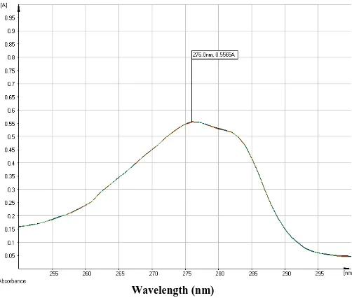
Fig. 1: C-7 isolate and standard XNT thin layer chromatogram using
toluene-ethyl-acetate; 99:1 as eluent; (i) XNT reference, (ii) C-7 isolate.
Wavelength (nm) Fig. 2: C-7 isolate ultraviolet Spectrum.
In the IR spectrum (Fig. 3), it showed the absorption characteristic at 3681 cm-1 (-OH in alcohol and phenol), 2966 cm-1 (CH3 and -CH2 aliphatic), 2873 cm-1 (-CH3 and -CH2 aliphatic), 1725 cm-1 (substituted benzene ring), 1515 cm-1 (benzene ring in aromatic compounds) wave number. The finger print of isolate C-7 at wave number less than 1500.. This shows that the C-7 isolate contains those functional groups.
Cytotoxic Test
The cytotoxic test is based on the surviving pasted SRB pigmented cell protein after the sample was given (Vichai and Kirtikara, 2006). The surviving cells were pigmented with 0.4% SRB diluted in acetic acid 1%. The pigmented cells were then further diluted in Tris base to determine the optical density using
ELISA plate reader with 515 nm wavelength. The reading of the ELISA plate reader showing absorption values for each
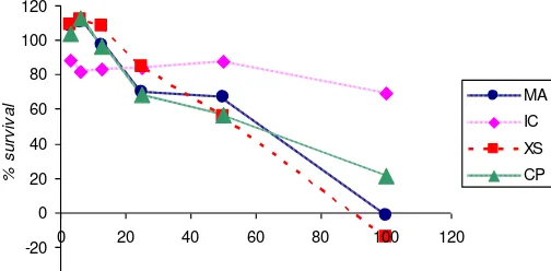
The results showed that increasing the concentration of the samples showed lower absorbance due to the smaller number of survival cells. The absorption values were taken from the number of cells remains after the test sample and cisplatin as the positive control. The greater the absorption value is, the greater the number of the living and SRB pigmented cells is; moreover, it can also be stated that the cytotoxic value is smaller if the absorption value is bigger. The absorption values, it can be determined the value of survival percentage which shows the cytotoxic effect.
Table. 3: Survival Percentage of XNT against T47D. Concentration
(µg/mL)
Survival Percentage
Cisplatin Essential oil Isolate XNT ref
100.000 21.186 -1.418 69.143 -13.897 results showed that increasing the concentration of the samples showed higher anticancer activity of cancer cells, as indicated by the survival percentage were lower. Best activity provided by xanthorrhizol reference, essential oil and xanthorrhizol isolated. According to the data of survival percentage, the smaller the survival percentage value is, the better the cytotoxic value will be. The increase of cisplatin, essential oil, isolate, and standard XNT concentration results in the increase of cytotoxic effect against
T47D breast cancer cells. Furthermore, the IC50 value for essential oil, isolate, and XNT reference were 55.503 µg/mL, 100 µg/mL, and 52.469 µg/mL, respectively according of the data. The small IC50 values indicate that the three tested substances possess strong toxicity against T47D breast cancer cells.
Fig. 4: Graph of survival percentage of essential oil, C-7 isolate, reference of
XNT, and Cisplatin. (MA ; essential oils, IC ; isolate compounds, XS : xanthorrizol reference, CP; cisplatin.
Other author (Cheah et al., 2006) reported that XNT inhibits the proliferation of the same human breast cancer cell line in this research, with an EC50 value of 1.71 µg/ml. In addition, xanthorrhizol also play a role as antiproliperative against the others human breast cancer lines MDA-MB-231 with IC50 8.67 µg/mL (Cheah et al., 2009, Cheah et al., 2008). However, the mechanism of xanthorrhizol as antiproliperative is not published yet. Fig. 3: Infra Red Spectrum between C-7 isolate (left) from temulawak and xanthorrizol reference (right).
Table. 2: The Average Absorption Values of Cisplatin, Essential Oil, Isolate, and XNT.
Concentration (µg/mL) Average Absorption Values ± SD
Cisplatin Essential oil Isolate XNT Reference
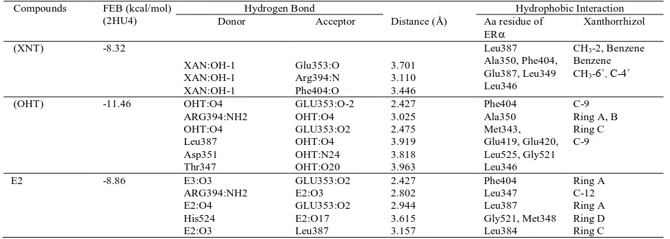
Table 4 Binding Interaction of xanthorrhizol (XNT), tamoxifen (OHT) and E2 (Estrogen) into ER. Compounds FEB (kcal/mol)
(2HU4)
Hydrogen Bond
Distance (Å)
Hydrophobic Interaction
Donor Acceptor Aa residue of
To know the mechanism, we tried to predict by using two methods of computational modeling; molecular docking simulation and pharmacophore modeling. The binding interaction of xanthorrhizol with human estrogen receptor-α can be explained through the following molecular docking simulation, while pharmacophore modelling could predict active chemical feature of xanthorrhizol that responsible for its biological activity.
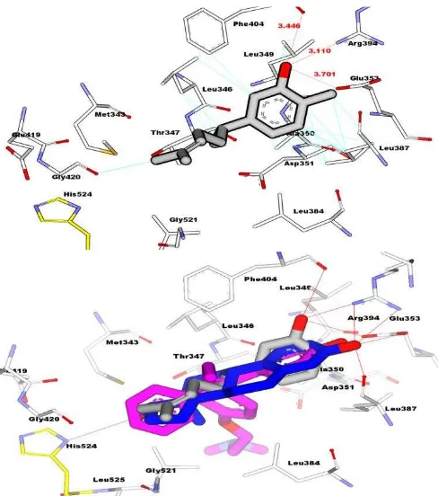
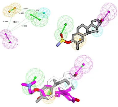
Fig 5 Interaction between XNT on the active side of HER-α (top) and overlay
binding interaction between xanthorrizol (grey carbon), tamoxifen (pink carbon), and E2 (white carbon) on the active side of HER-α (bottom). Red line: hydrogen bond interaction, blue line: hydrophobic interaction.
Interaction Study Result of XNT against HER-α by Computational Modeling
Molecular docking simulation was employed to predict interaction of xanthorrhizol into hER. Protein data bank of estrogen alpha (3ERT) was used with tamoxifen as crystal ligand. We docked xanthorrhizol, tamoxifen and E2 to clarify biding interaction into the site pocket of hER as shown in Table 4 and Fig. 5. The value of free energy binding (ΔG) and Ki of XNT were obtained -8.32 kcal/mol and 7.93 x 10⁻⁷, respectively.. The molecular docking simulation shows that XNT has less activity compared than tamoxifen . As shown Fig. 5, Hydroxyl groups of xanthorrhizol strongly formed hydrogen bond with Arg394 and Glu353 as mention E2 and tamoxifen that formed same interaction with same residue as shown in table 4.
Such as tamoxifen, XNT has formed a strong hydrophobic interaction with hER. Phe404, Le346, and Leu387 predominantly produced hydrophobic interaction with xanthorrhizol as shown in Table 4. It might explain that 1-OH and alkyl chain of xanthorrhizol that the hydroxyl group has played an
important role as chemical features responsible for their activity. XNT has hydrogen bond with Glu353 and also forms hydrogen bond with Arg394, but unfortunately, XNT loss interactions with residues of His524. Glu353, Arg394, and His524 are important residue since E2 formed four hydrogen bonds between hydroxyl groups of E2 (Table 4) and residues of side Glu353, Arg394, His524. His524 is important in binding of copep (coprotein peptida) into H12, because H524 shows a conserved hydrogen bond from E2(O17) to His524(N) during MD simulation (Celik et al., 2007). This residue is also role in building zipper network H3 and H11 with the others residues (K531, E419, and E339) (Gangloff et al., 2001).
The presence of a network zipping H3 and H11 stabilized conformation of agonist. This zipper network confirms that H12 is placed in the “mouse trap”, as consequence it can not reach the antagonist position on the hER LBD surface when H11 is not allowed to relax (Celik et al., 2007). However, when antagonist (4-OHT) is placed, this network zipper is lost. In this study, the presence XNT in binding pocket also has eliminated this network zipper as shown Fig. 6.
Fig 6. Zipper network were built by His524, Lys531, Glu419, and Glu339 on
Table 6. Training set for HipHop2.
No. Structure IC50
(nm)
Principal MaxOmitFeat
1 OH
O N
4 1 0
2
O N
OH
NN
+N
-1 2 0
3
O N
OH
Cl
4 1 0
4
OH S
O N
HO
O
7.8 1 1
The unleashing of zipper network in antagonist interaction is speculated as a consequence of the rotation of His524, hence Glu339 and Lys531 are positioned too far away from each other to interact. As shown in Table 4, hydrogen bond distance between OHT and important residues (Arg394 and Glu353) was smaller than hydrogen bond distance between XNT and residues. In addition, hydrophobic interaction of OHT-hER LBD was more strongly than XNT- hER LBD. It was reason why XNT was less active than OHT. Based on molecular docking results, we postulated that 1-OH and alkyl chain of xanthorrhizol has been responsible into the activity. To test of our postulates, we created pharmacopohore modelling using HipHop running. HipHop running would identify important chemical features based on most active compounds against MCF7. We created two HipHop with different training set consisted by using catalyst embedded Discovery Studio 2.5. HipHop1 model was built by training set (Table 5) that consisted of six compounds as shown in Fig. 7. Four compounds were set as active and two compounds were set as moderate active using Principal and MaxOmitFeat of HipHop parameters (Barnum et al., 1996). 2-ethylE2-3-O-sulfamate (Newman et al., 2004) (1), raloxifen derivate (Grese et al., 1996)
(2), and two arilbenzothiophene (Grese et al., 1997) (3 and 4) derivates were set as most active compounds.
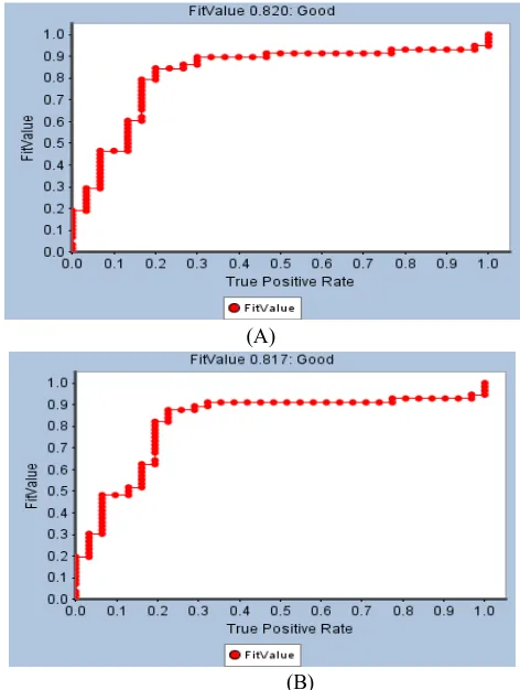
Model HipHop2 (Figure 8) was created by four compounds of tamoxifen derivatives (Chao et al., 2006) compounds (Table 6). Feature of best HipHop models were used to provide the pharamacophoric features of xanthorrhizol. Best HipHop models successful were selected based on ROC analysis. The both of models consisted the same features Hydrogen Bond Donor (HBD), Hydrogen Bond Acceptor (HBA), Hydrophobic (HY), and Ring Aromatic (RA), however different position, angle, and torsion. HipHop1 models (was shown in Fig. 8a that has ROC value 0.82 (Fig. 7a), while HipHop2 has ROC value 0.817 (Fig 7b). Xanthorrhizol missed two features (RA and HBD) of HipHop1 as shown in Figure 8.
(A)
(B)
Fig 7. Received operator curves (ROC) conducted for pharmacophore models:
(A) HipHop1 and (B) HipHop2 (numbers of active are 35 of 60 molecules). As predicted of molecular docking results, only 1-OH and tail of 1’-alkyl were mapped by HBA and HY, respectively.
Figure 8 ilustrated that HBA, RA, and HY of HipHop2 successfully mapped 1-OH, benzene ring, and 1’-alkyl of xanthorrhizol, respectively. However, xanthorrhizol missed HBD features of HipHop2. On the other hand, in molecular docking results might show that RA only play role as hydrophobic interaction. The molecular docking results were in accordance with pharmacophore mapping results.
alkyl chain of xanthorrhizol were two chemical features that have an important role in biological activity as partial antagonist hER. Ring aromatic of xanthorrhizol also contributed in molecular interaction with hER. To improve activity of xanthorrhizol, we have suggestion that modified 6-methyl and attachment some
HBD groups in 1’ and 2’ of alkyl chain may allow increased
activity against MCF7 human breast cancer lines.
CONCLUSIONS
Based on these studies, it is known that essential oil,
temulawak’s rhizome isolate and pure xanthorrhizol possess
cytotoxic effects on T47D breast cancer cells with IC50 concentration, 55.503 µg/mL, 100 µg/mL and 52.469 µg/mL, respectively. The increased essential oil, isolate and xanthorrhizol
concentration of temulawak’s rhizome can result in cytotoxic
increase. The xanthorrhizol mechanism as a medicine for breast cancer in that xanthorrhizol is hypothesized as partial estrogen antagonist is justifiable based on its competitive characteristic versus tamoxifen (OHT-200) which was located on the active side of HER-α.
ACKNOWLEDGMENT
We gratefully acknowledge to Prof. Habibah A Wahab from USM for the support of the computational laboratory facilities, and thanks to Yaya Rukayadi for supporting the XNT reference, thanks to Ambarsari ayuningtyas, Angga Gegana P and Yuni Fitria for preparing sample and analysis
REFERENCES
Agarwal G, Pradeep PV, Aggarwal V, Yip CH, Cheung PS. Spectrum of breast cancer in Asian women. World J Surg 2007;31(5): 1031-40.
Barnum D, Greene J, Smellie A, Sprague P. Identification of Common Functional Configurations Among Molecules. Journal of Chemical Information and Computer Sciences 1996;36(3): 563-71.
Brooks BR, Bruccoleri RE, Olafson BD, States DJ, Swaminathan S, Karplus M. CHARMM: A Program for Macromolecular Energy, Minimization, and Dynamics Calculations. J Comput Chem 1983;4(2): 187-217.
Celik L, Lund JD, Schiott B. Conformational dynamics of the estrogen receptor alpha: molecular dynamics simulations of the influence of binding site structure on protein dynamics. Biochemistry 2007;46(7): 1743-58.
Chao EY, Collins JL, Gaillard S, et al. Structure-guided Fig 8. (A) HipHop2 model generated by HipHop module of Catalyst in DS 2.5 Package with view distance inter -features (B) HipHop2 aligned on 2Ete2MATE
synthesis of tamoxifen analogs with improved selectivity for the orphan ERRgamma. Bioorg Med Chem Lett 2006;16(4): 821-4.
Cheah Y, Nordin F, Sarip R, et al. Combined xanthorrhizol-curcumin exhibits synergistic growth inhibitory activity via apoptosis induction in human breast cancer cells MDA-MB-231. Cancer Cell International 2009;9(1): 1.
Cheah YH, Nordin FJ, Tee TT, Azimahtol HLP, Abdullah NR, Ismail Z. Antiproliferative Property and Apoptotic Effect of Xanthorrhizol on MDA-MB-231 Breast Cancer Cells. Anticancer Research 2008;28(6A): 3677-89.
Cheah YH, Azimahtol HLP, Abdullah NR. Xanthorrhizol Exhibits Antiproliferative Activity on MCF-7 Breast Cancer Cells via Apoptosis Induction. Anticancer Research 2006;26(6B): 4527-34.
Chung WY, Park JH, Kim MJ, et al. Xanthorrhizol inhibits 12-O-tetradecanoylphorbol-13-acetate-induced acute inflammation and two-stage mouse skin carcinogenesis by blocking the expression of ornithine decarboxylase, cyclooxygenase-2 and inducible nitric oxide synthase through mitogen-activated protein kinases and/or the nuclear factor-kappa B. Carcinogenesis 2007;28(6): 1224-31.
Eiler S, Gangloff M, Duclaud S, Moras D, Ruff M. Overexpression, purification, and crystal structure of native ER alpha LBD. Protein Expr Purif 2001;22(2): 165-73.
Farnsworth NR. Biological and phytochemical screening of plants. J Pharm Sci 1966;55(3): 225-76.
Gangloff M, Ruff M, Eiler S, Duclaud S, Wurtz JM, Moras D. Crystal structure of a mutant hERalpha ligand-binding domain reveals key structural features for the mechanism of partial agonism. J Biol Chem 2001;276(18): 15059-65.
Gauthier S, Caron B, Cloutier J, et al. (S)-(+)-4-[7-(2,2-dimethyl-1-oxopropoxy)-4-methyl-2-[4-[2-(1-piperidinyl)-
ethoxy]phenyl]-2H-1-benzopyran-3-yl]-phenyl
2,2-dimethylpropanoate (EM-800): a highly potent, specific, and orally active nonsteroidal antiestrogen. J Med Chem 1997;40(14): 2117-22.
Grese T, Cho s, Bryant HU, et al. Synthesis and Pharmacology of 2-Alkyl Raloxifene Analogs. Bioorganic & Medicinal Chemistry Letters 1996;6(2): 201-06.
Grese TA, Cho S, Finley DR, et al. Structure-activity relationships of selective estrogen receptor modulators: modifications to the 2-arylbenzothiophene core of raloxifene. J Med Chem 1997;40(2): 146-67.
Hwang JK, Shim JS, Baek NI, Pyun YR. Xanthorrhizol: a potential antibacterial agent from Curcuma xanthorrhiza against Streptococcus mutans. Planta Med 2000;66(2): 196-7.
Ismail N, Pihie AH, Nallapan M. Xanthorrhizol induces apoptosis via the up-regulation of bax and p53 in HeLa cells. Anticancer Res 2005;25(3B): 2221-7.
Jordan VC. Molecular mechanisms of antiestrogen action in breast cancer. Breast Cancer Res Treat 1994;31(1): 41-52.
Lupien M, Jeyakumar M, Hebert E, et al. Raloxifene and ICI182,780 increase estrogen receptor-alpha association with a nuclear compartment via overlapping sets of hydrophobic amino acids in activation function 2 helix 12. Mol Endocrinol 2007;21(4): 797-816.
Lin CY, Strom A, Vega VB, et al. Discovery of estrogen receptor alpha target genes and response elements in breast tumor cells. Genome Biol 2004;5(9): R66.
Morris GM, Goodsell DS, Halliday RS, et al. Automated docking using a Lamarckian genetic algorithm and an empirical binding free energy function. Journal of Computational Chemistry 1998;19(14): 1639-62.
Newman SP, Leese MP, Purohit A, et al. Inhibition of in vitro angiogenesis by 2-methoxy- and 2-ethyl-estrogen sulfamates. Int J Cancer 2004;109(4): 533-40.
Qin Z, Kastrati I, Chandrasena RE, et al. Benzothiophene selective estrogen receptor modulators with modulated oxidative activity and receptor affinity. J Med Chem 2007;50(11): 2682-92.
Shiau AK, Barstad D, Loria PM, et al. The structural basis of estrogen receptor/coactivator recognition and the antagonism of this interaction by tamoxifen. Cell 1998;95(7): 927-37.
Sanner MF. Python: a programming language for software integration and development. J Mol Graph Model 1999;17(1): 57-61.
Sidik R, Wardoyo MM. Temulawak. Jakarta: Phyto Medika, 1992.
Vichai V, Kirtikara K. Sulforhodamine B colorimetric assay for cytotoxicity screening. Nat Protoc 2006;1(3): 1112-6.
Wagner HS, E.M. B, Zgainski. Plant drug Analysis. Berlin Springer Verlag, 1984.
How to cite this article: