Research Journal of Pharmaceutical, Biological and Chemical
Sciences
Interaction of Aspartyl-Dipeptides Derivatives with Metabotropic Glutamate
Receptor (mGluR) Using Molecular Docking Simulation.
Muchtaridi Muchtaridi*, Siti Farhain Binti Amir, Wiwiek Indriyati, and Ida Musfiroh.
Department of Phar,aceutical Anlysis and Medicinal Chemistry, Faculty of Pharmacy, Universitas Padjadjaran, Jl. Jatinangor KM 21-Sumedang.
ABSTRACT
Aspartame (L-alpha-aspartil-L-phenylalanine-1-methyl ester) known as a molecule that plays role in sweet taste and the derivatives of aspartile-dipeptide is also known to have the potential as a sweetener. The object of this study was to know the interaction of the aspartyl-dipeptides derivatives against metabotropic glutamate receptors (mGluR). This target protein can induce active synapse signal for a sweet taste. The study has been carried out to observe the interaction of ligand and target protein using molecular docking simulation. The interaction of the aspartyl-dipeptides derivatives connected with Lys409 through hydrogen bond interaction. The carboxylate group might be responsible in the sweetening interacted with Ser186, Thr188, Asp318, Lys409, and Arg78, and N-H group interact with Ser186 and Asp318 favorably by forming hydrogen bond. The benzene ring could interact with Tyr74 and Tyr236 with hydrophobic pocket at the binding site.
Keywords:Aspartame, aspartile-dipeptide, docking, sweetener.
INTRODUCTION
Aspartame or aspartyl-phenylalanin methyl ester is an artificial sweetener that has a degree of sweetness about 180-200 times more than sucrose and can be metabolize by the body as big as 3.97 Kkal/g [1, 2]. The derivatives of aspartil-dipeptide can also be used as sweeteners and is commonly used in products in the market, especially in food products or sugar-free products [1].
The use of aspartame have side effects due to the metabolism of aspartame in the body such as toxicity from methanol which is one of the product from the process of metabolism, the increase in concentration of phenylalanin (Phe) in plasma and aspartic acid can increase the transportation of amino acid into the brain and will change the neurochemical in the brain, as well as induce the effect of epilepsy and brain tumor [3, 4].
Many studies have been done on the use of aspartame in various species such as human, rat, mouse and rabbit [5-7]. However, not many studies were made on aspartame interaction as target site, which is sweet receptor.
The discovery of the sweet receptors has open new perspectives for designing new sweeteners that require explanation on the mechanism of interaction between the sweetener and sweet receptor. T1R2-T1R3 receptors are activated by the sweet molecules which are Class C, G-protein coupled receptor (GPCR) with the sequence homology of mGluR receptors (metabotropic Glutamate receptors). Metabotropic glutamate receptors are homodimeric while T1R3 receptors are heterodimeric [8]. Previous study [9-11], the structure and activity analysis (QSAR) was used a derivative-dipeptide aspartil derivatives and lead structure 3-amino acid suksinamate suggested that sweet receptors shows some activity with the molecule [12].
The previous study, the molecular docking process has been conducted on three compounds that have the potential to give sweet taste that are neotame, superaspartam, dan SC-45647 [13, 14]. The results showed that proposed active conformations of the high-potency sweeteners neotame, superaspartame, and SC-45647 could interact favorably in this binding site, forming ion pairs or ionic hydrogen bonds with His-163, Glu-318, and His-407, in addition to hydrophobic interactions with numerous nonpolar side-chains [13]. Here we docked forty compounds of the aspartyl-dipeptides and derivatives to know the interaction against sweetening receptor. This study was expected to give information on planning the production of aspartyl-dipeptides that is safe to use by knowing the place of the amino acid interaction from the various kind of derivatives.
MATERIALS AND METHOD
Materials
This research used LBD (ligand binding domain) of Structure of 3D LBD(ligand binding domain) metabotropic Glutamate receptor (mGluR) (PDB id : 1EWK[15]) structure with 2.2 Å resolution [15]. In this crystal structure (1EWK), glutamate acid is complexed with mGluR. The crystal structures were selected should have best resolution or lower resolution value, and also have R-free and R-value lower than 0.25 [16]. The 3D-structures of ligands were constructed using Hyperchem 7, then were optimized using Austin Model 1 (AM1) [17].
Molecular Docking Simulation
The ligands and proteins were prepared by AutoDockTools (ADT). Ligand and protein available in the PDB structure were converted to PDBQ and PDBQS format by adding charges, hydrogens and assigning ligand flexibility. Kollman charges and solvation parameter were assigned using default value to the protein while Gasteiger charges were added to each ligand. A grid box of 60 x 60 x 60 points, with a spacing of 0.375 Å and a precise coordinate 11.407, 13.031 and 12.342 along the x, y and z axes pertaining the center of the active site was built around the binding region. Population size of 150 and 250 000 energy evaluations were used for 50 search runs via Lamarckian Genetic Algorithm (LGA) [17, 21]. The molecular docking result were analyzed based on the lowest free energy binding chosen from the most populated cluster and saved in dlg file for visualization. Aspartame was used as control docking that imposed against mGluR (PDB ID:1EWK)[15].
RESULTS AND DISCUSSION
The compounds of aspartyl-dipeptide derivatives that have known their degree of sweeteness (high, medium and low) are choose based on availability of optimize geometry process. The degree of sweetness is correlated with relative sweetness (RS), where RS is sweetness power (SP) relative to sucrose. Log RS and SP (Log 1+RS) value are converted into 103-Log RS. Lower the value of 103-Log RS, higher the degree of sweeteness [9, 11, 18].
Observation of Target Protein Metabotropic Glutamate Receptor (mGluR)
The observation of crystal structure of receptor have been done to identify the essential of amino acid favorably interact with the ligands and this is important for contribution of a sweet taste.
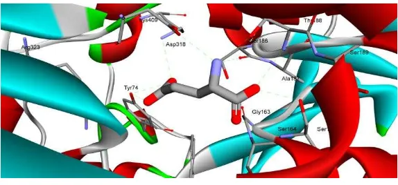
Figure 1: The interaction of amino acid residue on catalytic side of mGluR receptor with model ligand of glutamic acid (bold stick). This figure was visualized by Discovery Studio 3.5.
The data showed that the essential residues of amino acid (Tyr74, Arg78, Ser164, Ser165, Ser186, Thr188, Tyr236, Glu292, Gly293, Asp318, Arg323 and Lys409) that interact with glutamate acid natural ligand before re-docking as shown in Fig. 1. In previous study, the site active of hT1R2 receptor are Tyr103, Asp142, Asn143, Ser144, S165, Ala166, Ileu167, Ser168, Tyr215, Arg270, Val272, Val274, Phen275, Ser301, Glu302, Ser303, Asp305, Thr326, Arg378, Lys379, Ser380, and Arg383 [22, 23]. Zhang et al. (2009) found the
hydrophobic site in hT1R2 include Asp43, Val64, Ileu67, Tyr103, and Lys65 of the upper lobe, and Pro277, Lys279, and Val309 of the lower lobe [12].
Tabel 1: Hydrogen Bond Interaction of Glutamic Acid with mGluR.
Amino acid residue atom Ligand Atom Distance (Å)
TYR74:OH their population with 50 times running at different dimension of grid box.
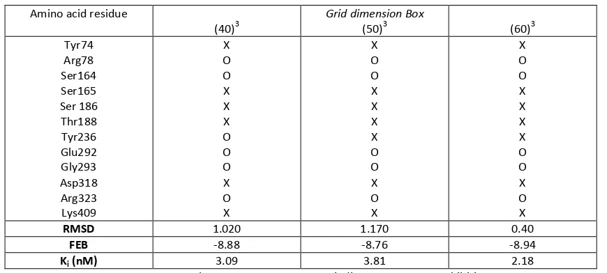
Table 2: Re-docking result simulations of amino acid with variance dimension of Grid Box.
Amino acid residue
X= present; O = absent; FEB = Free energy Binding; Ki = Constant Inhibition
Different dimension of grid box was started with narrower one and further with larger one thus all the target amino acid could be interacted with the ligand as shown in Table 2. The results also showed that value of Ki relatively did not change with increasing size of dimension and keep in scale on nanomolar value.
However, no all of hydrogen bond interaction could be produced as crystal form, the value of RMSD still in the range not higher than 2 Å. There was still the interaction of essential amino acid with the ligand even in increasing dimension of grid box. The amino acid Ser165, Ser186, Thr188 and Lys409 had hydrogen bond interaction to the ligand in all the dimension (40)3, (50)3 dan (60)3 point, 0.375 Å apart and their distance was lower than 3 Å which suited with hydrogen bond distance requirement. According to Kunishima et al. (2000),
the most important hydrogen bond to the ligand of glutamate was interaction with amino acid Tyr74 thus the simulation docking result showed the amino acid that had the interaction was amino acid Tyr74, Arg78, Ser164, Ser165, Ser186, Thr188, Tyr236, Glu292, Gly293, Asp318, Arg323 and Lys409. This amino acid became the main reason in choosing the most suited dimension of grid box [15].
Simulations docking of aspartyl-dipeptide derivatives
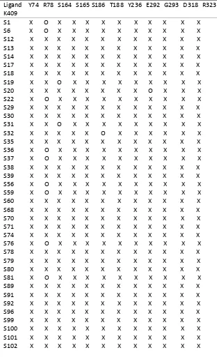
of these ligands have aromatic ring and aliphatic chain that have influence on interaction bond between ligand and with the receptor at binding site. Almost all of the ligand of ligand-aspartyl dipeptide derivatives had capable to generate the interaction with the target amino acid residues. Some ligands was not able to produce bond interaction with the amino acid residues Arg78 as shown Table 3 due to the position of Arg78 bit away from the main dimensions of the ligand binding site, such as ligand of S1, S6, S18, S22, S36, S37, S56, S59, S76, S81 and Q84 (See in Supplementary). Ligand of S19 and S31 also did not have bond interaction with Ser164 amino acid residues. S32 did not have binding interactions with amino acid Thr188, while S20 did not interact with amino acid of Gly293.
Table 3: Amino acid residues that interact with the ligand of aspartyl-dipeptide derivatives.
Ligand Y74 R78 S164 S165 S186 T188 Y236 E292 G293 D318 R323 K409
S111 X X X X X X X X X X X X S112 X X X X X X X X X X X X S113 X X X X X X X X X X X X S132 X X X X X X X X X X X X Q83 X X X X X X X X X X X X Q84 X O X X X X X X X X X X Q85 X X X X X X X X X X X X Q86 X X X X X X X X X X X X Q88 X X X X X X X X X X X X Q90 X X X X X X X X X X X X Q91 X X X X X X X X X X X X Q104 X X X X X X X X X X X X Q111 X X X X X X X X X X X X
Explaination: X= presence of interaction of amino acid, O = absent of interaction of amino acid
The main group of carboxyl group, carbonyl group, carboxylic group, NH groups and some derivatives that have the benzene ring. Molecular docking simulation is performed in previous studies also suggested that bond interactions with the receptor structure has the same features that are carboxyl groups, two hydrophobic substituents, and three polar NH. These groups were responsible for sweet taste inducer. Nature of the binding site of mGlu receptors have a very high polar characteristic due to a lot of hydroxyl groups [24]
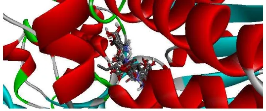
The overall result of aspartyl dipeptide derivatives could be concluded that carboxylic group (COO-) of aspartyl-dipeptide derivatives had a bonding interaction with Lys409 amino acids. Carbonyl group interacted with amino acid Arg323 and the carboxyl group had interactions with amino acids Ser186, Lys409 and Arg78. While, the benzene ring of the compounds formed hydrophobic bond interaction with amino acid Tyr74 and Tyr236. In addition, some of derivatives that have benzene ring make interaction bondingwith amino acid Trp110 which is not an amino acid target. a Groups of NH of this derivatives formed hydrogen bon interaction with amino acid Ser186, and Asp318. Molecular docking results of superaspartame with mGlu receptors showed that carboxylate groups have bonding interactions with amino acid His163 and His407 in the surface of LB1. Glu318 in the surface of LB2 make bonding interaction with polar groups of –NH and CH2 groups of the ligand bonded to amino acid Ser164 and Ser292 [24]. Figure 2 showed that the overlay of all ligands against binding pockets of mGlu receptor in the same place. Overlay of several ligands exhibit different ligand conformation yet still be able to bind and interact with the essential amino acid residues.
Figure 2: The results showed that the overlay of ten ligands interacted with target amino acid.
chelates and acidity. Changes in these properties can affect the biological activity of compounds [27]. However, the addition of a methyl group at position N various derivatives leads to loss of hydrogen bonds and weak intermolecular bonds between molecules that cause less soluble in polar solvents.
However, hydrophobic interaction had an important role to stabilize the protein conformation, for the lipid transportation by plasma proteins. Molecular docking results showed that there were some hydrophobic interactions on aspartyl- dipeptide derivatives which have chiefly benzene group. It was instrumental forming a more stable complex between the ligand and the target protein. Ligands that have a phenyl group, aromatic and aliphatic group formed a hydrophobic bond with amino acid Tyr74. Hydrophobic bonding was due to polar region of the ligand with non-polar regions of protein targets. Formation of hydrogen bonds and hydrophobic an important factors for sweet taste.
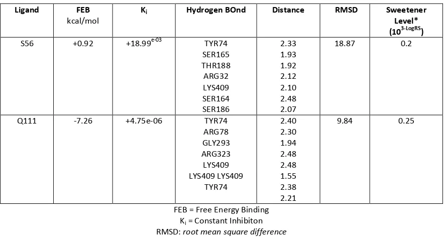
Table 3 Docking result of ligand that has highest the degree of sweetness.
Ligand FEB
kcal/mol
Ki Hydrogen BOnd Distance RMSD Sweetener
Level* (103-LogRS)
S56 +0.92 +18.99e-03 TYR74 SER165 THR188 ARG32 LYS409 SER164 SER186
2.33 1.93 1.92 2.12 2.10 2.48 2.07
18.87 0.2
Q111 -7.26 +4.75e-06 TYR74 ARG78 GLY293 ARG323 LYS409 LYS409 LYS409
TYR74
2.40 2.30 1.94 2.48 2.48 1.55 2.38 2.21
9.84 0.25
FEB = Free Energy Binding Ki = Constant Inhibiton
RMSD: root mean square difference
* Sweetener level was declared by LOG RS (Relative sweetness), Log 1 +RS have been converted to 103-Log RS
There was different in value for FEB and Ki value of the ligand S56 and Q111. The value of FEB of the ligand S56 was 0.92 kcal/mol, ligand S56 was +0.92 kcal/mol and ligand Q111 was -7.26 kcal/mol. Binding affinity of the ligand Q111 to the target protein was much stronger other than ligand S56 even they have the same high degree of sweetness. While, Ki value showed minimum concentration needed to work effectively on
target protein. The Ki value of ligand Q111 with the value of 4.75 10-06 much smaller than S56 with 18.99 10-03.
Figure 3 : The interaction of amino acid residue on catalytic side of mGluR receptor with Q111 compound (bold stick and black carbon). This figure was visualized by Discovery Studio 3.5. Green line: hydrogen bond interaction,
The higher means that the higher affinity for the target protein due to relatively stronger the bond. While, the smaller value was the value of Ki much better. However, this factor did not considered as a major
factor as not all drugs require the lowest energy to bind to the receptor, but the conformation is one of the factor of solubility due to capability to make hydrogen bond [28].
As shown in Fig. 3, the amine group of Q111 could interact well with Asp318. The carbocylate acid group formed hydrogen bond with Thr188, Asp318, and Ser186. The aromatic ring group was closed to Tyr74, Ser164, and Val167. Asp318, Thr188, and Gln211 formed hydrogen bod network to stabilize the interaction of ligand and receptors eith recognition of negatively charged groups of ligands[29]. Therefore, this network was closed with carboxylate acid groups of ligands.
CONCLUSION
The conclusion can be drawn that the computational docking studies were able to see the interaction of amino acid residues in various derivative of aspartyl-dipeptide. Q111 gave the best interaction that have smallest FEB. Carboxilate acid, carbonyl, amine, and benzene ring of aspartyl-dipeptide derivatives might play important role in binding interaction against mGlu receptor. The carboxylate group of the ligand derivatives interact with amino acid Lys409. The carbonyl group ligand derivatives interact with amino acid Arg323 as NH group interacts with amino acid Ser186 and Asp318. Q111 contained benzene ring that have hydrophobic interaction with amino acid residues Tyr 74 and Tyr 236.
REFERENCES
[1] Yang Q, The Yale journal of biology and medicine 2010;83:101-108. [2] Donato K, Hegsted DM.Proc Nat Acad Sci 1985;82:4866-4870. [3] Lean ME, Hankey CR. BMJ 2004;329:755-756.
[4] Yilmaz S, Ucar A, Cytotech (2014).
[5] Humphries P, Pretorius E, Naude H, Eur J Clin Nutr 2007;62:451-462.
[6] Ranney RE, Oppermann JA, Muldoon E, McMahon FG. J Toxicol Environ Health 1976;2:441-451. [7] Magnuson B, Williams GM. Environ Health Persp 2008116:A239-240.
[8] Pin JP, Galvez T, Prezeau L. Pharmacology & therapeutics 2003;98:325-354 [9] Laszlo T, Lupescu I. Groposila-Constantinescu, ARKIVOC X (2005) 254-271.
[10] Afantitis A, Melagraki G, Sarimveis H, Koutentis PA. J. Markopoulos, O. Igglessi-Markopoulou, QSAR & Combinatorial Science 2006;25 :928-935.
[11] Laszlo T, Lupescu I. Groposila-Constantinescu, ARKIVOC XIII 2006;pp.22-40.
[12] Zhang F, et al. Proceedings of the National Academy of Sciences 2010;107:4752-4757. [13] Eric Walters D. Pure Appl. Chem. 2002;74 :1117–1123.
[14] Wermuth CG. The Practice of Medicinal Chemistry, Elsevier Science, 2011. [15] Kunishima N, et al. Nature 2000;407:971-977.
[16] E.N. Brown, S. Ramaswamy, Acta Crystallogr D Biol Crystallogr 63 (2007) 941-950. [17] M. Muchtaridi, K. Lestari, Int J Pharm Pharmac Sci 6 (2013) 795-800.
[18] van der Heijden A, Brussel LBP, Peer HG. Chemical Senses 1979;4:141-152.
[19] Morris GM, Goodsell DS, Halliday RS, Huey R, Hart WE, Belew RK, Olson AJ. J Comput Chem 1998;19 :1639-1662.
[20] Morris GM, Huey R, Olson AJ, Curr Protoc Bioinformatics Chapter2008; 8 :14. [21] Morris GM, Lim-Wilby M. Methods Mol Biol 2008;443:365-382.
[22] Li X, et al. Chem Senses 2011;36:453-475. [23] Temussi PA. FEBS Letters 2002;526:1-4.
[24] Walters DE. Homology-based model of the extracellular domain of the taste receptor T1R3, Pure and Applied Chemistry, 2002, p. 1117.
[25] Valiana T. Creating of The Features of Pharmacophore of Aspartil-dipeptida Derivatives as Sweetener., Faculty of Pharmacy, Universitas Padjadjaran, Jatinangor, 2013.