www.elsevier.com/locate/jinsphys
Native and heterologous neuropeptides are cardioactive in
Drosophila melanogaster
Erik Johnson
a, John Ringo
a, Harold Dowse
a, b,*aDepartment of Biological Sciences, 5751 Murray Hall, University of Maine, Orono, ME 04469-5751, USA bDepartment of Mathematics and Statistics, University of Maine, Orono, ME 04469-5751, USA
Received 20 September 1999; accepted 11 January 2000
Abstract
Nine neuropeptides isolated from Drosophila melanogaster and five neuropeptides, previously isolated from the CNS of Limulus with antisera to FMRFamide-related peptides, were tested for their effects on the myogenic heart of Drosophila melanogaster. Of the native peptides, TDVDHVFLRF-NH2(Dromyosuppressin), DPKQDFMRFamide, and PDNFMRFamide significantly slowed the
heart. Of the Limulus peptides, DEGHKMLYFamide (LP1) increased heart rate significantly, GHSLLHFamide (LP2) and PDHHMMYFamide (LP3) decreased the heart’s rate, while DHGNMLYFamide (LP4) and GGRSPSLRLRFamide (LP5) had no effect at the concentrations we employed. Dromyosuppressin, DPKQDFMRFamide, and PDNFMRFamide from Drosophila, and LP2 and LP3 from Limulus, which belong to a novel group of peptides structurally unrelated to FMRFamide, are among only a very few substances from within the general group of neuropeptides and neurohormones known to slow the heart of Drosophila, and as such offer an important tool for investigating the molecular mechanisms underlying the control of the pacemaker.2000 Published by Elsevier Science Ltd. All rights reserved.
Keywords: Cardiac pacemaker; Drosophila; Heartbeat; Limulus; Neuropeptides
1. Introduction
The Drosophila heart offers an excellent system for the elucidation of cardiac physiology at the molecular level, owing to the variety of genetic and molecular tools available. It is a simple tube found in the posterior por-tion of the abdomen, and it is continuous with a contrac-tile dorsal vessel which terminates in the head region Rizki, 1978; Curtis et al., 1999). The cardiac pacemaker, located in the most caudal portion of the heart proper, is myogenic (Rizki, 1978; Dowse et al., 1995). The ion channels central to the pacemaker are under scrutiny (Dowse et al., 1995; Johnson et al., 1998) and the overt effects of neurohormones such as serotonin, octopamine, and norpinephrine on heartbeat have been investigated (Johnson et al., 1997; Zornik et al., 1999).
The discovery of the molluscan peptide,
Phe-Met-* Corresponding author. Tel.:+1-207-581-2536; fax:+ 1-207-581-2537.
E-mail address: [email protected] (H. Dowse).
0022-1910/00/$ - see front matter2000 Published by Elsevier Science Ltd. All rights reserved. PII: S 0 0 2 2 - 1 9 1 0 ( 0 0 ) 0 0 0 4 3 - 3
Arg-Phe-NH2(FMRFamide), has led to the identification
of a number of N-terminally extended FMRFamide-related peptides (Price and Greenberg, 1989) with wide-spread phylogenetic distribution. One effect of a subset of these peptides is modulation of heartbeat (Review: Ga¨de et al., 1997). A number of native neuropeptides have been isolated from Drosophila based on similarity to FMRFamide (Nambu et al., 1988; Schneider and Taghert, 1988; Nichols, 1992; McCormick and Nichols, 1993; Nichols et al. 1995, 1997).
Nichols, 1992). Hewes et al. (1998), tested eight of these peptides, synthesized from predicted sequences, on the neuromuscular junction (NMJ) of Drosophila larval muscle. All but one were excitatory at the level of the NMJ, and there was no evidence of synergy when the peptides were injected in combination. This led these workers to propose a functional redundancy among the peptides encoded by this gene. They cautioned, however, that for evolutionary conservation of these peptides to have occurred, differential effects in other tissue targets is likely (Hewes et al., 1998). We measured the response of the Drosophila pupal heart to these and other peptides in the hope of discovering differential effects. Any dif-ferences between the response of the heart and that of the NMJ, or differences in response among the peptides within the cardiac system, would reveal functional diver-sity among the peptides, a critical step in a search for receptors and modes of action. Cardioinhibitory peptides have also been isolated from Limulus polyphemus; these peptides affect the modulation of this animal’s neuro-genic heart (Gaus et al. 1993, 1994). We tested these substances as well for activity in the Drosophila heart.
2. Materials and methods
2.1. Stocks and rearing
All flies tested were Canton-S and were raised on a standard cornmeal-molasses-malt-agar medium. Propi-onic acid was added to control mold. Flies were cultured in 250-ml bottles at 25°C, 12:12 LD.
2.2. Recording
Heartbeat was recorded at 25°C by placing an early pupa (P1; Ashburner, 1989) on a glass slide set in the light beam of an Olympus binocular compound micro-scope. At this early stage the pupa is nearly transparent, easily passing a light beam. A drop of distilled water placed on the slide concentrated the light passing through the animal. The heart was centered in the field of view. A phototransistor positioned at the center of the exit pupil of one of the eyepieces served to detect movements of the organ through the pupa. Microscope stage temperature was held constant by a Sensortek TS-4 unit. The signal was pre-amplified with a 7TS-41C oper-ational amplifier circuit, and then conditioned with a low-pass electronic filter (World Precision Instruments, LPF-30; 200 Hz cutoff) and amplified by a Grass 79D polygraph. The data were recorded directly by a 486i computer through a Metrabyte DAS8 analog to digital converter at 100 Hz.
2.3. Data analysis
The data were analyzed with our own software. Rate was estimated by Maximum Entropy Spectral Analysis (MESA) (Ulrych and Bishop, 1975; Dowse and Ringo 1989, 1991). Autocorrelation analysis (Chatfield, 1980; Dowse and Ringo, 1989) provided a test of significance for the MESA peaks. Regularity of heartbeat was esti-mated from the autocorrelation function by determining the numerical value of the second peak. As the envelope of the autocorrelation function decays as a direct func-tion of the regularity of periodicity (Chatfield, 1980), this provides a numerical measurement of the strength of the signal (see Johnson et al., 1998 for details). We term this correlation coefficient the “rhythmicity” of the signal. Records with low rhythmicity scores had some or all of the following irregularities: intervals of arrhythmicity (i.e. widely varying intervals between beats), skipped beats, varying frequency or amplitude, and occasional total cessation of cardiac activity. Means of rate and rhythmicity before and after treatment were compared using t-tests.
2.4. Injection of peptides
2.4.1. Preparation of neuropeptides and injection techniques
All peptides were dissolved in Ca2+-free Drosophila
Ringer solution (Ashburner, 1989) because Ca2+
increases heart rate. Injections were done dorsally in the caudal end of the animal near the heart, through needles pulled from capillary glass on a Narishige PB-7 unit. The tips were adjusted to a 3-µm aperture with a Narishige microforge. Needle placement was by micromanipulator. The substances were metered by a World Precision Instruments Picopump expressing a 0.4-s pulse of N2.
The volume dispensed at the instrument setting was esti-mated to be 2±1 nl by measuring the image on a video monitor which had been calibrated with a stage micrometer; the droplet was assumed to be nearly spheri-cal. Once set, the injection volume did not vary percep-tibly when multiple droplets were measured.
2.4.2. Substances injected, concentrations, and dilution factor
Substances injected are listed in Table 1 and II with their abbreviations. All peptides were injected at four dosage concentrations: 1 mM, 100 µM, 10 µM, and 1
Table 1
Effects of native neuropeptides on Drosophila heart rate (HR) and regularity (r)a
Peptide Rate before Rate after %DHR r before r after %Dr
Saline control 2.41±0.1 2.4±0.1 21.0 0.55 0.52±0.07 25.0
TDVDHVFLRF-NH2 2.32±0.1 1.64±0.04 229.2† 0.68±0.06 0.51±0.03 225.4
(Dromyosuppresin)
SAPQDFRS-NH2 2.43±0.2 2.48±0.06 +1.6 0.78±0.09 0.58±0.2 225.1
MDSNFVIRF-NH2 2.30±0.1 2.4±0.09 22.7 0.69±0.9 0.56±0.1 218.8
PDNFMRF-NH2 2.65±0.05 1.54±0.1 241.8† 0.52±0.2 0.44±0.1 216.0
SPKQDFMRF-NH2 2.5±0.06 2.41±0.1 23.6 0.50±0.1 0.46±0.1 24.8
SDNFMRF-NH2 2.54±0.1 2.44±0.1 23.9 0.60±0.1 0.52±0.1 212.5
TPAEDFMRF-NH2 2.39±0.1 2.22±0.1 27.0 0.54±0.09 0.47±0.1 214.4
DPKQDFMRF-NH2 2.61±0.1 1.47±0.1 243.6† 0.51±0.08 0.33±0.1 235.0
DPKQDFMRF-OH 2.45±0.05 2.37±0.05 +2.4 0.51±0.09 0.48±0.1 26.6 SVQDNFMHF-NH2 2.46±0.08 2.37±0.05 23.6 0.62±0.1 0.60±0.1 22.9
aFive individuals were tested at every dosage. The results for the highest dosage are reported. Means before and after each injection were
compared by t-test.†Indicates the change was significant at the P,0.05 level.
1994), hearts were isolated and perfused with seawater containing the desired concentration. Based on our esti-mates of hemolymph volume in Drosophila pupae and published values for a wide range of insects (Jones, 1977), we estimate a total dilution factor of approxi-mately 200:1 for the injected agents.
2.5. Recording protocol
Heartbeat was recorded for 30 s after allowing 120 s for the animal to acclimate to 25°C. Within 3 min of this recording, the animal was injected. A second 30-s recording was made 120 s after injection. This interval is sufficient to allow for even diffusion throughout the animal (Johnson et al., 1998), but we also did systematic test recordings at 0, 300, and 600 s post injection (data not shown), and the results were not significantly differ-ent from those taken from recordings done at 120 s. These results also indicate that injection near the heart did not cause transient effects that were a result of an initial pulse of concentrated peptide.
3. Results
3.1. Effects of neuropeptides on heart rate and rhythmicity
3.1.1. Effects of Drosophila neuropeptides
TDVDHVFLRFamide (Dromyosuppresin) (Fig. 1), PDNFMRFamide, and DPKQDFMRFamide (Fig. 2) from Drosophila slowed the heart significantly at one or more dosages. A 50-µM dose of Dromyosuppresin caused a reduction in rate that was approximately 50% of maximum (EC50). EC50s for PDNFMRFamide and
DPKQDFMRFamide were approximately 80 µM and 0.3µM, respectively. Table 1 summarizes heart rates and
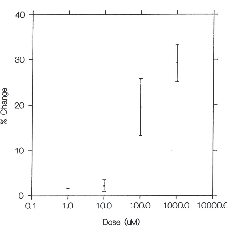
rhythmicity for compounds tested at the highest dosage, 1 mM. Rhythmicity was significantly reduced by DPKQDFMRFamide even at very low concentrations, down to 10 µM. Fig. 3 illustrates dose–response curves for Dromyosuppresin (Fig. 3a) and DPKQDFMRFamide (Fig. 3b).
3.1.2. Effects of Limulus neuropeptides
Limulus peptides LP1 (Fig. 2), LP2 (Fig. 2), and LP3
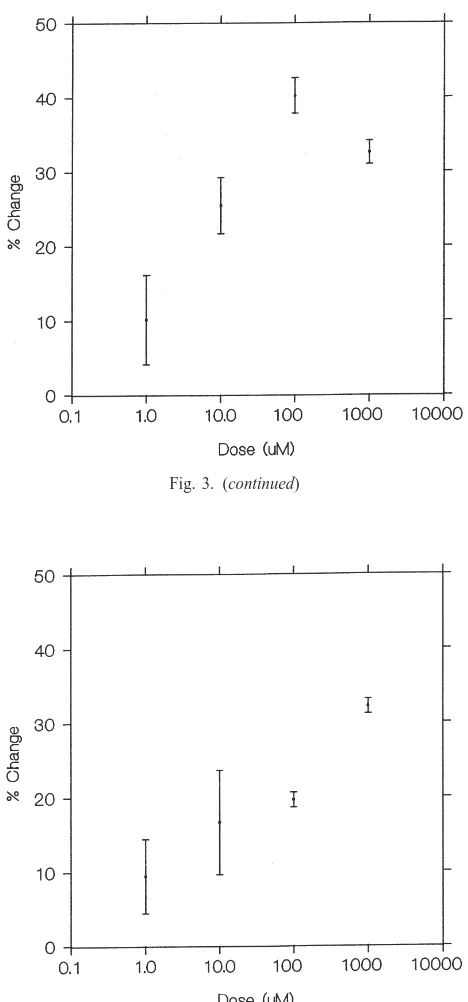
significantly altered heart rate. LP1 increased heart rate significantly (P,0.05) at relatively high doses (1 mM– 100µM) (EC50<10µM). Except at one dose, 100 µM,
the regularity of the rhythmicity was unaffected by LP1. Peptides LP2 and LP3 significantly lowered heart rate at a number of doses. A 10-µM dose of LP2 was maxi-mally effective, with higher doses saturating (EC50<4 µM ). LP2 did not affect the rhythmicity of the heart at any of the doses tested. LP3 was more effective than LP2 in decreasing the heart rate. This peptide produced a significant lowering of rate at concentrations as low as 1 µM (EC50<5 µM). LP3 affected the rhythmicity
adversely while lowering rate at the 1023M dosage. This
was the only concentration at which rhythmicity was affected by LP3. LP4 and LP5 had no effect on rate, but LP4 lowered the rhythmicity score significantly at 1023
M. Table 2 summarizes heart rates and rhythmicity for these peptides. Dose–response curves are shown for LP1 (Fig. 3c) and LP2 (Fig. 3d).
4. Discussion
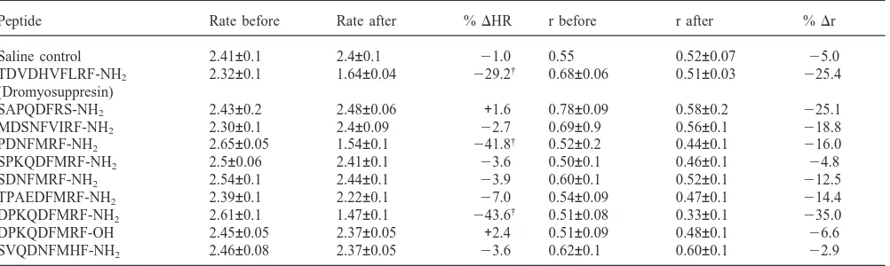
Fig. 1. Effects on the heartbeat of a Drosophila melanogaster pupa of injection of peptide TDVDHVFLRF-NH2(Dromyosuppresin) at a
concentration of 100 µM. The Upper panel depicts the spectrum derived from 30 s of heartbeat recorded optically and digitized at 100 Hz. Maximum Entropy Spectral Analysis was employed for this esti-mation of heat rate. The small peaks are harmonics of the major fre-quency. The middle panel depicts the autocorrelation function derived from the data. The lower panel shows the 30 s of raw data as recorded directly from the optical system. The time-scale bar indicates 5 s. (a) This set of analyses shows a wild-type heart beating at a rate of approximately 2.3 Hz. The heart is beating regularly as indicated by the slow decay of the autocorrelation function and observation of the time series from which the spectra and autocorrelograms were derived. (b) This series was derived from data from the same fly after injection of Dromyosuppresin. Note that the rate has decreased to approximately 1.7 Hz.
NMJ are inactive in the heart. Thus the prediction that differential functional effects might be found among these peptides is borne out here. We note an important parallel finding in our results: Hewes et al. (1998) tested two forms of DPKQDFMRF, the amide and the hydroxylated form. They found that the 2OH species did not have any effect, while the amide was active, which we find also to be true in the heart.
It is unclear, based on the results reported here, whether these peptides are acting on neuronal tissue or directly on the myocardium. The Drosophila heart is innervated (Miller 1985, 1994), and our injections were done in intact flies, leaving open the possibility of neural
Fig. 1. (continued)
modulation. Neuronal synapses were found to be potentiated by DPKQDFMRFamide (Hewes et al., 1998), and neurosecretory nerve termini in the heart could have been stimulated by the active peptides to release cardioinhibitory substances. On the other hand, Hewes et al. (1998), when testing the FMRFamide gene peptides, found a general increase in spontaneous muscle activity after perfusion with several of the peptides. This response was independent of the nervous system, occur-ring in muscles not being directly stimulated, and twitches were out of synchrony with the stimulating sig-nal when present (Hewes et al., 1998). Additiosig-nally, or alternatively, these peptides could be targeting neurohae-mal organs like the corpora cardiaca to release secondary inhibitory substances. This last possibility seems unlikely, as when we do long-term recordings initiated at injection time, any changes appear almost immedi-ately after injection and persist unaltered for up to 10 min.
Fig. 2. Effects of injection of neuropeptides on heartbeat in
Droso-phila pupae. Only the raw optical data are shown in this figure. (a and
b) Native Drosophila neuropeptide DPKQDFMRFamide at a concen-tration of 1025M. (a) The heart of this fly is beating at a rate of 2.41
Hz. The heart is beating regularly with a rhythmicity score of 0.6. (b) Data from the same fly after injection of the amide. Note that the rate has decreased to approximately 1.33 Hz. The regularity of the beat of this heart has deteriorated owing to the treatment as indicated by the reduction of the rhythmicity score to 0.394. (c and d) Limulus peptide LP1 (DEGHKLMLYFamide) at a concentration of 100µM. (c) Raw optical data from a wild-type heart beating at a rate of approximately 2.5 Hz. The heart is beating regularly. (d) Data from the same fly after injection of LP1. Note that the rate has increased to approximately 3.2 Hz. The regularity of the beat has deteriorated somewhat. (e and f)
Limulus peptide LP2 (GHSLLHFamide). Data as above for a fly before
(e) and after (f) injection with a 10µM concentration of this substance. The beat is slowed by this treatment from about 2.5 Hz to about 1.1 Hz. Regularity was not significantly affected at any dose by LP2.
Among native peptides, only DPKQDFMRFamide low-ered the strength of the rhythmicity significantly while slowing the heart, and only at one concentration. Dro-myosuppressin and PDNFMRFamide left the rhyth-micity unchanged.
If the peptides are acting on the myocardium directly, what is their mechanism of action? In Manduca sexta, it was determined that cardioacceleratory peptides (CAPs) isolated from this organism (Tublitz and Truman, 1985; Huesmann et al., 1995), act through an inositol tri-phosphate pathway to increase Ca2+efflux from the
sar-coplasmic reticulum (Tublitz, 1988). Work will need to be done to determine the mechanism in Drosophila, but this forms a starting, testable hypothesis. Our
prelimi-Fig. 3. Dose–response curves, expressed as % change at each dose, for neuropeptides found effective in altering heart rate (either by an increase or a decrease) at one or more dosages. (a) TDVDHVFLRF-NH2 (Dromyosuppressin). (b) DPKQDFMRF-NH2. (c)
DEGHKMLYF-NH2(LP1). (d) GHSLLHF-NH2(LP2).
Fig. 3. (continued)
nary results show that serotonin (5-HT) works through a similar pathway (E. Johnson, unpublished)
Fig. 3. (continued)
Fig. 3. (continued)
Table 2
Effects of Limulus neuropeptides on Drosophila heart rate (HR) and regularity (r)*a
Peptide Rate before Rate after %DHR r before r after %Dr
DEGHKMLYF-NH2(LP1) 2.43±0.09 3.22±0.07 +28† 0.64±0.1 0.258±0.1 272†
GHSLLHF-NH2(LP2) 2.71±0.1 1.86±0.3 231† 0.48±0.09 0.47±0.2 24
PDHHMMYF2NH2(LP3) 2.58±0.08 1.75±0.2 232† 0.75±0.1 0.23±0.2 270†
DHGNMLYF-NH2(LP4) 2.61±0.1 2.56±0.1 +6 0.45±0.2 0.19±0.1 +9
GGRSPSLRLF-NH2(LP5) 2.76±0.1 2.68±0.1 22 0.55±0.1 0.603±0.2 25
a†Five individuals were tested at every dosage. The results for the highest dosage are reported. Statistics as in Table 1.
in the Drosophila heart (Johnson et al., 1998): a voltage-gated Ca2+ channel sensitive to ω-Conotoxin VIIC, the
K+channels encoded by the genes Shaker (Catsch, 1944) and ether-a-go-go (Kaplan and Trout, 1968), and the Ca++-activated K+channel encoded by slowpoke (Elkins et al., 1986). Any of these channels could be targets for pharmacological modification. We are presently attempting to delineate these possibilities.
TDVDHVFLRFamide (Dromyosuppressin) is a myoinhibitory peptide encoded by a separate gene, and strictly speaking is a FLRFamide-related peptide (Nichols, 1992). It is found in brain and gut (McCormick and Nichols, 1993). It has been previously found to slow the Drosophila heart (R. Nichols, pers. communication). Its mechanism for slowing the heart remains to be inves-tigated.
Limulus has a neurogenic pacemaker, with the beat
originating in a ganglion innervating the heart (Prosser and Brown, 1961). This conclusion rests largely on elec-trophysiological and surgical evidence (Prosser and Brown, 1961; Miller, 1985). There is an initial sharp spike of depolarization, followed by a prolonged period of inactivity between beats (Lang, 1971a). Recordings taken from the associated ganglion show trains of action potentials synchronous with the beat (Lang, 1971a), similar to the very well investigated pattern seen in the lobster, Homarus (Anderson and Cooke, 1971).
In contrast, the heartbeat of Drosophila is myogenic (Rizki, 1978; Dowse et al., 1995; Gu and Singh, 1995). TTX, which stops all sodium-dependent action potentials by selectively blocking Na+ channels (Barchi, 1988), fails to affect heart rate even in high doses (Dowse et al., 1995; Gu and Singh, 1995; Johnson et al., 1998). The mutation paralytictemperature sensitive, which causes
reversible paralysis at high temperatures as a result of blocked sodium-dependant action potentials (Wu and Ganetzky, 1980; Kauvar, 1982; Jackson et al., 1984), encodes a subunit of a Na+ channel (Loughney et al., 1989). This mutation has no effect on the heart at any temperature (Dowse et al., 1995). Additionally, the elec-trocardiogram is typical of myogenic hearts (Rizki, 1978; Johnson, unpublished observations).
car-dioactive in a myogenic system. LP2 and LP3 (Gaus et al. 1993, 1994), isolated from Limulus, are cardioinhibi-tory in both animals. LP1 is a potent excitacardioinhibi-tory peptide in Drosophila, but has the opposite effect in Limulus, in which it inhibits heart rate (Gaus et al. 1993, 1994). There are other examples of such differential effects. Serotonin, is an inhibitory transmitter in the Limulus heart (Pax and Sanborn, 1969), yet in Drosophila, it is excitatory (Johnson et al., 1997). Acetylcholine (ACh), which slows vertebrate hearts, also slows the heart of
Hyalophora cecropia (McCann, 1969), but accelerates
the heart of Drosophila (Johnson et al., 1997) (however note Zornik et al., 1999). We find that the action of ACh requires an intact nervous system (unpublished data), so such variability is to be expected given the far upstream action of this substance.
A commonality of mechanism may reside in a myog-enic component of the Limulus heartbeat. Removal of the cardiac ganglion in this and simultaneous treatment with proctolin shift the mode of beat in this organism from a fast simultaneous contraction that involves the entire myocardium to a peristaltic wave, typical of insects (Watson and Hoshi, 1985; review: Lang, 1971b). The similarity of response to these neuropeptides in both systems establishes an hypothesis that at some funda-mental level the mechanisms of pacemaking and pace-maker control in both Drosophila and Limulus are essen-tially similar. Tests at similar developmental stages in both organisms will be needed before this hypothesis can be explored further.
5. Uncited references
Trautwein and Osterrieder, 1986 is not cited in the text.
Acknowledgements
This work was made possible, in part, by a grant from the American Heart Association. We thank Dr Paul Taghert for supplying the Drosophila neuropeptides, and Dr Gabrielle Gaus for the Limulus neuropeptides.
References
Anderson, M., Cooke, I.M., 1971. Neural activation of the heart of the lobster Homarus americanus. Journal of Experimental Biology 55, 449–468.
Ashburner, M., 1989. Drosophila, A Laboratory Manual. Cold Spring Harbor Press, Cold Spring Harbor, New York.
Barchi, R., 1988. Probing the molecular structure of the voltage-depen-dent sodium channel. Annual Review of Neuroscience 11, 455– 495.
Catsch, A., 1944. Eine erbliche sto¨rung des bewegungsmechanismus
bei Drosophila melanogaster. Zeitschrift Independent Abstracten Vererbung 82, 64–66.
Chatfield, C., 1980. The Analysis of Time Series. Chapman and Hall, London.
Curtis, N.J., Ringo, J., Dowse, H., 1999. Morphology of the pupal heart, adult heart, and associated tissues in the fruit fly Drosophila
melanogaster. Journal of Morphology 240, 225–235.
Dowse, H.B., Ringo, J.M., 1989. The search for hidden periodicities in biological time series revisited. Journal of Theoretical Biology 183, 487–515.
Dowse, H.B., Ringo, J.M., 1991. Comparisons between “periodog-rams” and spectral analysis: apples are apples after all. Journal of Theoretical Biology 148, 139–144.
Dowse, H.B., Ringo, J.M., Power, J., Johnson, E., Kinney, K., White, L., 1995. A congenital heart defect in Drosophila caused by an action potential mutation. Journal of Neurogenetics 10, 153–168. Elkins, T., Ganetzky, B., Wu, C.-F., 1986. A Drosophila mutation that
eliminates a calcium-dependent potassium current. Proceedings of the National Academy of Sciences USA 83, 8415–8419. Ga¨de, G., Hoffmann, K.-H., Spring, J., 1997. Hormonal regulation in
insects: facts, gaps, and future directions. Physiological Review 77, 963–1032.
Gaus, G., Doble, K.E., Price, D.A., Greenberg, M.J., Lee, T.D., Bat-tele, B.-A., 1993. The sequences of five neuropeptides isolated from Limulus using antisera to FMRFamide. Biological Bulletin 184, 322–329.
Gaus, G., Casaretto, M., Stieve, H., 1994. Cardioinhibitory peptides from Limulus polyphemus: modulation of the neurogenic heart. Journal of Comparative Physiology B164, 191–194.
Gu, G.-G., Singh, S., 1995. Pharmacological analysis of heartbeat in
Drosophila. Journal of Neurobiology 28, 269–280.
Hewes, R.S., Snowdeal III, E.C., Saitoe, M., Taghert, P., 1998. Func-tional redundancy of FMRFamide-related peptides at the
Droso-phila larval neuromuscular junction. Journal of Neuroscience 18,
7138–7151.
Huesmann, G.R., Cheung, C.C., Loi, P.K., Lee, T.D., Swiderek, K.M., And, N.J., Tublitz, K.M., 1995. Amino acid sequence of CAP2b,
an insect cardioacceleratory peptide from the tobacco hawkmoth
Manduca sexta. Federation of European Biochemical Societies
Let-ters 371, 311–314.
Jackson, F.R., Wilson, S., Stricharz, G., Hall, L., 1984. Two types of mutants affecting voltage-sensitive sodium channels in Drosophila
melanogaster. Nature 308, 189–191.
Johnson, E., Ringo, J.M., Dowse, H.B., 1997. Modulation of
Droso-phila heartbeat by neurotransmitters. Journal of Comparative
Physi-ology B167, 89–97.
Johnson, E., Ringo, J.M., Bray, N., Dowse, H., 1998. Genetic and pharmacological identification of ion channels central to the
Droso-phila cardiac pacemaker. Journal of Neurogenetics 12, 1–24.
Jones, J., 1977. The Circulatory System of Insects. Charles C. Thomas, Springfield, Illinois.
Kaplan, W., Trout, W., 1968. The behavior of four neurological mutants of Drosophila. Genetics 61, 399–409.
Kauvar, L., 1982. Reduced [H3]-tetrodotoxin binding in the napts para-lytic mutant of Drosophila. Molecular and General Genetics 187, 172–173.
Lang, F., 1971a. Intracelluar studies on pacemaker and follower neu-rones in the cardiac ganglion of Limulus. Journal of Experimental Biology 54, 815–826.
Lang, F., 1971b. Induced myogenic activity in the neurogenic heart of
Limulus polyphemus. Biological Bulletin 141, 269–277.
Loughney, K., Kreber, R., Gantezky, B., 1989. Molecular analysis of the para locus, a sodium channel gene in Drosophila. Cell 58, 1143–1154.
McCormick, J., Nichols, R., 1993. Spatial and temporal expression identify Dromyosuppressin as a brain-gut peptide in Drosophila
melanogaster. Journal of Comparative Neurology 338, 279–288.
Miller, T.A., 1985. Structure and physiology of the circulatory system. In: Kerkut, G.A., Gilbert, L.I. (Eds.), Comprehensive Insect Physi-ology, Biochemistry, and PharmacPhysi-ology, vol. 3. Pergamon Press, Oxford, pp. 289–353.
Miller, A., 1994. The internal anatomy and histology of the imago of Drosophila melanogaster. In: Demerec, M. (Ed.), Biology of Drosophila, Facsimile edition. Cold Spring Harbor Laboratory Press, Cold Spring Harbor, USA, pp. 420–480.
Nambu, J.R., Murphy-Erdosh, C., Andrews, P.C., Feistner, G.J., Scheller, R.H., 1988. Isolation and characterization of a Drosophila neuropeptide gene. Neuron 1, 55–61.
Nichols, R., 1992. Isolation and structural characterization of
Droso-phila TDVDHVFLRFamide and FMRFamide-containing neural
peptides. Journal of Molecular Neuroscience 3, 213–218. Nichols, R., McCormick, J., Lim, I., Caserta, L., 1995. Cellular
expression of the Drosophila melanogaster FMRFamide neuropep-tide gene product DPKQDFMRFamide. Journal of Molecular Neu-roscience 6, 1–10.
Nichols, R., McCormick, J., Lim, I., 1997. Multiple antigenic peptides designed to structurally related Drosophila peptides. Peptides 18, 41–45.
Pax, R.A., Sanborn, R.C., 1969. Cardioregulation in Limulus. III. Inhi-bition by 5-hydroxytryptamine and antagonism by bromlysergi acid delthylamide and picrotoxin. Biological Bulletin 132, 392–403. Price, D.A., Greenberg, M.J., 1989. The hunting of FaRPs: the
distri-bution of FMRFamide-related peptides. Biological Bulletin 177, 198–205.
Prosser, C.L., Brown, F.A. Jr., 1961. Comparative Animal Physiology. Little and Brown, New York.
Rizki, T.M., 1978. The circulatory system and associated cells and tissues. In: Ashburner, M., Wright, T.R.F. (Eds.), The Genetics and Biology of Drosophila, vol. 2B. Academic Press, New York, pp. 397–452.
Schneider, L.E., Taghert, P., 1988. Isolation and characterization of a
Drosophila gene that encodes multiple neuropeptides related to
Phe-Met-Arg-Phe-NH2(FMRFamide). Proceedings of the National
Academy of Sciences USA 85, 1993–1997.
Tublitz, N., 1988. Insect cardioactive neuropeptides: peptidergic modu-lation of the intrinsic rhythm of an insect heart is mediated by inositol 1,4,5-triphosphate. Journal of Neuroscience 8, 4394–4399. Tublitz, N.J., Truman, J.W., 1985. Insect cardioactive peptides. I. Dis-tribution and molecular characteristics of two cardioacceleratory peptides in the tobacco hawkmoth Manduca sexta. Journal of Experimental Biology 114, 365–379.
Ulrych, T., Bishop, T., 1975. Maximum entropy spectral analysis and autoregressive decomposition. Review of Geophysics and Space Physics 13, 183–200.
Watson III, W.H., Hoshi, T., 1985. Proctolin induces rhythmic contrac-tions and spikes in Limulus heart muscle. American Journal of Physiology 249 (4/2), R490–R495.
Wu, C.-F., Ganetzky, B., 1980. Genetic alteration of nerve membrane excitability in temperature-sensitive paralytic mutations of
Droso-phila melanogaster. Nature 286, 814–816.