INDEX Page-1.
S.N.( Page Numbers) Title of Paper and Names of Authors
(514-520)
Study of Cancer causing Food Product material Analysis by using UV Spectroscopy
Palanti Sasi, Dr.V.Ravichandiran and M.Sumithra (521-530)
Areca Catechu Husk Fibers and Polypropylene Blended Nonwovens for Medical Textiles.
V.Bhanu Rekha, K.Ramachandralu, Vishak S (531-534)
Isolation and screening of microorganism producing urate oxidase from poultry waste soil.
Sankari N, M.Vijayalakshmi, M.Deecaraman (535-544)
The Efficacy of Taraxacum officinale Leaves Extract in Regulate Apoptosis, RARß2 gene and Sox2 expression on Primary Culture Hum Cervical Cancer Stem Cells.
Ketut Edy Sudiarta, Satuman, Wibi Riawan, Ketut Gede Muliartha†, Karyono Mintaroem, Aulanni’am Aulanni’am, Mulyohadi Ali (545-550)
Preparation and Characterization of Nanoparticles for Dissolution Rate Enhancement of Ibuprofen
Ahmad Gazali Sofwan Sinaga, Karsono and Edy Suwarso (551-557)
Analysis of Total Protein and Non Protein Nitrogen in Coconut Water and Meat (Cocos Nucifera L.) by using Kjeldahl Method
Siti Morin Sinaga, Linda Margata and Jansen Silalahi (558-561)
Antimicrobial activity of salicylaldimine Schiff bases
M. Abirami and V. Nadaraj (562-568)
Studies on Dapsone in transition metal complexes
A,Vijayalakshmi, B. Chithra and P. Balaramesh (569-580)
Stability Indicating Method Development and Validation of Bosentan in Bulk Drug and Formulation by Rp-Hplc Method
S.H.Rizwan, V.Girija Sastry, Q.Imad (581-594)
Design and development of fast disintegrating dosage form of taste masked lornoxicam
Kinjal R. Shah, Tejal A. Mehta (595-601)
Effect of inhibitor protein kinase A (PKA) on Leishmaniatropica promastigotes viability, infectious ability and differentiation
Mohamed Anas AL Moalem, Chadi Soukkarieh, Mahmoud Kweider (602-607)
Quality Assessment and Detection of Adulteration in Buffalo Milk Collected From Different Areas of Gandhinagar by Physico-Chemi Method
Jivraj Makadiya and Astha Pandey (608-613)
Expression of Leishmania tropica pdI-2 gene in both promastigote and amastigote phases
Dina Ali, Chadi Soukkarieh, and Mahmoud Kweider (614-621)
Zebrafish Parkinson’s Model: Rotenone decrease motility, Dopamine, and increase α-synuclein Aggregation and Apoptosis of Zebraf Brain
INDEX Page-2.
N.( Page Numbers) Title of Paper and Names of Authors
(670-677)
Synthesis of Magnetite Nanoparticles for Arsenic Removal from Ground Water Pond
Karthik Rajendran, Gopal Samy Balakrishnan, Jegatheesan Kalirajan (678-690)
Evaluation of Ficus glomerata extract as potential anticancer agents and prevents the genetic toxicity induced by benzo(a)pyrene in ma mice
Khaled Mahmoud, Wagdy K.B. Khalil, Omeima Abd Elmoniem,Karima F. Mahrous (691-695)
Cytotoxicity of Biologically Synthesized Silver Nanoparticles from Citrus Lemon Against some Cell Lines
Raju Nalvolthula, Ramchander Merugu, Jahnavi Alwala, M.P.Pratap Rudra (696-701)
Comparison of Antimicrobial Activity of Red Betel (Piper Crocatum Ruiz & Pav) Leaves Nanoparticle and Powder Ethanolic Extract again Methicillin Resistant Staphylococcus Aureus
Karsono, Popy Patilaya, Nur Azisah, Nerdy (702-708)
The Effectiveness of EM4 Addition into Biofilter to Reduce of BOD, COD and MPN Coliform of Hospital Wastewater
Pitriani (709-712)
Phytochemical Analysis on Leaf Extract of Celosia argentea Land its Efficacy of Antioxidant and Anti Bacterial Activity
Preethi.Jand Saranya.V.T.K. (713-724)
Formulation Development and Evaluation of Rosiglitazone Maleate Sustained Release Tablets using 32 Factorial Design
Raghavendra Kumar Gunda (725-729)
Abutilon indicum (Linn.) Sweet leaves, a natural source of saponin : a specrtophotometric assay
Ranjana singhand Vijay d. Mendhulkar (730-736)
Phylogenetic Analysis of Bacteria Associated with AscidianPhallusia julineaandInhibitory Activity of Extracellular Protein Against Clinic Isolate
Rina A. Mogea, Suharjono, Tri Ardyati, SetijonoSamino (737-740)
Nursing and midwifery students: A study about learning preferences
Ivan Elisabeth Purba (741-749)
The Accuracy of API 20E and PCR Using 16s rRNA Gene for Characterization of Escherichia coli Strains Causing Urinary Tract Infection in Damascus-Syria
Sawsan shbeb and shady soukarieh (750-770)
Biosynthesis, Characterization, Antimicrobial Activity Of Copper Nanoparticles Using Fresh Aqueous Ananas Comosus L. (Pineappl Extract
International Journal of
Pharm
Tech Research
CODEN (USA): IJPRIF, ISSN: 0974-4304 Vol.8, No.4, pp 653-665, 2015
Effect of Alginate Chitosan Ratio on the Swelling,
Mucoadhesive, and Release of Ranitidine from Spherical
Matrices of Alginate-Chitosan
Anayanti Arianto
1, Hakim Bangun
1*, Urip Harahap
1, and Syafruddin Ilyas
21
Faculty of Pharmacy, University of Sumatera Utara, Jl. Tridharma No. 5,
Kampus USU, Medan, Indonesia;
2
Department of Biology, Faculty of Mathematics and Natural Sciences, University of
Sumatera Utara, Jl. Bioteknologi No. 1, Kampus USU, Medan, Indonesia
Abstract: Alginate (Alg) is an anionic biopolymer and Chitosan (Ch) is a cationic biopolymer. Alg and Ch interact each other by electrostatic interaction. The purpose of this study was to prepare and to determine the effect of Alg Ch ratio (1:0; 1:1; 1:3; 1:5; 0:1; 3:1; 5:1) on the swelling, mucoadhesive, release of ranitidine HCl (RH), drug-matrix interaction, conductivity, and thermal properties of Alg-Ch matrices. The preparation of spherical matrices containing RH was performed by incorporating various ratio of sodium Alg and Ch with the addition of starch mucilage as binding agent. The compact mass was molded to form
spherical matrices. The formulated matrices were charaterized for swelling, in vitro drug
release, kinetics of drug release, mucoadhesive, and conductivity properties. The drug-matrix interaction was assessed by Fourier Transform Infrared Spectroscopy (FT-IR) and Differential Scanning Calorimetry (DSC) studies. The drug release and swelling properties of matrices were investigated in simulated gastric fluid (SGF). The average diameter of prepared spherical matrices was 8.8 mm. The results showed that the swelling, RH release, mucoadhesive force, and the conductivity of the matrices was dependent the Alg Ch ratio. The swelling and mucoadhesive force were highest, while the RH release and the conductivity were lowest on the Alg-Ch matrices (1:1). FT-IR and DSC studies showed that no interaction between RH and Alg-Ch matrix, but there was an interaction between Alg and Ch. The RH release from Alg-Ch matrices followed Higuchi model. According to Korsmayer-Peppas model, the release exponent (n) values was found in ranged of 0.554– 0.657, indicating the drug release mechanism was the anomalous transport. The n value of Alg-Ch (1:1) matrices was the highest. The DSC study showed that the exothermic peak of Alg-Ch matrices influenced by the Alg-Ch ratio.
Key words: Alginate-chitosan; swelling; release; mucoadhesive; ranitidine; DSC; FTIR.
Introduction
Alg is an anionic copolymer consisting of residue β-D-mannuronic acid and α-L- guluronic in bond
1,4.1 The most important advantage of Alg as a matrix for the controlled release formulation is because Alg is
biodegradable and biocompatible.2 Ch is a cationic polimer, non- toxic, biocompatible, easily biodegradable,
and mucoadhesive.3Alg and chitosan form electrostatic interaction between –COOH groups of Alg and -NH2
recently, we studied the comparison of swelling, mucoadhesive, and release of RH from sperical matrices of
Alg, Ch, Alg-Ch, and calcium Alg-Ch.6 We found that the spherical of Alg-Ch matrices possess the highest
swelling degree and mucoadhesive force, and gave the most extended of drug release in SGF than other formulated matrices. Accordingly, in this research we studied further about the effect of Alg Ch ratio on the swelling, mucoadhesive and drug release from Alg-Ch spherical matrices.
RH was used as drug model. RH is a drug that commonly used to treat duodenal ulcer disease, gastric ulcer, and gastric acid hypersecretion conditions. RH acts by reducing gastric acid secretion. The biological elimination half-life of RH is short, 2.5-3 hours. RH is absorbed in the upper part of the small intestine and
shows a low bioavailability that is 50%.7-8 The absorption will be reduced due to the decomposition and
metabolism of ranitidine by microbes in the colon which causes the low bioavailability of RH9.
The purpose of this study was to prepare and to compare the swelling, mucoadhesive, and RH release properties from spherical matrices which prepared in various Alg Ch ratio in order to obtain the optimal Alg Ch ratio for the preparation of the gastroretentive drug delivery systems of RH, so that the efficacy of the RH could be improved. In this paper, the effect of Alg-Ch ratio on the swelling, RH release, drug release kinetics, mucoadhesive properties, and the conductivity of matrices will be discussed. Furthermore, the FT-IR and DSC data will also be discussed.
Methods
Materials
Sodium Alg 500-600 cP was purchased from Wako Pure Chemical Industries, Ltd., Japan, and Ch was from Funakoshi Co., Ltd., Japan. RH (ranitidine HCl) was obtained from PT. Mutifa, Medan. Hydrodrochloric acid, calcium chloride-dihydrate, and sodium chloride all were the product of Merck.
Preparation of spherical matrices
The formula of various ratio Alg-Ch matrices containing RH are listed in Table 1. The preparation of
spherical matrices containing RH was conducted with the same procedure with that we reported previously.6
Sodium Alg, Ch, and RH were homogenized in a mortar with the addition of starch mucilage as binding agent to form a compact mass. The compact mass was divided in to ten parts and each part was molded to be a spherical matrix. Then, the obtained spherical matrices were dried at room temperature.
Determination of drug release
The release of RH from spherical matrix was tested using the USP paddle method dissolution tester at
50 rpm in the 900 ml medium of SGF (pH 1.2) at 37±0.5⁰C, as previous report.6 Five milliliters of aliquot was
withdrawn at predetermined time. The dissolution test was done for 10 hours. At determined time, samples were taken 5 ml and replaced by 5 ml of fresh SGF. RH concentrations were analyzed by using UV spectrophotometer at 224.6 nm.
The obtained dissolution data were plotted as percent cumulative drug release versus square root of time according to Higuchi equation.
Q= Kt½...(1)
Table 1. The composition of spherical matrix containing ranitidine HCl for ten spherical matrices
Hakim Bangun et al/Int.J. PharmTech Res. 2015,8(4),pp 653-665. 655
The release data were treated by the Rigers and Peppas equation.10 Then, the equation was treated
logarithmically to determine the value of release exponent, n; the value of n indicates the drug release mechanism.
= Ktn ...(2)
This equation in logarithm form:
Log Mt - M = Log K + n Log t ...(3)
Determination of swelling properties
The swelling and erosion properties of spherical matrices were observed using USP dissolution tester at
50 rpm in the 900 ml medium of SGF (pH 1.2) at 37ºC as previous procedure.6 At a certain time intervals, the
spherical matrices were withdrawn and rolled on a tissue paper to remove the excess of water and the matrices were weighed. The swelling and erosion of matrices were determined based on the change of diameter and matrices weight as
...(4)
w1 :initial weight of spherical matrices; w2: weight of spherical matrices after immersed in the medium
In vitro evaluation of mucoadhesive force
In vitro bioadhesion study was done by using rabbit stomachs with the modification of DuNouy
tensiometer as we reported previously.6 The platinum-iridium ring of a tensiometer was replaced by a spherical
matrix that hanged using a spun cotton to the arm of the tensiometer. The experiment was done at 37⁰C. The
stomachs tissue were used immediately at this study. The stomachs were rinsed with saline solution to replace the stomachs content. Before the measurement the spherical matrices and the fresh stomachs were immersed in SGF (pH 1.2) for 15 minutes. The arm of tensiometer was lowered until the spherical matrix came in proper contact with the stomach tissue and it was kept as for 15 minutes. Then, the knob of tensiometer was moved upward direction slowly until the spherical matrix was completely detached from the stomach tissue. During the
experiment the stomachs tissue were wetted by dropping the SGF solution. The width (cm2) of each spherical
matrix contact with the stomach tissue during the experiments was measured. The force of mucoadhesive force
was obtained in dyne/cm2.
Determination of conductivity and zeta potential
The conductivity of spherical matrices of various Alg Ch ratio without RH were determined in water
and 0.1 N HCl solution by using Beckman Coulter DellsaTMNano.
Thermal Analysis
The thermal analysis of the matrices of Alg, Ch, Alg-Ch in various ratio, and Alg-Ch containing RH were observed by using a Differential Scanning Calorimetry (SDT Q 600). The samples were heated from 35ºC until 600ºC at rate of 10ºC/min under constant purging of nitrogen.
Fourier transformed infra-red spectroscopy (FTIR)
IR spectra were obtained by using a Shimazu IR Prestige-21 spectrometer. The pulverized samples was
micronized with KBr powder and measured in the ranged of 4000-400 cm-1.
Results and Discussion
Preparation of large spherical matrices of Alg-Ch containing RH
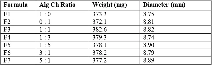
spherical matrices were similar each other. The photograph of initial and swollen (after 10 hours in SGF) of various spherical matrix containing 168 mg RH (Figure 1) and its spesifications is listed in Table 2.
A (initial) B (initial) C (initial) D (initial) E (initial)
A (swollen) B (swollen) C (swollen) D (swollen) E (swollen)
Figure 1. Photograph of initial and swollen spherical matrices of Alg-Ch containing RH A:Alg-Ch (1:1), B: Alg-Ch(1:3); C: Alg-Ch (1:5), D: Alg-Ch (3:1), and E: Alg-Ch (5:1).
Table 2. Specifications of spherical matrices containing RH
Formula Alg Ch Ratio Weight (mg) Diameter (mm)
F1 1 : 0 373.3 8.75
F2 0 : 1 372.1 8.81
F3 1 : 1 382.6 8.82
F4 1 : 3 379.3 8.74
F5 1 : 5 378.1 8.90
F6 3 : 1 378.2 8.79
F7 5 : 1 377.2 8.89
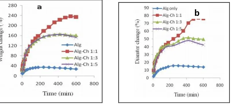
Effect of Alg Ch ratio on the swelling properties of spherical matrices of Alg-Ch
Effect of the increase of sodium Alg
The effect of the increase of Alg Ch ratio is shown in Figure 2. The swelling properties were observed based on the increase of matrices weight (Figure 2a) and matrices diameter (Figure 2b) in SGF. The matrices
that only made of chitosan dissolved in SGF, as we reported in our study previously.6 But, the increase of Alg
Ch ratio or the increase of sodium Alg amount in the matrices caused the degree of swelling decreased. In the medium of SGF the sodium Alg was changed to alginic acid which was insoluble in SGF, thereby the swelling degree decreased. The monomer of alginic acid is mannuronic acid and guluronic acid; the pka of mannuronic
acid is 3.38 and the pka of guluronic acid is 3.65.11 Therefore, alginic acid was mostly in unionized form in
SGF, so that the electrostatic repulsion among the carboxylate groups of alginic acid decreased and resulting the swelling decreased.
Effect of the increase of chitosan
Hakim Bangun et al/Int.J. PharmTech Res. 2015,8(4),pp 653-665. 657
Alg and Ch can interact through the carboxylate groups of Alg and amine groups of Ch to form Alg-Ch complex. The swelling Alg-Ch matrices in SGF was thought to be due to the electrostatic repulsion among the positive charge of protonated amin groups, and the osmosis occurred because of the high osmotic pressure of the matrices as a result of the protonization of amine groups of Ch in acid medium. .
The swelling property of Alg-Ch matrices is suitable for the preparation gastroretentive formulation of RH. The swelling of matrices result in the increase of spherical matrices diameter, thereby it will be prevented the exit of matrices from stomach through the pylorus and consequently the matrix will be retained in stomach for longer period of time.
Figure 2. Effect of the increase of sodium Alg amount on the swelling of Alg- Ch spherical matrices in SGF at 37ºC (n=3). (a) weight change; (b) diameter change
Figure 3. Effect of the increase of chitosan amount on the swelling of Alg-Ch spherical matrices in SGF at 37ºC (n=3). (a) weight change; (b) diameter change
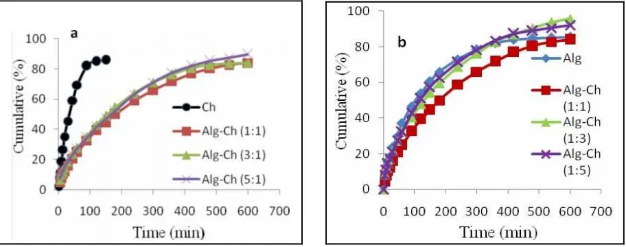
Effect of Alg-Ch ratio on the RH release from Alg-Ch spherical matrices
Effect of the increase of sodium Alg
As shown in Figure 4 (a), the release of RH from the Ch matrices only was very fast but with the addition of Alg the release became slower. The release of RH decreased with the increase of sodium Alg or with the increase Alg Ch ratio. The RH release from Alg-Ch (1:1) was slower than the release of RH from Alg-Ch (3:1) and Alg-Ch (5:1) matrices. The drug release from Alg-Ch (3:1) matrices was not significant different statiscally (p>0.05) with that from Alg-Ch (5:1).
b
Effect of the increase of Ch
The release of RH decreased with the increase of Ch amount in the matrices or decreased with decreased Alg Ch ratio. The release of RH was slower from Ch (1:1) than that of from Ch (1:3), Alg-Ch (1:5), and Alg matrices only. The release of RH from Alg-Alg-Ch (1:3), Alg-Alg-Ch (1:5), and Alg matrices only was not significant different (p>0.05). The release of RH from Al-Ch (1:1) matrices was more sustainable was thought to be due to the value of the tortuosity of the capillaries system was higher in this matrices. The formation of Alg-Ch matrices at 1:1 ratio was at the equivalent of the weight ratio between Alg and Ch. As a result, the ionic interaction between Alg and Ch caused the cross-linked of polymers and resulting the higher of the tortuosity of the matrices. According to the second form of Higuchi equation, the drug release decreases
with the increase of tortuosity.12Meng at al.13, observed that the polyblend solution viscosity of Alg and Ch
(1:1) reached to the highest at the composition polyblend of (1:1). The increase of viscosity of the matrices was also thought to be responsible to the sustained release of RH from Alg-Ch (1:1) matrices.
Figure 4. Effect of the increase of sodium Alg (a) and the increase of Ch (b) on the release of RH from Alg-Ch matrices in SGF in SGF at 37ºC (n=3). (a) Alg increase; (b) Ch increase
Drug release kinetics
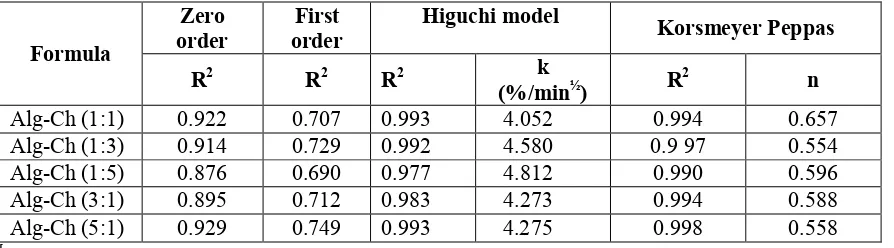
The drug release data were analyzed for determination of the kinetics and mechanism of drug release from the matrices. For Higuchi model, the data were treated with Equation (1). It was found that the release of RH from all of the Alg-Ch matrices to the SGF was dependent on square root of time. When the cumulative percent of drug was plotted versus the square root of time, a linear relationship was obtained with the
correlation coefficient (R2) close to 1 (Figure 5 and Table 3). The comparison of the kinetic parameters of RH
release from different Alg Ch ratio of Alg-Ch matrices based on zero order, first order, Higuchi model, and
Korsmeyer-Peppas model plots is also listed in Table 3. The correlation coefficients (R2) are higher in Higuchi
model in all ratio of Alg-Ch matrices (close to 1) compared to zero order and first order.
In controlled or sustained release formulations, diffusion, swelling, and erosion are the three most
important rate controlling mechanism. The drug release from the polymeric system is mostly by diffusion and is best described by Fickian diffusion. But, in the case of formulations containing swelling polymers, other processes in addition to diffusion play an important role in exploring the drug release mechanism. These
processes include relaxation of polymer chains, imbibitions of water causing the polymers to swell.14 Therefore,
the release data were further treated by Eq. (2) given by Ritger and Peppas, or also called the Power low.10 The
release mechanism of RH from the matrices was characterized by determination of n value. For the spheres when n takes the value 0.43 indicates Fickian diffusion and for the value 0.43 < n < 0.85 indicates the
anomalous transport drug release. Value of n 0.85 indicates the Case II transport.12
The Korsmeyer-Peppas plot of the release of RH from the matrices is showen in Figure 6. Log cumulative of drug released was plot against log t and straight line was obtained. The slope of the line is n. To
determine of n exponent of the portion of the release curve the Mt/M∞ < 0.6 was used. The value of n with
Hakim Bangun et al/Int.J. PharmTech Res. 2015,8(4),pp 653-665. 659
ranged of 0.554–0.657, indicating anomalous transport. The highest value was found in Alg-Ch (1:1) matrices, it was 0.657. It was due to the swelling degree of Alg-Ch (1:1) matrices in SGF was higher than other matrices.
Figure 5. Higuchi plot of RH release from different Alg Ch ratio of Alg-Ch spherical matrices in SGF at 37ºC (n=3). (a) Increase of Alg (b) Increase of Ch.
Figure 6. Korsmeyer-Peppas plot of RH release from different Alg Ch ratio of Alg-Ch matrices in SGF at 37º. (a) Increase of Alg (b) Increase of Ch.
Table 3. The comparison of the kinetic parameters of RH release from different Alg Ch ratio of Alg-Ch matrices based on zero order, first order, Higuchi model, and Korsmeyer-Peppas model plot.
Zero order
First order
Higuchi model
Korsmeyer Peppas Formula
R2 R2 R2 k
(%/min½) R
2
n
Alg-Ch (1:1) 0.922 0.707 0.993 4.052 0.994 0.657
Alg-Ch (1:3) 0.914 0.729 0.992 4.580 0.9 97 0.554
Alg-Ch (1:5) 0.876 0.690 0.977 4.812 0.990 0.596
Alg-Ch (3:1) 0.895 0.712 0.983 4.273 0.994 0.588
Alg-Ch (5:1) 0.929 0.749 0.993 4.275 0.998 0.558
[
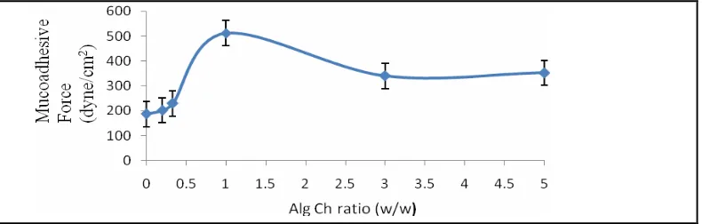
Effect of Alg Ch ratio on mucoadhesive force of spherical Alg-Ch matrices
The mucoadhesive force of various ratio of the spherical matrices of Alg-Ch is shown in Figure 7. The ratio of Alg to Ch influenced the mucoadhesive strength of the matrices. It was found that the highest mucoadhesive force was in Alg-Ch (1:1) among various ratio of Alg Ch matrices examined. The mucoadhesive force of the matrices increased with increasing of ratio Alg to Ch and reached maximum at
b
a
the ratio Alg-Ch was 1:1. Then, the mucoadhesive force decreased with increasing of the ratio of Alg to Ch. The mucoadhesive force of Alg-Ch (1:1) was higher than other matrices was due to the swelling degree of Alg Ch (1:1) was higher than other matrices. The swelling of the matrices cause the contact area of the matrices to stomach mucosa become larger and as a result the more intimate contact of matrices to the stomach mucosa. The mucus lining of the stomach is rich in mucin, which contain an oligosaccharide chain
with terminal sialic acid. According to Chikering at al.15, polyanions, especially polymer bearing carboxylic
groups and high charge density, serve as powerful “ligands” for mucin and are called mucoadhesive polymers, for example Alg. In this experiment the potential zeta of Alg-Ch (1:1) matrices obtained was -1.89 mV. Therefore, the bioadhesion mechanism between Alg-Ch matrix and mucus was not due to the electrostatic force between Alg-Ch matrix and sialic acid.
Figure 7. Effect of Alg Ch ratio on the mucoadhesive force of the Alg-Ch spherical matrices (n=3).
During the experiment, the Alg-Ch matrixes hydrated and swelled and contact on the surface of mucus, and as a result the matrices adhere to the mucosal surface. The formation of hydrogen-bonds between the functional groups of the Alg-Ch polymers and mucosa layer may plays an important role. The bioadhesion of Alg-Ch matrices gives several advantages such as longer gastric residence time and improves drug absorption and bioavailability.
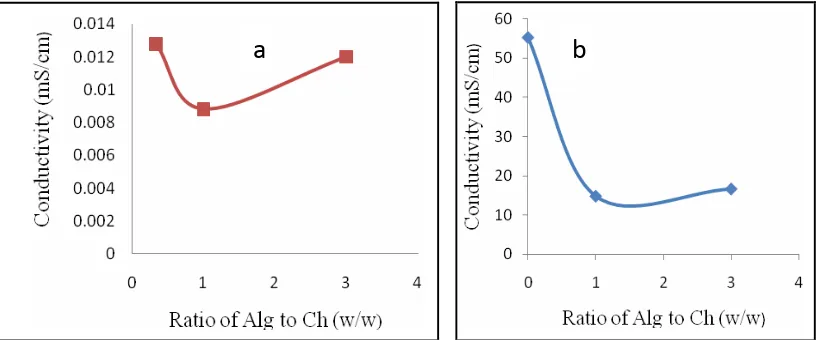
Conductivity of spherical Alg-Ch matrices
The conductivity of the matrices was measured without RH. The conductivity of Alg matrices and Ch matrices in water were found 0.3148 and 0.0094 mS/cm, respectively. While, the conductivity of Alg matrices and Ch matrices in the medium 0.1N HCl solution were obtained much higher than in water, it was 55.1473 and 57.6874 mS/cm, respectively. The effect of Alg-Ch ratio on the conductivity of Alg-Ch matrices in the medium of water and 0.1N HCl solutions is showed in Figure 9a and 9b, respectively.
In the Alg-Ch matrices, the amino groups of Ch were neutralized by the formation of complex with carboxylic groups of Alg, leading to decrease the conductivity caused by decreasing the concentration of overall ions present in matrices. The conductivity decreased with the increase of Alg in Alg-Ch matrices, then after achieving the equivalent point, an increase of conductivity was observed with the increased of Alg (carboxylic
ions) ions added in the matrices.16 Among the tested matrices, the lowest conductivity was observed in
Hakim Bangun et al/Int.J. PharmTech Res. 2015,8(4),pp 653-665. 661
Figure 8. Effect of Alg-Ch ratio on the conductivity of Alg-Ch matrices in water (a) and in the 0,1 N HCl soluction (b)
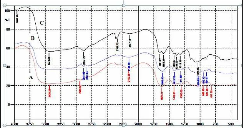
Fourier transform infrared spectroscopy (FT-IR)
The FT-IR spectrum of sodium Alg, Ch, and Alg-Ch (1:1) matrices is shown in Figure 9. In the IR
spectrum of Alg matrix we can observe the symmetrical streching of -COO-groups of alginate at 1411.89 cm-1
and the asymmetrical streching of -COO- groups at 1620.21 cm-1 (Figure 9A). In the Ch matrix the absorption
band of the carbonyl (C=O) streching of secondary amide at 1651.07 cm-1 and the absorption band of (-NH2)
groups at 1589.34 cm-1 (figure 9 C). The spectrum of Alg-Ch matrix shows that the absorption band of the
symmetrical streching of -COO-groups of alginate at 1411.89cm-1 and the asymmetrical streching of -COO
-groups at 1620.21 cm-1 of Alg shiffted to 1415.75 cm-1 and 1608.63 cm-1 (Figure 9 B) and the absorption band
at the carbonyl (C=O) streching of Ch at 1651.07 cm-1 and the absorption band of -NH2 groups at 1589.34 cm-1
of Ch disappeared. These results indicate that the carboxylic groups (-COO-) of Alg associate with amino
groups (NH3+) of Ch through electrostatic interaction to form the polyelectrolyte complex of Alg-Ch.17 For the
other matrices, i.e, Alg-Ch (1:3, Alg-Ch (1:5), Alg-Ch (3:1), and Alg-Ch (5:1) gave the similar absorption peak with Alg-Ch (1:1), the spectra are not shown here.
Furthermore, the spectra of RH and matrix Alg-Ch containing RH is shown in Figure 10. The FT-IR spektrum of RH (Figure 10 A) shows that absorption bands at 3217.27cm-1 and 3417.86 cm-1 show the streching vibration of N-H groups. The absorption peak at 3024.38 cm-1 and 3089.96 cm-1 show to the furan ring. The characteristic peaks at 2468.88 cm-1 is due to amino tertiery groups and the absorption peak at 1616.35 cm-1 is due to the C=C groups. The other characteristic peaks is at 13.88.75 cm-1 and 1573.91 cm-1 are assigned to the NO2 groups, 1134.14 cm-1 and 1238.30 cm-1 are due to C-N groups. This results is similar
to results obtained by previous workers18-20.
The FT-IR spectrum of Alg-Ch 1:1 matrix containing RH (Figure 10 B) shows the similar
characterisctis peaks to the spectra of RH matrix. The presence of the characterictic peaks of RH proves that there is no change of functional groups of RH. This results showed that no interaction between RH and Alg-Ch matrices.
Figure 9. The FT-IR spectra of Alg (A), Alg-Ch (1:1) (B), and Ch (C) matrices.
Figure 10. The FT-IR spectra of RH (A) and Alg-Chi (1:1) matrix containing RH (B)
Differential Scanning Calorimetry
Thermogram of DSC were studied in order to investigate the possibility the interactions between Alg and Ch and between RH and Alg and CH. The comparison of thermogram of Alg, Ch, and Alg-Ch (1:1) matrices is shown in Figure 11. The thermogram of Alg showed the endothermic peak at 95.08ºC and the
exothermic peak at 248ºC. The similar result was observed by previous worker.21 The endothermic peaks are
correlated to loss of water associated to hydrophilic groups of polymers, while the exothemic peaks are resulted from degradation of polyelectrolytes due to degradation and depolymerazation reactions most probably due to
the partial decarboxylation of the carboxylic groups and oxidation of polymers.21 The thermogram of Ch
.
matrices shows the a broad curve and the a small peak of exothermic peak 330.78 ºC, while the endothermic
peak was not observed. Previous workers,21 observed the exothemic peak of Ch was at 311 ºC. The thermogram
Hakim Bangun et al/Int.J. PharmTech Res. 2015,8(4),pp 653-665. 663
The DSC thermogram of pure RH shows the sharp endothermic peak at 152.47 ºC indicates the melting point of RH and exothermic peak at 184.08 ºC related to the decomposition of RH. (Figure 11). Figure 12 shows the DSC thermogram of Alg-Ch (1:1) matrix containing RH, the sharp endothermic peak at 154.36 ºC related to the melting point of RH and the exothermic peak at 186.23 ºC. The appearance of of endothermic and exothermic peaks of RH in Alg-Ch (1:1) containing RH shows that there is no interaction between RH and Alg-Ch matrix.
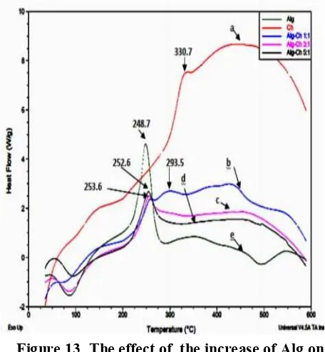
Effect the of the increase of Alg on DSC thermogram is shows in Figure 13. The DSC thermogram of Alg-Ch (3:1) shows the exothermic peak at 253.56 similiar to and DSC curve of Alg-Ch matrices (5:1) shows the exothermic at 252.68 ºC.
The effect of increase of Ch is shown in Figure 14. The DSC curve of Alg-Ch (1:3) shows the exothermic peak at 290.97 ºC and the DSC curve of Alg-Ch (1:5) shows at 301.78 ºC. The exothermic peak of the matrices which higher content of Ch such Alg-Ch (1:5) is higher than Alg-Ch (1:3).
Figure 11. The DSC thermogram of Alg (a), Figure 12. The DSC thermogram of pure (b) Alg-Ch (1:1), and (c) Ch. RH (a) and (b) Alg-Ch (1:1) containing RH
Conclusions
We studied the effect of Alg Ch ratio on the swelling, mucoadhesive, and RH release from Alg-Ch matrices. The Alg-Ch ratio influenced the swelling, mucoadhesive force, and RH release properties. AlgCh (1:1) matrices showed the highest swelling and mucoadesive properties, and gave the RH release followed a sustained release type with Higuchi model drug release in the medium of SGF. Accordingly, Alg-Ch (1:1) is potential to be developed to be a gastroretentive matrices for RH and other anti ulcer drugs.
Acknowledgement
The work was funded by Directorate General of Higher Education of Indonesia (DIKTI) through the scheme of the Fund of Posgraduate Team (Hibah Tim Pascasarjana) 2013-2014.
References
1. Haug A., Larsen, B. and Smidsrod, O., Acta Chem. Scand., 1966, 20, 183.
2. Sachan N.K., Pushkar, S. and Jha, A., Sodium alginate: The wonder polymer for controlled drug
delivery, Journal of Pharmacy Research, 2009, 2(8), 1191-1199.
3. Felt O., Buri P. and Gurny R., Chitosan: A unique polysaccharide for drug delivery, Drug Dev Ind
Pharm, 1998, 24(11), 979-993.
4. Knil C.J., Kennedy J.F., Mistry J., Miraftab M., Smart G., Groocock, M.R. and Williams, H.J., Alginate
fibre modified with unhydrolysed and hydrolysed chitosan for dressings, Carbohydrate Polymers, 2004, 55, 65-76.
5. Kaban J., Bangun H. and Dawolo K. A., Preparation of polyelectrolyte complex alginate-chitosan
membrane (Pembuatan membran kompleks polielektrolit alginat-kitosan), Jurnal Sains Kimia, 2006, 10 (1), 10-16.
6. Arianto A., Bangun H., Harahap U., and Ilyas S., The comparison of swelling, mucoadhesive, and
release of ranitidine from spherical matrices of alginate, chitosan, alginate-chitosan, and calcium alginate-chitosan, Int. J. PharmTech Res. 2014, 6 (7), 2054-2063.
7. Tolman K.G., Gastrointestinal and liver drugs ( In: Remington: The Science and Practice of Pharmacy
(Gennaro A.R.), Lippincott Williams & Wilkins, Philadelpia, 2000, 1225.
8. Grant S.M., Langtry, H.D. and Brogden, R.N., Ranitidin an updated review of its pharmacodynamic
and pharmacokinetic properties and therapeutic use in peptic ulcer disease and other allied diseases,
Drugs,1989,37(6), 801-870.
9. Basit A.W. and Lacey, L. F., Colonic metabolism of ranitidin: Implications for its delivery and
absorption, International Journal of Pharmaceutics, 2001, 227(1-2), 157-65.
10. Peppas N.A., Bures, P., Leobandung, W. and Ichikawa, H., Hydrogel in pharmaceutical
formulations, European Journal of Pharmaceutics and Biopharmaceutics, 2000, 50, 27-46.
11. Donati I. and Paoletti, S., Materal Properties of alginate. In: Alginates: Biology and Applications
(Rehm, B H.A), Springer, New York, 2009, 21.
12. Sinko J.P., Martin's Physical Pharmacy and Pharmaceutical Sciences, Sixth Edition, Wlters Kluwer,
Philadelphia, 2011, 248-250.
13. Meng X., Tian F., and Yang J., Chitosan and alginate polyelectrolite complex membranes and their
properies for wound dressing application, J. Mater Sci: Mater Med, 2010, 21, 1751-1759.
14. Siepman J. and Peppas, N.A., Modeling of drug release from delivery systems based on hydroxypropyl
methylcellulose (HPMC), Advanced Drug Delivery Reviews, 2001, 48, 139-157.
15. Chikering D.E., Jacob, J.S., Desai, T.A., Harrison, M., Harris, W.P., Morcel, C.N., Chaturvendi, P. and Mathiwitz E., Bioadhesive microspheres: An in vivo transit and bioavailability study of drug loaded alginate and poly (fumaric-co-sebacic anhydrate) microspheres, J. Controlled Release, 1997, 48, 35-46.
16. Lucinda-Silva R M. and Evangelista R C., Studies on formation of complex coacervates between
chitosan and alginate during microparticles preparation, Acta Farm. Bonaerense, 2005, 24 (3), 366-70.
17. Silverstein M.R., Bassier C.G., and Morrill C.T., Spectrometric identification of organic compounds,
Forth Edition, John Willey & Sons Inc., New York, 1991, 121-128.
18. Dhankar V, Garg G, Dhamija K, and Awasthi R., Preparation, characterization, and evaluation of
Hakim Bangun et al/Int.J. PharmTech Res. 2015,8(4),pp 653-665. 665
19. Somasundaram R., Nandhakumar S., and Dhanaraju M.D., Development and in vitro evaluation of
biodegradable chitosan microspheres loaded with ranitidine and cross linked with glutaraldehyde, Int. J. PharmTech Res. 2011, 3 (1), 488-496.
20. Sarmento B., Ferreira D., Veiga F., and Rebeiro A., Characterization of insulin-loaded alginate
nanoparticles produced by ionotropic pre-gelation through DSC and FTIR studies, Cabohydrate Polymers, 2006, 66, 1-7.
21. Soares J.P., Santos J.E., Chierice E.T, and Cavalheiro, E.T.G., Thermal behaviour of acid and its
sodium salt, Electica, 2004, 29 (2), 57-63.