Abnormalities in the Thalamus and Prefrontal Cortex
during Episodic Object Recognition in Schizophrenia
Stephan Heckers, Tim Curran, Donald Goff, Scott L. Rauch, Alan J. Fischman,
Nathaniel M. Alpert, and Daniel L. Schacter
Background: Many patients with schizophrenia demon-strate memory deficits. We studied patterns of brain activity during episodic recognition of new and previously seen three-dimensional objects.
Methods:We used15O positron emission tomography to study regional cerebral blood flow in eight normal sub-jects and nine patients with schizophrenia during a visual object recognition task.
Results: In comparison with control subjects, patients with schizophrenia showed less regional cerebral blood flow increases in the pulvinar region of the right thalamus and the right prefrontal cortex during the recognition of new objects and significantly greater left prefrontal cortex regional cerebral blood flow increases during the recog-nition of previously seen objects. Patients with schizophre-nia exhibited alarm rates to new objects similar to those of control subjects, but significantly lower recognition rates for previously seen objects.
Conclusions:Schizophrenia is associated with attenuated right thalamic and right prefrontal activation during the recognition of novel visual stimuli and with increased left prefrontal cortical activation during impaired episodic recognition of previously seen visual stimuli. This study provides further evidence for abnormal thalamic and prefrontal cortex function in schizophrenia. Biol Psychi-atry 2000;48:651– 657 © 2000 Society of Biological Psychiatry
Key Words: Positron emission tomography, cognitive deficits, memory, pulvinar
Introduction
S
chizophrenia is associated with a wide range of cog-nitive deficits. In particular, patients with schizophre-nia often present with impaired attention and memory, abnormal language, and disturbed executive functions (Goldberg and Gold 1995). Memory function was one of the first cognitive abilities to be studied in schizophrenia (Hull 1917), and episodic memory was found to be particularly impaired (Aleman et al 1999; Goldberg and Gold 1995). Structural and functional brain abnormalities proposed to explain memory deficits in schizophrenia include abnormalities of the prefrontal cortex, medial temporal lobe, thalamus, and cerebellum.We have recently provided evidence that schizophrenia is associated with an impaired recruitment of the hip-pocampus during episodic memory retrieval (Heckers et al 1998, 1999). Whereas normal subjects activated a frontal– temporal network to retrieve previously studied words, schizophrenic patients failed to recruit medial temporal lobe structures but showed robust and even increased activation of prefrontal regions. Furthermore, schizo-phrenic patients performed less accurately and did not activate medial temporal lobe structures when using a semantic encoding strategy.
Here we tested the hypothesis that schizophrenia is associated with performance deficits and abnormal brain activation patterns during the recognition of visually presented objects. We studied object recognition in schizo-phrenia for three reasons: First, schizoschizo-phrenia is associated with impairments of nonverbal memory (Aleman et al 1999; Heinrichs and Zakzanis 1998). Second, previous studies in schizophrenic patients have demonstrated im-paired recognition of visually presented objects (Aleman et al 1999; Clare et al 1993). Third, neuroimaging studies of object recognition in normal subjects have shown activation of brain regions implicated in the pathogenesis of schizophrenia: the bilateral medial temporal lobes and prefrontal areas during the recognition of previously seen objects and the thalamus, prefrontal, and medial temporal lobe areas during the recognition of new objects (Schacter et al 1995, 1997, 1999; Uecker et al 1997). We hypothe-sized that schizophrenia is associated with impaired
rec-From The Psychotic Disorders Unit (SH, DG) and The Psychiatric Neuroimaging Research Group (SH, SLR), Department of Psychiatry, and The Positron Emission Tomography Laboratory, Division of Nuclear Medicine, Department of Radiology (SLR, AJF, NMA), Massachusetts General Hospital, Boston; the Department of Psychology, Case Western Reserve University, Cleveland, Ohio (TC); and the Department of Psychology, Harvard University, Cambridge, Massachusetts (DLS).
Address reprint requests to Stephan Heckers, M.D., Massachusetts General Hospital East, Dept of Psychiatry, CNY-9132, Building 149, 13th Street, Charlestown MA 02129.
Received September 13, 1999; revised February 8, 2000; accepted April 28, 2000.
© 2000 Society of Biological Psychiatry 0006-3223/00/$20.00
ognition of previously presented objects and that abnormal brain activation during the recognition of previously seen or new objects would involve three regions of interest, i.e., the prefrontal cortex, the medial temporal lobe, and the thalamus.
Methods and Materials
Subjects
Nine patients with schizophrenia and eight normal subjects were studied. The schizophrenia patients were recruited from an outpatient mental health clinic in Boston, Massachusetts and diagnosed according to DSM-IV criteria (American Psychiatric Association 1994) by an experienced clinician (DG). Control subjects were recruited by advertisement and did not report any history of psychiatric disorders as assessed by a structured clinical interview (Spitzer et al 1991). All subjects provided written informed consent. Subjects were excluded if they had a history of neurological or medical illness, current substance abuse, or lifetime substance dependence. The study was ap-proved by the Human Subjects Committee of the Massachusetts General Hospital and the Central Office Research Review Committee of the Commonwealth of Massachusetts, Department of Mental Health.
The schizophrenic subjects were chronic, stable outpatients (mean duration of illness was 16.266.1 years). Mean scores on the positive, negative, and global scales of the Positive and Negative Syndrome Scale (PANSS) were 12.263.6, 21.366.3, and 27.765.1, respectively. All subjects were right-handed and male, and the two groups were matched for age (normal subjects 42.964.8 years, schizophrenic patients 41.166.3 years). The control subjects had a higher educational status [control subjects 15.8 6 2.8 years, schizophrenic patients 12.8 6 1.3 years,
t(15)52.9,p5.01], but mean parental educational status was not significantly different [control subjects 13.3 6 2.4 years, schizophrenic patients 12.360.3 years,t(14)51.1,p5.29]. All patients were treated with typical neuroleptics (mean chlor-promazine equivalent dose 7256 708 mg/day). Three patients were treated with benztropine (1–2 mg/day).
Experimental Design
Subjects viewed stimuli presented in the center of a computer screen positioned about 50 cm in front of their eyes. Stimuli were presented for 4.5 sec and the screen was blank for 0.5 sec between stimuli. The stimuli were line drawings (300 3 300 pixels) of novel three-dimensional objects (“possible objects”; Schacter et al 1991; Williams and Tarr 1997). A plus sign was used in the fixation condition.
The positron emission tomography (PET) experiment con-sisted of eight PET scans, collected during one scanning session. There were four experimental conditions (Fixation, View, Rec-ognition Old, RecRec-ognition New) and two runs per condition. For scans 1 and 8 (Fixation), subjects were instructed to fixate on a crosshair displayed on the screen. During scans 2 and 7 (View), subjects were instructed to view line drawings of novel three-dimensional objects.
Before scan 3, subjects were given a study list of 48 novel three-dimensional objects, presented twice in randomized order on the computer screen. The first and last objects on the list were nontested fillers. To encode the stimuli, subjects were instructed to decide whether each object could be best used as a tool (e.g., scooping, cutting, or pounding) or for support (e.g., stepping, sitting, or leaning on it). Subjects indicated their choice by pressing one of two buttons on a keypad. Scan 3 was started 5 min after completion of the second presentation of the study list. During scans 3– 6, subjects were presented either with objects previously studied (Recognition Old) or with novel objects (Recognition New). During each scanning block 24 objects were presented. Six buffer items (two identical to and four different from the target type) were presented in a random sequence before and after 12 target objects (either old or new objects). Subjects were asked to perform a recognition task by pressing one button for old objects and another for new objects. The type of recognition scans was pseudorandomized: the first and second as well as the third and fourth scan were always different from each other. The four possible scan sequences were counterbalanced for the first eight subjects in each group. Objects were rotated through the new, old, and passive viewing conditions so that each object appeared in each condition two or three times across each subject group.
PET Facilities and Data Acquisition
PET CAMERA. Positron emission tomography data were
acquired with a Scanditronix PC4096 (General Electric, Milwau-kee) 15-slice, whole-body tomograph. The slice geometry con-sists of contiguous slices with center-to-center distance of 6.5 mm (axial field equal to 97.5 mm) and axial resolution of 6.0 mm full-width half maximum. Images were reconstructed using a computed attenuation correction and a Hanning-weighted recon-struction filter set to yield 8.0-mm in-plane spatial resolution full-width half maximum. Additional corrections were made to account for photon absorption, scatter, and dead time effects.
IMAGE ACQUISITION. Subjects were positioned in the
scanner with an individually molded thermoplastic mask to minimize head motion. Head alignment was made, relative to the canthomeatal line, to ensure maximal coverage of prefrontal areas and complete coverage of the medial temporal lobes. Transmission measurements were made using an orbiting pin source. Subjects underwent eight 1-min scans and inhaled (15O)CO
2 gas beginning 30 sec after the initiation of the task.
Each scan was followed by a 10-min wash-out period.
Data Analysis
BEHAVIORAL DATA. We analyzed the effects of group,
condition, and run on the accuracy of old/new recognition judgments with a repeated measures analysis of variance (ANOVA) using subject as a random effect. Where indicated by significant effects, we performed post-hoc two-tailedttests.
PET DATA. All images were corrected for interscan
Talairach as described previously (Alpert et al 1993). Images were smoothed with a 2D Gaussian filter of width 15 mm full-width half maximum.
Statistical analyses were performed with SPM96 (Wellcome Dept. of Cognitive Neurology, London, UK). We used the random effects kit within SPM96 to collapse data of each condition into a single file. The data were then modeled with explanatory variables for group and condition. Main effects and interactions were assessed usingtstatistics subsequently trans-formed intozscores. Considering that a mixed effects model is appropriate to study population differences and that we had strong localizing hypotheses (for the prefrontal cortex, the temporal lobe, and the thalamus) we thresholded parametric maps at an uncorrectedp,.001 (i.e.,z.3.09). To obviate bias, we have listed all activations withz.3.09 in Tables 1–3, but indicated in italics those areas that were not considered as regions of interesta priori.
Results
Behavioral Data
Both groups responded with “old” more often to previ-ously seen objects (control subjects: 87%, schizophrenic patients: 61%) than to new objects (control subjects: 20%, schizophrenic patients: 26%) [main effect of condition: F(1,15) 5 120.1, p , .0001]. There was a significant group-by-condition interaction [F(1,15)59.2,p,.005] because the hit rate (i.e., “old” judgments to previously presented objects) was significantly different between the two groups (ttest,p5.005) but the false alarm rate (i.e., “old” judgments to new objects) did not differ (ttest,p5 .55). No other main effects or interactions were significant.
PET Data
We focused our analysis on two contrasts to assess changes in regional cerebral blood flow (rCBF) during episodic recognition of new objects (contrast: New–Old) and of previously seen objects (contrast: Old–New). To confirm the overall pattern of activation associated with the tasks, results were followed up by contrasts that compared the “New” and “Old” conditions with the two low-level baseline conditions in this experiment, “View” and “Fixation,” respectively.
CONTROL SUBJECTS. Recognition of new objects
(New–Old) was associated with rCBF increases in four regions of the right hemisphere: the posterior thalamus, the dorsolateral prefrontal cortex (area 10), the amygdala, and the posterior parahippocampal gyrus (Table 1). Similar rCBF increases of the posterior right thalamus were also seen in the contrasts New–View (22,228, 4; z5 3.42) and New–Fixation (22,230, 4;z53.84). Prefrontal areas
8 and 9 on the right and areas 10 and 47 on the left also showed rCBF increases in the contrast New–View.
The recognition of old objects (Old–New) increased rCBF in right inferior prefrontal area 47 and in anterior cingulate cortex. Right prefrontal areas 9 and 10 also showed rCBF increases in the contrast Old–View.
PATIENTS WITH SCHIZOPHRENIA. Recognition of
new objects (New–Old) was associated with rCBF in-creases in the posterior cingulate/precuneus region (Table 2). In addition, the contrast New–Fixation revealed rCBF increases in the anterior cingulate cortex.
Recognition of old objects (Old–New) was associated with rCBF increases in left prefrontal area 8. Similar rCBF increases in left area 8 (22, 30, 36;z5 3.39) and more widespread increases in right prefrontal areas 9/10 (10, 50, 16;z53.86) and 9/46 (46, 32, 28;z53.39) were seen in the contrast Old–View. In addition to prefrontal areas, rCBF increases in the contrast Old–New were also found in right parietal area 39 and the cerebellum.
BETWEEN-GROUP COMPARISONS. We focused our
between-group comparisons on the two contrasts of inter-est (New–Old and Old–New; Table 3) and included only
Table 1. Brain Regions with Significant rCBF Increases in Control Subjects
Region (Brodmann areas) Zscore Coordinates
New–Old
R thalamus 3.46 22,230, 4
R prefrontal (10) 3.26 12, 56, 20
R amygdala 3.21 24,24,212
R parahippocampal gyrus 3.15 12,232,24
Old–New
R prefrontal (47) 3.73 46, 18,24
R anterior cingulate(24) 3.14 2, 6, 32
The numbers in parentheses refer to Brodmann areas. Coordinates (in mm) refer to the three axes (x, y, z) of the Talairach and Tournoux (1988) brain atlas. The maximum excursion (z) is reported for each activation. All activations withz.
3.09 (p,.001) are listed. Post hoc findings are in italics. rCBF, regional cerebral blood flow; R, right.
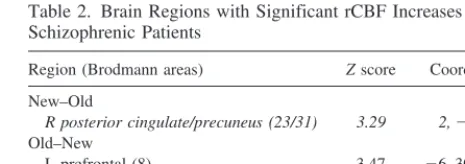
Table 2. Brain Regions with Significant rCBF Increases in Schizophrenic Patients
Region (Brodmann areas) Zscore Coordinates
New–Old
R posterior cingulate/precuneus (23/31) 3.29 2,248, 24 Old–New
L prefrontal (8) 3.47 26, 30, 36
R parietal (39) 3.43 40,266, 16 Cerebellum 3.24 24,258,28
The numbers in parentheses refer to Brodmann areas. Coordinates (in mm) refer to the three axes (x, y, z) of the Talairach and Tournoux (1988) brain atlas. The maximum excursion (z) is reported for each activation. All activations withz.
those areas for which we had found a condition effect in the previous within-group analyses.
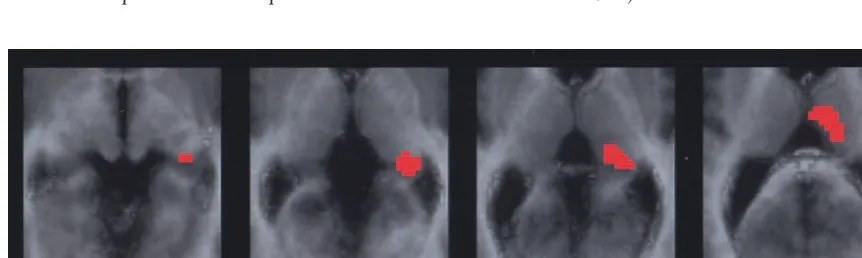
Recognition of new objects (New–Old) was associated with different patterns of rCBF changes in the two groups: control subjects showed greater rCBF increases in the right prefrontal cortex (area 10) and right posterior thala-mus, and patients with schizophrenia showed greater rCBF increases in the right posterior cingulate/precuneus region. Similarly, greater thalamic rCBF increases in the control group were also found in the two contrasts New–View (22, 234, 4; z 5 2.40) and New–Fixation (20, 236, 4; z 5 3.21). Further analysis at a lowerzscore (z.2.33,p, .01) revealed that the rCBF differences in the contrast New–Old extended anteriorly into the thalamus, but not posteriorly into the hippocampus (Figure 1).
Recognition of old objects (Old–New) was associated with greater rCBF increases in left prefrontal area 8 in the patients with schizophrenia. Greater prefrontal rCBF
in-creases in the schizophrenia subjects was also seen in the contrast Old–View (22, 30, 36;z52.90).
Discussion
Patients with schizophrenia showed attenuated activation of the right thalamus and right prefrontal cortex during the recognition of new objects and greater left prefrontal cortical activation during the recognition of previously seen objects. Patients with schizophrenia produced false alarm rates to new objects similar to the control subjects but significantly lower recognition rates for previously presented objects. These data provide evidence that schizophrenia is associated with decreased right hemi-spheric thalamic and prefrontal activation during the recognition of novel visual stimuli and with increased left prefrontal cortex activation during impaired episodic rec-ognition of old visual stimuli. We will first consider the PET data of the control group and then discuss the relevance of our findings to schizophrenia.
Recognition of novel objects was associated with an activation of the pulvinar region in the right posterior thalamus. This is consistent with the thalamic activation during the recognition of novel objects observed using a similar experimental paradigm (Uecker et al 1997). Pre-vious PET and functional magnetic resonance imaging studies have reported activation of thalamic nuclei, includ-ing the pulvinar, durinclud-ing tasks that modulated attention and arousal, especially when responding to visually presented stimuli (Corbetta et al 1991; Heinze et al 1994; Kinomura et al 1996; LaBerge and Buchsbaum 1990; Portas et al 1998b; Vandenberghe et al 1997). Furthermore, electro-physiological studies in the behaving monkey have dem-onstrated a modulation of neural activity in pulvinar nuclei during the recognition of visually presented objects (Pe-tersen et al 1985). The thalamic activation seen in the
Figure 1. Positron emission tomography (PET) statistical map comparing the contrast (New–Old) between control subjects and schizophrenic patients. The PET image is coregistered with an average normal magnetic resonance image, transformed to Talairach space. The images are horizontal slices through the thalamus and medial temporal lobes atz50 and 4, 8, and 12 mm above the anterior commissure–posterior commissure line. Compared with the control group, the right thalamus was significantly less activated (p,.01,
z.2.33) in the schizophrenia group, with a maximum excursion (z53.30) in the pulvinar (at coordinates 22,234, 4). Table 3. Brain Regions with Significant Differences of rCBF
Increases in the Two Groups
Region (Brodmann areas) Zscore Coordinates
New–Old
Control subjects.schizophrenic patients
R Prefrontal (10) 4.01 12, 58, 20
R Thalamus 3.30 22,234, 4
Schizophrenic patients.control subjects
R posterior cingulate/precuneus (23/31) 3.32 2,248, 24 Old–New
Control subjects.schizophrenic patients No regions
Schizophrenic patients.control subjects
L Prefrontal (8) 4.01 26, 30, 36
The numbers in parentheses refer to Brodmann areas. Coordinates (in mm) refer to the three axes (x, y, z) of the Talairach and Tournoux (1988) brain atlas. The maximum excursion (z) is reported for each activation. All activations withz.
contrast New–View in our experiment is most likely related to attentional modulation, because the stimuli were comparable (novel stimuli), but the instructions required greater attention to new objects during the recognition testing compared with passively viewed objects. The thalamic activation seen in the contrast New–Old indicates a greater involvement of the pulvinar when recognizing novel objects compared to previously seen objects.
We found activation of prefrontal areas during the recognition of old objects, which replicates the results of previous studies using a similar experimental design (Schacter et al 1995, 1997; Uecker et al 1997). We found the most significant rCBF increase during the Old–New contrast in right prefrontal area 47. Previous studies have shown that right inferior prefrontal areas contribute to episodic memory for visuospatial stimuli (Wagner 1999). During episodic retrieval, visuospatial representations of the target object may be maintained in working memory as part of the retrieval process (Wagner 1999). The magni-tude of right prefrontal activation, however, might not systematically vary with recognition performance (Wag-ner et al 1998).
The control subjects in our study showed medial tem-poral lobe activation only during the contrast New–Old. Two previous PET studies of episodic object recognition had reported medial temporal lobe activation during the recognition of old objects (Schacter et al 1995, 1997), and a more recent PET study (Schacter et al 1999) had reported medial temporal lobe activation for the recogni-tion of old as well as new objects. Our study, which employed an experimental design similar to the recent study by Schacter et al (1999), confirms their finding of medial temporal lobe activation in the contrast New–Old. It is not clear why we did not find medial temporal lobe activation during the recognition of previously seen ob-jects, but several differences in study design have to be considered: 1) our control subjects were more accurate in recognizing old objects than were those in Schacter et al’s experiment; 2) our subjects were given only one study exposure to target objects (compared to two), which was followed by four (compared to six) recognition test scans; 3) the target objects in each scan followed six buffer items (compared to one); and 4) our subjects were all male, whereas those in Schacter et al’s study were all female, and our subjects were older and had achieved a lower level of education than those in the Schacter et al experiment. Future studies should explore which of these differences are responsible for the contrasting patterns of results.
The patients with schizophrenia did not show tha-lamic activation during the recognition of novel objects, neither in the New–Old contrast nor in comparison with the two baseline conditions. This difference was signif-icant as a group-by-condition interaction for the
con-trasts New–Old and New–Fixation. This provides evi-dence for an impairment of thalamic function in schizophrenia during tasks that require subjects to pay attention to or recognize novel visual objects. This finding is of interest, because recent neuroimaging studies have demonstrated functional abnormalities, and some, but not all, have reported structural abnormalities of the thalamus in schizophrenia (Andreasen et al 1994, 1996; Arciniegas et al 1999; Buchsbaum et al 1996; Hazlett et al 1999). Specifically, thalamic glucose metabolism was reduced during a visual continuous performance task (Buchsbaum et al 1996) and a serial verbal learning task (Hazlett et al 1999). Furthermore, thalamic rCBF decreases in schizophrenia, in the con-text of an overall impaired prefrontal–thalamic– cer-ebellar circuitry, have been found during the recall of complex narrative material (Andreasen et al 1996). Our experiment provides the first evidence that thalamic function during the recognition of novel visual stimuli is impaired in schizophrenia.
semantic/phono-logical stimuli eliciting left prefrontal activation pat-terns (Wagner 1999). The lack of hemispheric asymme-try for the recognition of visually presented objects as seen in the patients with schizophrenia might indicate a different strategy, a failure of right prefrontal cortex recruitment, or a disinhibition of left prefrontal cortex activity that interferes with normal visuospatial memory processes. Interestingly, recent neuroimaging studies have provided evidence for an abnormality of left hemispheric activity during the performance of cogni-tive tasks in schizophrenia (Gur and Chin 1999).
The patients with schizophrenia did not show any medial temporal lobe activation during object recognition. Because our control group also did not show robust medial temporal lobe activation, we did not find significant group-by-condi-tion interacgroup-by-condi-tions. Previous studies have demonstrated that episodic recognition is associated with the recruitment of a frontal–temporal neural network. Data from this experiment provide evidence that the prefrontal component of this network is dysfunctional in schizophrenia but cannot speak to a possible involvement of the medial temporal lobe.
We have to consider several details of our study design that might limit the generalizability of our results. First, all subjects were male and all schizo-phrenic subjects were chronic, stable outpatients, treated with typical neuroleptics. We decided not to interfere with the current treatment of the patients, for practical, ethical, and scientific reasons. For example, discontinuation of neuroleptic medication might impair memory function in chronic schizophrenic patients (Gilbertson and van Kammen 1997). Second, we stud-ied small samples; however, we used a mixed effect model analysis to test for population inferences (Holmes and Friston 1998; Woods 1996). Third, we do not have information about brain structure in our subjects. It is possible that some of the functional abnormalities noted in our schizophrenia sample are attributable to structural differences (e.g., smaller tha-lamic or prefrontal cortex volumes); however, thalamus volume differences are not consistently found in schizo-phrenia (Arciniegas et al 1999; Portas et al 1998a) and a recent study by Hazlett et al demonstrated functional abnormalities of the thalamus without concurrent vol-ume loss in schizophrenia (Hazlett et al 1999).
In conclusion, we found evidence for abnormal tha-lamic and prefrontal cortical function during episodic object recognition in schizophrenia. Object recognition paradigms provide a tool to study thalamic and prefron-tal dysfunction in schizophrenia. Future studies are needed to elucidate the mechanisms underlying de-creased thalamic and abnormal (dede-creased right and increased left) prefrontal activation during object rec-ognition in schizophrenia.
SH was supported by a Young Investigator Award from the National Alliance for Research on Schizophrenia and Depression, the Clinical Investigator Training Program: Harvard/MIT Health Sciences and Tech-nology-Beth Israel Deaconess Medical Center, in collaboration with Pfizer Inc., and a mentored patient-oriented research award from the National Institute of Mental Health (Grant No. K23 MH01763-01).
The authors thank Dmitry Berdichevsky, M.S., Zakhar Levin, Sandra Barrow, Avis Loring, Steve Weise, Ed Amico, and Jessica Temkin for technical support.
References
Aleman A, Hijman R, de Haan EHF, Kahn RS (1999): Memory impairment in schizophrenia: A meta-analysis.Am J Psychi-atry156:1358 –1366.
Alpert NM, Berdichevsky D, Weise S, Tang J, Rauch SL (1993): Stereotactic transformation of PET scans by nonlinear least squares. In: Uemura K, Jones T, editors. Quantification of Brain Function. Tracer Kinetics and Image Analysis in Brain PET.Amsterdam: Excerpta Medica, 459 – 463.
American Psychiatric Association (1994): Diagnostic and Sta-tistical Manual of Mental Disorders,4th ed. Washington, DC: American Psychiatric Press.
Andreasen NC, Arndt S, Swayze VN, Cizadlo T, Flaum M, O’Leary D, et al (1994): Thalamic abnormalities in schizo-phrenia visualized through magnetic resonance image aver-aging.Science266:294 –298.
Andreasen NC, O’Leary DS, Cizadlo T, Arndt S, Rezai K, Boles Ponto LL, et al (1996): Schizophrenia and cognitive dysme-tria: A positron-emission tomography study of dysfunctional prefrontal-thalamic-cerebellar circuitry.Proc Natl Acad Sci U S A93:9985–9990.
Arciniegas D, Rojas DC, Teale P, Sheeder J, Sandberg E, Reite M (1999): The thalamus and the schizophrenia phenotype: Failure to replicate reduced volume. Biol Psychiatry 45: 1329 –1335.
Buchsbaum MS, Someya T, Teng CY, Abel L, Chin S, Najafi A, et al (1996): PET and MRI of the thalamus in never-medicated patients with schizophrenia. Am J Psychiatry
153:191–199.
Buckner RL, Koutstaal W, Schacter DL, Dale AM, Rotte M, Rosen BR (1998): Functional-anatomic study of episodic retrieval. II. Selective averaging of event-related fMRI trials to test the retrieval success hypothesis.Neuroimage7:163– 175.
Clare L, McKenna PJ, Mortimer AM, Baddeley AD (1993): Memory in schizophrenia: What is impaired and what is preserved?Neuropsychologia31:1225–1241.
Corbetta M, Miezin FM, Dobmeyer S, Shulman GL, Petersen SE (1991): Selective and divided attention during visual discrim-inations of shape, color, and speed: Functional anatomy by positron emission tomography.J Neurosci11:2383–2402. Frith CD, Friston KJ, Herold S, Silbersweig D, Fletcher P, Cahill
C, et al (1995): Regional brain activity in chronic schizo-phrenic patients during the performance of a verbal fluency task.Br J Psychiatry167:343–349.
Goldberg TE, Gold JM (1995): Neurocognitive deficits in schizophrenia. In: Hirsch SR, Weinberger DR, editors.
Schizophrenia.Oxford, UK: Blackwell Science, 146 –162. Gur RE, Chin S (1999): Laterality in functional brain imaging
studies of schizophrenia.Schizophr Bull25:141–156. Hazlett EA, Buchsbaum MS, Byne W, Wei T-S, Spiegel-Cohen J,
Geneve C, et al (1999): Three-dimensional analysis with MRI and PET of the size, shape, and function of the thalamus in the schizophrenia spectrum.Am J Psychiatry156:1190 –1199. Heckers S, Goff D, Schacter DL, Savage CR, Fischman AJ,
Alpert NM, et al (1999): Functional imaging of memory retrieval in deficit vs nondeficit schizophrenia. Arch Gen Psychiatry56:1117–1123.
Heckers S, Rauch SL, Goff D, Savage CR, Schacter DL, Fischman AJ, et al (1998): Impaired recruitment of the hippocampus during conscious recollection in schizophrenia.
Nat Neurosci1:318 –323.
Heinrichs RW, Zakzanis KK (1998): Neurocognitive deficit in schizophrenia: A quantitative review of the evidence. Neuro-psychology12:426 – 445.
Heinze HJ, Mangun GR, Burchert W, Hinrichs H, Scholz M, Mu¨nte TF, et al (1994): Combined spatial and temporal imaging of brain activity during visual selective attention in humans.Nature372:543–546.
Holmes AP, Friston KJ (1998): Generalisability, random effects and population inference.Neuroimage7:S754.
Hull CL (1917): The formation and retention of associations among the insane.Am J Psychol28:419 – 435.
Kinomura S, Larsson J, Gulyas B, Roland PE (1996): Activation by attention of the human reticular formation and thalamic intralaminar nuclei.Science271:512–515.
Kotrla KJ, Weinberger DR (1995): Brain imaging in schizophre-nia.Annu Rev Med46:113–122.
LaBerge D, Buchsbaum MS (1990): Positron emission tomo-graphic measurements of pulvinar activity during an attention task.J Neurosci10:613– 619.
Manoach DS, Press DZ, Thangaraj V, Searl MM, Goff DC, Halpern E, et al (1999): Schizophrenic subjects activate dorsolateral prefrontal cortex during a working memory task, as measured by fMRI.Biol Psychiatry45:1128 –1137. Petersen SE, Robinson DL, Keys W (1985): Pulvinar nuclei of
the behaving rhesus monkey: Visual responses and their modulation.J Neurophysiol54:867– 886.
Portas CM, Goldstein JM, Shenton ME, Hokama HH, Wible CG, Fischer I, et al (1998a): Volumetric evaluation of the thala-mus in schizophrenic male patients using magnetic resonance imaging.Biol Psychiatry43:649 – 659.
Portas CM, Rees G, Howseman AM, Josephs O, Turner R, Frith CD (1998b): A specific role for the thalamus in mediating the interaction of attention and arousal in humans. J Neurosci
18:8979 – 8989.
Schacter DL, Cooper LA, Delaney SM, Peterson MA, Tharan M (1991): Implicit memory for possible and impossible objects: Constraints on the construction of structural descriptions.J Exp Psychol Learn Mem Cogn17:3–19.
Schacter DL, Curran T, Reiman EM, Chen K, Bandy DJ, Frost JT (1999): Medial temporal lobe activation during episodic encoding and retrieval: A PET study. Hippocampus9:575– 581.
Schacter DL, Reiman E, Uecker A, Polster MR, Sheng Yun L, Cooper LA (1995): Brain regions associated with retrieval of structurally coherent visual information. Nature 376:587– 590.
Schacter DL, Savage CR, Alpert NM, Rauch SL, Albert MS (1996): The role of hippocampus and frontal cortex in age-related memory changes: A PET study. Neuroreport
7:1165–1169.
Schacter DL, Uecker A, Reiman E, Yun LS, Bandy D, Chen K, et al (1997): Effects of size and orientation change on hippocampal activation during episodic recognition: A PET study.Neuroreport8:3993–3998.
Spitzer RL, Williams JBW, Gibbon M, First MB (1991):
Structured Clinical Interview for DSM-III-R. Washington, DC: American Psychiatric Press.
Talairach J, Tournoux P (1988):Co-Planar Stereotaxic Atlas of the Human Brain.Stuttgart, Germany: Thieme.
Uecker A, Reiman EM, Schacter DL, Polster MR, Cooper LA, Yun LS, et al (1997): Neuroanatomical correlates of implicit and explicit memory for structurally possible and impossible visual objects.Learn Mem4:337–355.
Vandenberghe R, Duncan J, Dupont P, Ward R, Poline J-B, Bormans G, et al (1997): Attention to one or two features in left or right visual field: A positron emission tomography study.J Neurosci17:3739 –3750.
Wagner AD (1999): Working memory contributions to human learning and remembering.Neuron22:19 –22.
Wagner AD, Desmond JE, Glover GH, Gabrieli JDE (1998): Prefrontal cortex and recognition memory. Functional-MRI evidence for context-dependent retrieval processes. Brain
121:1985–2002.
Williams P, Tarr MJ (1997): Structural processing and implicit memory for possible and impossible figures.J Exp Psychol Learn Mem Cogn23:1344 –1361.