PHY SIOLOGY OF K
1BALANCE
Franklyn G. Knox
Depa rtm ent of Interna l Medicine a nd Depa rtm ent of Physiology a nd Biophysics, Ma yo Founda tion, Rochester, Minnesota 55905
T
his is an exercise involving the integration of a case presentation of diuretic-induced hypokalemia with the physiology of K1balance. Students are presented with a case presentation involving the development and correction of diuretic-induced hypokalemia. K1homeostasis, including the distribution of K1in the body and total body K1balance, is summarized. The renal handling of K1, including the filtered load, reabsorption by proximal segments of the nephron, and secretion by distal segments of the nephron, is outlined. The mechanisms for the control of the secretion of K1are discussed. Finally, an exercise involving integration of the case presentation with the physiology of K1balance is presented. This exercise works well in an interactive session with students. The exercise has proved highly useful in the presentation and understanding of basic concepts involved in the physiology of K1balance.AM. J. PHYSIOL. 275 (ADV. PHYSIOL. EDUC. 20): S142–S147, 1998.
Key words:renal potassium handling; hypokalemia
An exercise concerning the physiology of K1balance has been used and refined as part of the first-year curriculum in the Mayo Medical School. The exercise incorporates three major components: a case presenta-tion of a common clinical problem, total body K1 balance, and the renal handling of K1. The students extract data from each of these three elements to complete the exercise. The exercise is presented without the answers so that the table at the conclu-sion of the exercise can be used in an interactive fashion with students. Student feedback on this exer-cise has been very positive, with students comment-ing that ‘‘workcomment-ing with the numbers helped with an understanding of the concepts.’’
EXERCISE: INTEGRATION OF A CASE PRESENTATION OF DIURETIC-INDUCED HYPOKALEMIA WITH THE PHYSIOLOGY OF K1 BALANCE
The objective of this exercise is to apply the basic principles of the physiology of K1balance to a case
presentation describing the development and correc-tion of diuretic-induced hypokalemia.
Case pr esentation: Diur etic-induced hypokale-mia.A middle-aged man who weighed 20 lb over his ideal weight presented blood pressure elevated to 160/110 mmHg. He had no cardiovascular complica-tions of hypertension and normal levels of serum creatinine and K1 (4 meq/l). Hydrochlorothiazide (a diuretic that decreases Na1reabsorption in the distal nephron) at 25 mg twice daily was prescribed, with advice to return for reexamination in 1 mo.
He returned 1 mo later complaining of muscle cramps, frequent nighttime urination, and general weakness. His blood pressure was 150/90 mmHg, and the serum creatinine was still normal. However, the serum K1 concentration had decreased to 3.0 meq/l. A 24-h urine specimen contained 256 meq of Na1. Because the urinary excretion of Na1usually approximates the dietary intake, it was concluded that he was ingesting excessive Na1. A nutritionist instructed the patient in
a 90-meq Na1 diet with weight-reduction features. The hydrochlorothiazide was continued, but both 25-mg tablets were now to be taken in the morning, with none being taken at bedtime.
One month later, the patient’s blood pressure was 130/90 mmHg, he had lost 8 lb, and the serum K1 concentration was 4.0 meq/l. He was urinating only once or twice during the night, and the cramps in his legs were no longer troublesome.
K1HOMEOSTASIS
K1 is the major intracellular cation (1). A high level within cells is important for the optimal operation of many enzymatic processes and the control of cell volume. In addition, the establishment of a K1 concen-tration ([K1]) difference across the plasma membrane of the cell is important for the normal function of excitable tissues.
The extracellular fluid [K1] is a function of the total body K1content and the relative distribution of K1 between the extracellular and intracellular fluid com-partments. The total body K1 content depends on
both the dietary intake of K1 and its excretion from the body.
DISTRIBUTION OF K1 IN THE BODY
Virtually all K1 within the body is contained inside cells, with only 2% being located in the extracellular fluid space. The distribution of K1within the body is summarized in Fig. 1.
In the steady state, the intake of K1into the body must be matched by its excretion, a process occurring primarily via the kidneys. However, the renal re-sponses to fluctuations in K1 intake are not immedi-ate, and it takes several hours or even days for appropriate adjustments in K1 excretion to occur. Wide fluctuations in the [K1] of the extracellular fluid are prevented during the ‘‘lag period’’ by shifts of K1 between the intracellular extracellular compartments (extrarenal K1homeostasis).
The extrarenal handling of K1 is essential for the maintenance of plasma [K1] within a narrow range. This is especially true during situations in which the body is challenged with an acute load of K1. Thus,
FIG. 1.
with an acute K1 load (dietary or otherwise), K1 is rapidly taken up into cells (liver, skeletal muscle, adipose tissue). This prevents a large rise in plasma [K1]. Over the next several hours K1 is slowly released from these storage sites and excreted by the kidneys.
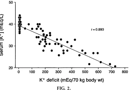
With potassium depletion, which occurs with diuretic therapy, the losses from the extracellular fluid are not fully compensated for by shifts from the tissue stores. A decrease in the plasma K1 from 4 to 3 meq/l corresponds to an,300-meq K1deficit as determined empirically (Fig. 2) (2). Note that if the loss of K1from the plasma were proportionate to the loss of total body K1, it would equal a total body K1 deficit of 1,000 meq.
RENAL K1 EXCRETION
The kidney can vary the amount of K1excreted in the urine over a wide range. The kidney is able to excrete large amounts of K1. Accordingly, with normal renal function and even in the face of a large dietary K1 intake, significant hyperkalemia is rarely seen. In contrast, the kidney is less well able to conserve K1 (fractional K1excretion can be reduced to,2% of the filtered load). Thus K1depletion and hypokalemia can result if K1intake is restricted.
The process of renal K1 excretion involves two general steps. First, 90% of the filtered load of K1is reabsorbed. This occurs in the proximal tubule and the loop of Henle. Second, K1 is secreted into the tubular fluid by the terminal portion of the distal
convoluted tubule and the cortical portion of the collecting duct. These general aspects of renal K1 handling are summarized in Fig. 3.
Note that ,90% of the filtered load of K1 is reab-sorbed before the K1secretory site. The amount of K1 appearing in the urine reflects in large part the secretion of K1 at the distal secretory sites. Several factors have been identified as important regulators of K1secretion and will be considered in detail below.
REABSORPTION OF FILTERED K1
Glomerulus.K1is a small cation and is not bound in any appreciable amount to plasma protein. As a result, the K1in the glomerular filtrate is essentially the same as in the plasma water. The filtered load of K1is then plasma [K1] (4.0 meq/l) multiplied by the glomerular filtration rate (180 l/day), or 720 meq/day.
Pr ox imal tubule. The proximal tubule (convoluted and straight) reabsorbs roughly 80% of the filtered load of K1. This fraction remains relatively constant under most conditions. Thus the proximal tubule does not normally contribute significantly to the regulation of urinary K1excretion.
FIG. 2.
Effect of K1depletion on plasma K1concentration [adapted fr om Ster ns et al. (2)].
FIG. 3.
The reabsorption of K1 by the proximal tubule probably involves both passive and active mecha-nisms. Under most conditions, K1 reabsorption is proportional to the amount of NaCl and fluid reab-sorbed.
Loop of Henle. Approximately 10% of the filtered load of K1is reabsorbed by the thick ascending limb of Henle’s loop. Under normal conditions, the reab-sorption of K1 is relatively constant, although this segment does have the capacity to increase K1 absorp-tion in response to an increased load.
SECRETION OF K1
Distal tubule and collecting duct. It is the distal convoluted tubule and the cortical portion of the collecting duct that primarily determine the amount of K1 appearing in the urine. The more terminal portions of the collecting duct can affect minor adjustments in urinary K1excretion.
Under most conditions (normal western diet), K1is secreted at these sites. However, with K1depletion, reabsorption of K1occurs. These two transport mecha-nisms are located in separate cells. The secretory cell is the ‘‘principal cell,’’ whereas the ‘‘intercalated cell’’ is the cell responsible for K1reabsorption.
K1secretion occurs by a two-step process. First, K1is brought into the cell across the basolateral cell mem-brane (Na1-K1-ATPase). Some K1recycles across this membrane, but a portion enters the tubule lumen by passive diffusion across the apical cell membrane. The amount of K1that either recycles back to the blood across the basolateral cell membrane or enters the tubule lumen is determined by the permeability of each membrane to K1and the respective electrochemi-cal gradients for K1. Normally, the apical cell mem-brane is more permeable to K1. In addition, as shown in Fig. 4, the electrical profile across the cell favors luminal K1entry.
REGULATION OF K1 EXCRETION
To understand the regulation of renal K1excretion, it is important to focus on the secretion of K1 by the terminal portion of the distal convoluted tubule and the cortical portion of the collecting duct (K1
secre-tory site). Several factors act at this site to regulate K1 secretion and thereby K1excretion.
Dietary K1. Urinary K1 excretion parallels dietary intake. With dietary K1 loading, K1 secretion is enhanced. This is the result of increased uptake of K1 into cells (via Na1-K1-ATPase) and is largely the result of changes in mineralocorticoid hormone levels (see Horm ones). Conversely, cellular uptake of K1 is reduced with K1 depletion, again in response to changes in mineralocorticoid hormone levels.
Plasma K1.As plasma K1is increased, K1secretion also increases, reaching a plateau at a plasma [K1] of ,6 meq/l. Because increases in plasma [K1] stimulate aldosterone secretion, the increased K1secretion can be attributed in part to mineralocorticoid-induced uptake of K1into the K1secretory cells. In addition, elevated plasma [K1] would also be expected to reduce the passive component of K1recycling across the basolateral cell membrane. Together, these effects increase K1excretion.
Hor mones.The most important hormone regulating K1 secretion by the terminal portion of the distal convoluted tubule and the cortical portion of the collecting duct is aldosterone. Aldosterone stimulates K1 secretion by increasing cellular uptake of K1via the Na1-K1-ATPase and by increasing the K1 permeabil-ity of the apical cell membrane of the K1 secretory
FIG. 4.
cells. Aldosterone also stimulates Na1reabsorption by these nephron segments. This, in turn, increases the transepithelial potential difference and further stimu-lates K1secretion.
Luminal flow rate. As the flow of tubular fluid past the K1 secretory site is increased, K1 secretion is increased. This flow-related increase in K1secretion is thought to result from the maintenance of a favorable cell-to-lumen concentration gradient for K1. Because the amount of K1secreted into the tubule lumen will be limited by this gradient, a fast flow rate (decreased contact time) will prevent luminal K1 from rising to levels that would subsequently limit further K1 secre-tion.
EFFECTS OF DIURETICS ON K1 EXCRETION
Diuretics commonly increase renal K1 excretion. Those diuretics that have their site of action proximal to the K1secretory site will increase urinary K1loss (e.g., osmotic diuretics, carbonic anhydrase inhibi-tors, loop diuretics, and thiazides). The increased K1 secretion results from the diuretic-induced increase in luminal fluid flow rate and delivery of Na1. In addition,
the loop diuretics inhibit K1reabsorption by the thick ascending limb. The K1-sparing diuretics (spironolac-tone, triamterene, and amiloride) prevent K1 loss in the urine through inhibition of its secretion.
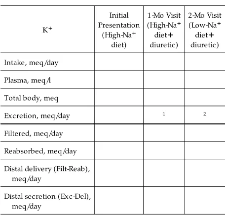
From the case presentation and the syllabus materials, complete the distribution of K1and the renal handling of K1for the patient’s initial presentation and at the 1-and 2-mo visits (Table 1). The answers to the exercise are found in Table 2.
DISCUSSION
At the initial presentation, the dietary intake of K1is 100 meq of K1 per day as derived from Fig. 1. The excretion of K1is 90 meq/day, also as derived from Fig. 1. The plasma K1is 4 meq/l as derived from both the case presentation and the syllabus. The total body K1 is 3,500 meq as derived from the information in Fig. 1, including both tissue stores and extracellular fluid. The filtered K1is 720 meq/day (as derived from the syllabus), where the filtered load is equal to 4 meq/l3180 l/day. The reabsorbed K1is equal to 90% of the filtered load shown in Fig. 3. The distal delivery is simply the filtered minus reabsorbed K1 at 72 meq/day. The distal secretion, therefore, is the ex-creted K1(90 meq/day) minus the delivered K1(72 meq/day), which is 18 meq/day.
TABLE 1
Integration of the case pr esentation with the physiology of K1balance: ex er cise
1Calculate average daily K1excretion to account for the decrease in total body K1.2Calculate average daily K1excretion to account for the increase in total body K1.
TABLE 2
Integration of the case pr esentation with the physiology of K1balance: Answers to the ex er cise
K1
Intake, meq/day 100 100 100
Plasma, meq/l 4.0 3.0 4.0
Total body, meq 3500 3200 3500
Excretion, meq/day 90 100 80
Filtered, meq/day 720 540 720
Reabsorbed, meq/day 648 486 648
At the 1-mo visit, the K1 intake continues at 100 meq/day. The excretion can be calculated from the K1 loss. The plasma K1concentration is 3 meq/l as obtained from the case presentation. This results from a decrease in total body K1of 300 meq as obtained from the information in Fig. 2. A 300-meq loss over a 30-day period represents a 10 meq/day increase in K1 excretion over the initial 90 meq/day. Thus the K1 excretion at the 1-mo visit has averaged 100 meq/day. The filtered K1 is now 540 meq/day, and the reab-sorbed K1 is 486 meq/day. The distal delivery is 54 meq/day, and, because the excretion is 100 meq/day, the distal secretion is 46 meq/day.
At 2 mo, this process is reversed. The K1 intake remains 100 meq/day, the excretion is calculated to be 80 meq/day, and the distal secretion now becomes 8 meq/day. Thus this exercise described the major role of distal secretion in determining final urinary excretion. In addition, it shows the marked effect of changes in Na1intake on the renal handling of K1in diuretic therapy. The high Na1 intake leads to in-creased delivery of Na1 and water to the distal nephron, where the luminal flow rate is further increased by the diuretic and leads to increased K1 secretion. The low Na1intake reduces the delivery of Na1and water to the distal nephron so that the effect of the diuretic on K1secretion is offset and K1balance is restored.
The case presentation is taken from an actual clinical case. However, the presentation has been simplified
by having the low-Na1 diet completely correct the hypokalemia. In the original case, the low-Na1 diet partially corrected the hypokalemia with a final serum K1 concentration of 3.5 meq/l. The simplification allows for a symmetrical calculation of the changes in distal K1 secretion. The exercise, of course, can be performed with the incomplete correction of the hypokalemia; however, this tends to complicate the calculations and interfere with the presentation of the basic principles.
A second simplification involves the attribution of the entire correction of the hypokalemia to changes in distal K1 secretion. In the presence of K1 conserva-tion, reabsorption of K1may also occur in the distal nephron segments. These two points can be brought out in the context of the discussion of Table 2 at the conclusion of the exercise. The exercise has been found to be useful in presenting and utilizing basic concepts involved in the physiology of K1balance.
Address reprint requests to the author.
Refer ences
1. Koeppen, B. M., and B. A. Stanton. Regulation of r enal physiology. In: Rena l Physiology(2nd ed.). St. Louis, MO: Mosby Year Book, 1997, chapt. 7, p. 117–133.