Achalasia: A Review of Etiology,
Pathophysiology, and Treatment
Nor Hedayanti, Supriono
Division of Gastroentero-hepatology, Department of Internal Medicine Faculty of Medicine, University of Brawijaya/Dr. Saiful Anwar Hospital, Malang
Corresponding author:
Supriono. Division of Gastroentero-hepatology, Department of Internal Medicine, Dr. Saiful Anwar General Hospital. Jl. Jaksa Agung Suprapto No. 2 Malang Indonesia. Phone/facsimile: +62-341-348265. E-mail: gastro_mlg@yahoo.com
ABSTRACT
Achalasia was a condition marked by peristaltic movement absent in lower esophageal sphincter and segment that hypertonic result in imperfect relaxation during food ingestion. Achalasia incidence did not differ between men and women, account for 1 in 100,000 people every year with prevalence of 10 in 100,000 people, unrelated
VSHFL¿FDOO\ZLWKHWKQLFDQGKDVLWVKLJKHVWLQFLGHQFHRQDJHJURXS
Based on its etiology, it was divided into primary and secondary Achalasia, while based on its motility, it was into hypermotil, hypomotil, and amotil achalasia. Until present, several therapeutic modalities were available to treat Achalasia, among them was pharmacology therapy, botulinum toxin injection via endoscopy, pneumatic dilatation, Heller myotomy surgery, and per oral endoscopy myotomy (POEM).
Keywords : achalasia, patophysiology, therapy
ABSTRAK
Akalasia merupakan suatu keadaan yang ditandai dengan tidak adanya peristaltik di esofagus bagian bawah dengan spingter esofagus bagian bawah (LES) yang hipertonik sehingga tidak terjadi relaksasi sempurna saat menelan makanan. Angka kejadian akalasia pada perempuan dan laki laki adalah sama, yaitu 1 dari 100.000 orang per tahun dengan prevalensi 10 pada 100.000 orang, tidak didapatkan pada ras khusus dan angka kejadian tertinggi pada usia 30-60 tahun.
Berdasarkan etiologi terdapat dua jenis akalasia, yaitu primer dan sekunder sedangkan berdasarkan motilitas terdapat tiga jenis akalasia, yaitu akalasia hipermotil, akalasia hipomotil, dan akalasia amotil. Hingga saat ini, terdapat beberapa modalitas terapi akalasia, antara lain intervensi farmakologi, injeksi toksin botulinum per endoskopi, dilatasi pneumatik, bedah Heller myotomy, dan Per Oral Endoscopy Myotomy (POEM).
Kata kunci :DNDODVLDSDWD¿VLRORJLWHUDSL
INTRODUCTION
$FKDODVLDZDV¿UVWO\GHVFULEHE\6LU7KRPDV:LOOLV
in 1674 after he use whale bone to dilate his patients esophagus because of lower esophageal sphincter relaxation failure. Achalasia was progressive idiopathic neural degeneration of Auerbach myenteric plexus,
result in food static during ingestion and esophagus dilatation. This condition will lead to several symptoms and complication, depend on its severity and duration.1,2
Some Literatures said that Achalasia was a primary
GLVRUGHURIHVRSKDJXVZLWKUHOD[DWLRQLQVXI¿FLHQF\
UDGLRJUDSKLF¿QGLQJVRIDSHULVWDOWLFHVRSKDJXVZLWK
minimal openings, called bird-beak sign.3
Incidence of Achalasia in men and women was similar, account for 1 from 100,000 people every year with prevalence of 10 in 100,000 people, did
QRWVSHFL¿FIRUVRPHHWKQLFVDQGKLJKHVWLQFLGHQFH
was found in 30-60 age groups. In US, 2000 cases of Achalasia was reported every year, mostly at 25-60 age groups, and rarely found in children. Otherwise, in Gastroentero-hepatology division, Internal Medicine Department, Faculty of Medicine, University of Indonesia/Dr. Cipto Mangunkusumo Hospital, 48 cases was found during 5 years period (1984-1988),
mostly with the same age groups.2,3Achalasia was
not a rare condition. This could be seen in middle-aged population, clinically marked with dysphagia, regurgitation, and epigastric discomfort. Clinically, Achalasia was divided into primary and secondary, based on its etiology.2,3
American collage Gastroenterology Clinical Guideline in Diagnosis and Management of Achalasia in 2013 recommended barium esophagogram to assess esophageal emptying, esophagogastric junction morphology both during gastric emptying and to evaluate its motility. Endoscopy procedure to see gastroesophageal junction and gastric cardia was recommended in all patients with Achalasia to eliminate the possibility of pseudoachalasia.3If
irreveribe functiona disorder happened, the main therapy for Achalasia was palliative treatment. Dominant symptoms of this disorder was progressive dysphagia, regurgitation, chest pain, and weight loss.3
ETIOLOGY
Achalasia was divided into primary and secondary Achalasia. In primary Achalasia, the exact etiology is unknown, probably caused by neutropic virus infection result in dorsal vagal nucleus lesion in brainstem and mesenteric ganglia in esophagus. Hereditary factor also has a role in this disorder. Otherwise, secondary Achalasia was caused by infection, intraluminar tumor such as cardia tumor, extraluminal pressure from pancreatic pseudocyst, anticholinergic drugs, or post vagotomi operation.2
PATHOPHYSIOLOGY
Esophagus main function is to deliver food ingested from faring to stomach by peristaltic movement ranged from 5-15 seconds. In upper and lower part of esophagus, a sphincter was important for gastric
DFLGUHÀX[EDUULHUWRHVRSKDJXVDQGQRUPDO\LQWRQLF
or constriction state, except during food ingestion. Esophagus mucosa was base and not suitable for acid from gastric secretion. Submucosal layer has secretory cells which produce mucus in order to help food movement during ingestion process.4
At normal state, esophagus shown two types of peristaltic: primary peristaltic and secondary peristaltic. Primary peristaltic was only a movement following previous peristaltic movement in faring and continuing to esophagus during ingestion process. This movement period start from faring to stomach was estimated for 8 to 10 seconds. But, food ingestion durin erect position result in faster food movement, about 5-8 seconds, because of gravity effect. If primary peristaltic movement fail to push all the food into stomach, secondary peristaltic movement than started. This movement start from mienteric nerve and several
UHÀH[HVIURPDIIHUHQWYDJDOQHUYHIURPHVRSKDJXVWR
medulla than back to esophagus.4
In the lower part of esophagus, about towo to
¿YHFHQWLPHWHUDERYHVWRPDFKHVRSKDJHDOFLUFXODU
muscle functioned as gastroesophageal sphincter. Anatomically, this sphincter was not differ to other esophagus parts. Physiologically, sphincter will have tonic constriction (with intraluminal pressure of 30 mmHg), differ from middle esophagus which in normal condition was in relaxation state. When peristaltic movement during ingestion process pass esophagus, receptive relaxation process will relax lower esophageal sphincter before peristaltic movement and allow food to enter stomach. Rarely, sphincter does not relax, so that the condition called Achalasia happened.4
CLINICAL MANIFESTATION
Dysphagia of both solid and liquid food was the most common symptoms of achalasia, followed by regurgitation, chest pain, nausea, vomiting, weight loss, and night cough. Otherwise, chest pain was the
PDLQ V\PSWRPV RI JDVWURHVRSKDJHDO UHÀX[ GLVHDVH
(GERD) caused by esophagus irritation from gastric acid. In patients with Achalasia, this symptoms could be caused by gastric acid retention or toxin produced by fermented lactate by bacteria in esophagus.3
dysphagia and regurgitation symptoms. In amotile Achalasia, there were failure of normal peristaltic so that esophagus was widely dilated in chronic Achalasia.5,6
There were several diagnostic modalities to evaluate
calcium channel blocker and long acting nitrate was effective to lower pressure in LES and sometimes reduce dysphagia, but did not improve its relaxation ability or peristaltic movement of LES.7
Because of longer transit time and delayed in esophagus emptying, which was a characteristic of Achalasia, absorption and effectivity of any oral drugs cannot be calculated. Those drugs was better used via sublingual route, such as nifedipine 10-30 mg sublingual for 30-45 minutes before meals and ISDN 5 mg sublingual 10-15 minutes before meals. Those drugs reduce pressure in LES for about 50%. Ong acting nitrate has a shorter time of action, 3-27 minutes, and showd a better clinical improvement in 53-87% case compared to nifedipine that works for 30-120 minutes with 0-75% effectivity.7
The main limit in those drugs is its short duration of action that only reduce some symptoms and reduce its
HI¿FDF\LQORQJWHUPXVH%HVLGHVLWVVLGHHIIHFWOLNH
peripheral edema, headache, and hypotension could be found in 30% patients.7
Drugs were used limited to patient in early condition without esophageal dilatation, or in patients
ZDLWLQJIRUGH¿QLWLYHWUHDWPHQWRUSDWLHQWLQKLJKULVN
that refuse to undergo invasive procedures. Drugs were also indicated in severe Achalasia with chest pain symptoms. Pharmacotherapy in this condition was
highly recommended.7
Endoscopic Botolium Toxin Injection
Toxin botulinum is a neuro toxin works to inhibit neurotransmitter in terminal cholinergic receptor. Botulinum toxin-A that used for Achalasia therapy works by breaks SNAP-25 protein molecule in presynaptic membrane, so that acethylcholine relese
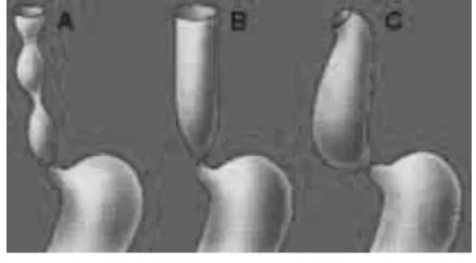
Figure 1. Types of Achalasia5
Achalasia, such as manometry, barium esophagogram,
esophagoduodenoscopy, and esophagus CT-scan.6
TREATMENT
Since 17th century, achalasia therapy was started.
6XUJHRQVKDYHPDGHVLJQL¿FDQWLPSURYHPHQWLQ$FKDODVLD
therapy.1 Otherwise, until now, none of achalasia therapy
could change its pathology and all treatment was just a palliative of its clinical manifestation3
Pharmacology
Based on Society of American Gastrointestinal and Endoscopic Surgeons in Guidelines for the Surgical Treatment of Esophageal Akalasiain 2009,the goal of pharmacotherapy was to help obstruction reduction and improve lower esophageal sphincter relaxation function. Several pharmacological agents such as
was blocked and inhibit acetylcholine exocytosis to synaptic area. This will result in transient muscle weakness by blocking cholinergic stimulation in LES (Figure 2).1
Botulinum toxin injection locally could reduce LES pressure and increase esophagus passive emptying. Toxin injected via sclerotherapy during endoscopy. In normal condition, 80 to 100 unit of Botulinum toxin-A was injected in each LES quadrant to reduce its pressure, increase esophageal opening, and improve esophageal emptying.1,8
Clinical effect from single injection was short term effect with relaps incidence more than 50% in 6 months. Otherwise, repeated injection could give more effect in 70-90% patients. A report showed that 21% of newly diagnosed Achalasia patients was treated using botulinum toxin as early modalities with injection duration of 6 months. Good response from botulinum toxin injection was found in patients aged less than 50 years old and patients with severe Achalasia.1,8
This therapy was safe, only 10% complained chest
SDLQDIWHULQMHFWLRQEXWPRVWO\GLGQRWUHTXLUHVSHFL¿F
therapy. But, repreated injeaction could make myotomy procedure during surgery seems harder because of adhesion of muscular layer and increase perforation possibility in mucosa. Regarding to patient safety, botulinum toxin injeaction could be given if no other choice present or if surgery correction contraindicated, and in patients with survival rate of 2 years.1
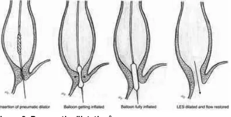
Pneumonic Dilatation
Dilatation procedure was firstly done to treat achalasia. This principle of therapy is to weakend
/(6E\WHDULQJLWVPXVFOH¿EHUUDGLDOO\IURPLQVLGH
esophagus. Endoscopic dilatation was believed as the most safe no surgical therapy for achalasia. Pneumatic dilator were preffered than stiff dilator in achalasia therapy because of its effect that widening and break muscle fibre in LES. Several pneumatic dilator
DYDLODEOHWRGD\KDYHWKHVDPHHI¿FDF\DQGVDIHW\EXW
only few data could support it.1
Dilatation treatment was slowly reduce its symptoms. The most simple treatment to done is Hurst plugging procedure, made from mercury-contained rubber, have four different size in F (French) scale. It works principally based on gravitation using the smallest radius plug to the biggest. Its effectivity only 50% with no relaps case, 35% with relaps, and 15% failure to respond.2
The most recommended procedure was LES dilatation using pneumatic dilator. This procedure
was used in recent 30 years with the most succesfull rate of 75-85% after repeated dilatation. Complication
VXFKDVHVRSKDJHDOUHÀX[RUHVRSKDJHDOSHUIRUDWLRQ
were rarely found. This special technique was not only based on it size, but also based on its duration of LES dilatation, range from seconds to 5 minutes. Balloon
Figure 3. Pneumatic dilatation9
was dilated perfectly in short time to get optimum LES dilatation. In 60 seconds, balon was reexpanded in several minutes, and in each procedure was using maximum two ballon (Figure 3).2
The use of dilator made from polyethylene has a succesfull rate of 93% with lower complication during 4 years follow up. Fluoroscopy guided endoscopy was effective to dilate balloon. Pneumatic dilatation was done using endoscopy in sedated patients, result in 60% reduce in LES pressure and 79% symptos resolution. A report from Cleaveland showed that 41% of new cases was treated with pneumatic therapy, result in clinical response of 86% and 54% esophagus emptying. 1,11
Pneumatic Dilatation vs. Botulinum Toxin Injection
In randomized study involving 42 patiens, botulinum injection and pneumatic dilatation was happened in 70% and 32%, respectively, in 12 months. A Cochrane database review in sic studies involving 178 patients showed statistically indifferent after four weeks of intervention. Three research, including reviewer, showed a remisiion of about 33 cc per 47cc after pneumatic injection. This also happened in 11 of 43 patients after both botulinum injection and pneumatic dilatation. Relative risk was 2,67. This showed that pneumatic dilatation was more effective than botulinum injection in long term Achalasia3
Laparoscopic Heller Myotomy (LHM)
invasive concept similar to open-surgery concept. This
PHWKRGZDVKDYLQJORWRIEHQH¿WEHFDXVHRILWVVKRUWHU
length of stay in hostpital, less pain, smaller operation scar, and faster recovery time. Laparoscopic approach was safe and effective in achalasia therapy, although
VRPHWLPHVDQDWRPLFDOYDULDWLRQPDGHLWVOLJKWGLI¿FXOW10
A meta-analysis that compare short term and long term effect of LHM and balloon dilatation via endoscopic was done. From 16 studies consist of 590 LHM procedure and balloon dilatation via endoscopy with follw up in 12, 24, and 60 months, it can be concluded an OR of 2,2 in 12 months, 5,06 in 24 months, and 29,83 in 60 months of LHM superiority as the main choice of therapy. Besides, only 18 patients reported having redilatation after LHM procedure, while 110 patients in balloon dilatation, even 36 among them should undergo myotomy as its retreatmen.12
LHM was found to be effective with long term outcome. Most of its risk came from general anesthesia procedure. Esopohageal perforation is one of the complication, but could be detected during operation and repaired without serious further complication.9
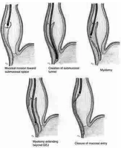
Peroral Endoscopy Myotomy (POEM)
POEM is a technique involving tunneling between esophageal muscle to treat achalasia, especially with Chagas disease. This procedure was done via endoscopy so that no exterior incision was made. POEM was effective to cure dysphagia symptom in Achalasia. After this procedures, achalasia was have Eckaradt score 0 or 1. In a study using barium intake showed a complete emptying in 90% and found to have lower esophageal pressure. Sphincter pressure were reduced after this procedure, but not as dramatically as patients undergo LHM procedure. This procedure was done via endoscopy in general anesthesia. A tunnel was made below esophageal deeper layer until
UHDFK/(6(VRSKDJHDOPXVFOH¿EHUDQGJDVWULFFDUGLD
then resected during endoscopy. Early result of this procedure was promising. But, esophageal damage risk still possible and there were no long term experience result of this therapy. GERD risk after procedure which needed a lifetime antacid was happened in 32% patients. POEM does not cure Achalasia, but this was a palliative procedure.13
POEM was fortly done in September 2008 in our institution. POEM with the longest follow up, 2
\HDUVZDV¿UVWO\UHSRUWHGLQ2FWREHU$OORYHU
the world, there have been 900 case that successfully treated with POEM. One of its contraindication was general anesthesia intolerance.13
Figure 4. POEM procedure13
CONCLUSION
There were several types of Achalasia based on its etiology and motility. Modality choices to treat Achalasia was more various in recent years. But, there were no therapy yet to change its pathology, so that those therapies were just palliative therapies.
REFERENCES
1. Ahmed A. Achalasia: what is the best treatment? Ann Afr Med 2008;3:141-8.
2. Bakry HA. Akalasia. In: Sudoyo A, Setyohadi B, Alwi I, Simadibrata M, Setiati S, et. al. Buku Ajar Ilmu Penyakit Dalam. 4th ed. Jakarta: Interna Publ 2014.p.1743.
3. 0LFKDHO)9DH]L-RKQ(3DQGRO¿0DUFHOR)9HOD0)$&*
clinical guideline: diagnosis and management of achalasia. Am JGastroenterol 2013;108:1238-49.
4. David C, Sabiston. Buku Ajar Bedah. 2nd ed. Jakarta:ECG
1995:II.p.460- 72.
5. Schulz G. Disturbances of the motor function of the esophagus [cited 2015 March 21]. Available from: URL: http:// www. achalasia.eu
6. Kurniawan A, Simadibrata M, Yuriandro P, Chen LK. Approach for diagnostic and treatment of achalasia.Indones J Gastroenterol Hepatol Dig Endosc2013;14:109-16.
7. Stefanidis D, Richardson W, Farrell T. Guidelines for the surgical treatment of esophageal achalasia.Society of American Gastrointestinal and Endoscopic Surgeons [serial online] [cited 2015 Feb 15] Available from: URL:http://www. sages.org/publications/guidelines/guidelines-for-the-surgical-treatment-of-esophageal-achalasia/.
9. Friedel D, Modayil R, Iqbal S, Grendell JH, Stavropoulos SN. Per-oral endoscopic myotomy for achalasia: An American perspective. World J Gastrointest Endosc 2013:9;420-7. 10. Pasricha PJ, Ravich WJ, Hendrix TR. Intrasphincteric
botulinum toxin laparoscopic heller myotomy versus endoscopic balloon dilatation for the
11. treatment of achalasia. N Engl J Med 1995;332:774-8. 12. Gideon RM, Castell DO, Yarze J. Prospective randomized
comparison of pneumatic dilatation technique in patients with idiopathic achalasia. Dig Dis Sci 1999;44:1853-57.
13. Campos GM, Vittinghoff E, Rabl C, Takata M, Gadenstätter M, Lin F, et al. Endoscopic and surgical treatments for achalasia: a systematic review and meta-analysis.Ann Surg2009:249:45-57.