Original Research Article
Early menarche and high parity contribute to better sexual-quality of
life in perimenopausal women
Lusia Asih Wulandari
1, Sutyarso
2, Mohammad Kanedi
2*
INTRODUCTION
Perimenopause, that so-called menopausal transition, is defined as the period (2-8 years) preceding menopause and the one-year period after final menses, resulting from the loss of ovarian follicular activity.1 [The loss of ovarian follicular activity causing some biological changes including the decrease in circulating estrogen levels. Estrogen deficiency accounts for irregular menstruation, diminished vaginal lubrication, changes in the vascular, muscular, and urogenital systems, and also alterations in mood, sleep, and cognitive functioning, influencing sexual function both directly and indirectly.2 Among postmenopausal women both physiological and psychological changes above significantly associated
with changes in sexual desire that lead to decreased sexual satisfaction.3 There are at least six types of menopausal symptoms that usually associated with the
decrease in women’s quality of life including vasomotor
symptoms (hot flushes), sleep disturbances, psychological symptoms (depressive symptoms, anxiety, and mood disturbances) urogenital problems (urinary incontinence and vaginal atrophy), sexual function effects (dyspareunia and decreased libido), and muscular and joint problems.4
The prevalence of female’s sexual function disorder, in
fact, vary between countries because of many factors that influence. Nevertheless the prevalence of female’s sexual dysfunction (FSD) increased significantly with age5 and
ABSTRACT
Background: It was well known that physiological, psychological, as well as sociocultural are the factors that contribute to female sexual dysfunction. This study aimed to find out whether sexual function of women at perimenopausal age correlated with their ontogenetic factors, such as the age at menarche and parity.
Methods: Women (n=80) from nine villages in district of Lampung Tengah, Lampung Province, Indonesia aged 40-50 years who meet inclusion criteria participated in the study. Predesigned questionnaire was used to assess socio-demographic characteristics, and the female sexual function index (FSFI) was used to score sexual function of the respondents.
Results: None of the 80 respondents smoke, consumes alcohol, and has medical records. Based on the FSFI scores, except for one participant, all respondents suffered from sesual dysfunction with the average of total score 18.77. By using median score (18.52) the respondents were dichotomized into two categories, high and low sexual dysfunction. The results of Chi-square analysis and logistic regression showed that respondents with the characteristics of age at menarche <15 years and parity >4 children have better sexual-quality in comparison to those with the age at menarche >15 years and the parity <4 children.
Conclusions: It can be concluded that early menarche and high parity might contributed to better sexual-quality of life in perimenopausal women.
Keywords: Female sexual dysfunction, Menarche, Parity, Perimenopause, Menopause
1Midwifery Academy of Patriot Bangsa Husada, Lampung Tengah, Lampung, Indonesia
2Department of Biology, Faculty of Mathematics and Sciences, University of Lampung, Bandar Lampung, Indonesia
Received: 01 April 2017
Accepted: 28 April 2017
*Correspondence:
Dr. Mohammad Kanedi, E-mail: [email protected]
Copyright: © the author(s), publisher and licensee Medip Academy. This is an open-access article distributed under the terms of the Creative Commons Attribution Non-Commercial License, which permits unrestricted non-commercial use, distribution, and reproduction in any medium, provided the original work is properly cited.
usually high in in peri- and postmenopausal women. In women with Kurdish culture from western Iran, for example, the prevalence of FSD up to 75.7% in perimenopausal women aged 40-50 years.6 Due to high
prevalence and lead to detrimental effects on women’s
quality of life, female sexual dysfunction is a significant public health problem that need serious attention from multidisciplinary expertise and practioners including psychosexual counselor, sexologist, therapist and the physician.7
There are many factors known to be associated with FSD that may lead to menopausal symptoms worse including physical and metabolic diseases, habits and life style.8 Diabetes is one among the metabolic syndromes that affect FSD. Prevalence of sexual function disorders among women with type-2 diabates was significantly higher in menopausal women (63.9%) as compared with nonmenopausal women (41.0%).9 Another metabolic syndrome that was suggested to increase the prevalence of FSD is the high levels of triglycerides.10 Smoking,
alcohol consumption, and physical activitiies are three among examples of habits and life style that allegedly influence prevalence of FSD in menopausal women. Smoke for example may cause early onset of menopause and high incidence of osteoporosis.11 The onset of perimenopause and the degree of the symptoms among women are varied (between 40-60 years) depended on biological, psychological, and socio-cultural back ground of the women.12,13
In spite of the metabolism, habits and lifestyle factors, there are other questions that are not less interesting to be asked: whether the FSD in menopausal women is influenced by their ontogenetic factors, such as the age at menarche and parity? The phenomenon of a shift in age at menarche has long been studied, but its impact on the prevalence of FSD has not been known.14 Sexual-quality of life among women after childbirth has also been studied, but what influence the frequency of births on the prevalence of sexual disorders is still need in-depth study.15
METHODS
Research design
The study was carried out in the district of Lampung Tengah, Lampung Province, Indonesia lasted three months (August-October 2015). The study area included nine villages situated at the coordinates of 5° 0' 39.2" S, 105° 17' 4.1" E. At the time of survey done the targeted villages were inhabited by 35.976 people. Inclusion criteria of the study were women aged 40–50 years, having spouse, settled resident, and willing to be respondent. Based on the criteria, there were 5.358 women included as the study population.
Sample size was determined using absolute precision formula from Lemeshow et al as follows.16
Where
n' = sample size with finite population correction, N = population size,
Z = Z statistic for a level of confidence,
P = expected prevalence or proportion (in proportion of one), and
d = precision (in proportion of one).
Given the study population (N) was 5358 women, the Z-value was set at 1.96, the expected prevalence (P) was set at 0.594, and precision (d) was at 0.1 so that the sample size (n′) is 79.66, or rounded to 80.
Data collection
To assess sexual dysfunction of the respondent a multidemensional self-report measure of sexual functioning, the FSFI (female sexual function index) that has been modified into Indonesian version, was used. The FSFI consists of 6 basic domains in female sexual dysfunction such as desire, subjective arousal, lubrication, orgasm, satisfaction and pain. Each domain consists two to four items so that the total items is 19. A predesigned questionnaire was used to collect basic demographic data of the participant. Factors associated with perimenopause include age at menarche, child-bearing age, number of children, workload, smoking, consuming alcohol, contraceptive use, medical history were asked. Along with the FSFI and demographical data questionnaires mentioned above, the clear definitions of terms and instructions for self-completion, as well as informed consent were also given to the participants. All respondent were informed that the participation is voluntary and any kind of refusal will not result in any consequence and all the data regarding the respondent private information will kept secret.
Data analysis
Both descriptive and inferential statistics used in data analysis and the significance level was set at p<0.05 for all test. For univariate analysis, results were expressed as counts and proportions (%) for categorical variables, and mean for continuous variables. Whereas chi-square test were performed for bivariate categorical comparisons. The strength of relationship between each of the dependent variables and the independent variables that were statistically significant in the bivariate analysis was evaluated using logistic regression analysis. The multivariate analysis results were expressed in terms of odds ratio (OR) together with its 95% CI.
RESULTS
Socio-demographic characteristics
participants resulted from descriptive analysis were presented in Table 1.
FSD situation of the respondents
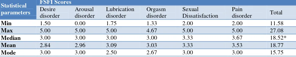
The depiction of sexual dysfunction among respondents based on the FSFI scores was presented in Table 2. Referring to the FSFI scores tabulated in the table, important aspects that can be described are as follows. Types and levels of sexual disorders experienced by study participants is very diverse, ranging from the most severe (Min=11.58) to normal (Max=27.08). However, of
the 80 respondent’s only one participant who is actually
normal, that is the respondent with the maximum score. The remaining 79 respondents included in the category of suffering from sexual disorders (Mean=18.77).
As suggested by Wiegel et al the optimal cut-off score for differentiating women with and without sexual dysfunction is 26.55.17Women with a total score ≥26.55 categorized as normal, and those with a total score <26.55 categorized as sexual dysfunction. Considering that this study sample consisted of women who are in perimenopausal age, and it is evident from Table 2 that only one of 80 participants without sexual dysfunction, using the Wiegel's cut-off score (26.55) is not relevant. Instead, the median score of total FSFI (Med=18.52) was used as the cut-off score.
By using the median value (18.52) as the cut-off score, respondents of the study can be divided into two categories: perimenopausal women with high FSD and preimenopausal women with low FSD.The prevalence of FSD in participants belong to high FSD and low FSD is shown in Table 3.
Table 1: Socio-demographic characteristics of the respondents.
Variables Respondent (n=80)
n %
Age at Menarche
< 15 years 44 55
≥ 15 years 36 45
Workload
Unemployed 44 55
Employed 36 45
Parity
≤ 4 children 61 76.2
> 4 children 19 23.8
Child-bearing Age
>40 years 15 18.8
≤40 years 65 81.2
Use Contraceptives
Yes 56 70
No 24 30
Alcohol Consumption
Yes 0 0
No 80 100
Smoking
Yes 0 0
No 80 100
Medical History
Yes 0 0
No 80 100
Table 2: Description of respondent sexual dysfunction based on the total FSFI scores and its domains.
Statistical parameters
FSFI Scores
Desire disorder
Arousal disorder
Lubrication disorder
Orgasm disorder
Sexual Dissatisfaction
Pain
disorder Total
Min 1.50 0.00 1.75 1.33 2.00 2.00 11.58
Max 5.00 5.00 5.00 4.67 5.00 5.00 27.08
Median 3.00 3.00 3.00 3.00 3.33 3.67 18.52*
Mean 2.84 2.96 3.09 3.03 3.33 3.53 18.77
Mode 3.00 3.00 2.50 2.67 3.00 3.00 15.75
Table 3: Prevalence of FSD in respondent categorized as high and low FSD using median (18.52) as the cut-off score.
FSD categories FSFI scores Prevalence (n=80)
n %
High FSD ≤18.52 41 51,2
Low FSD >18.52 39 48,8
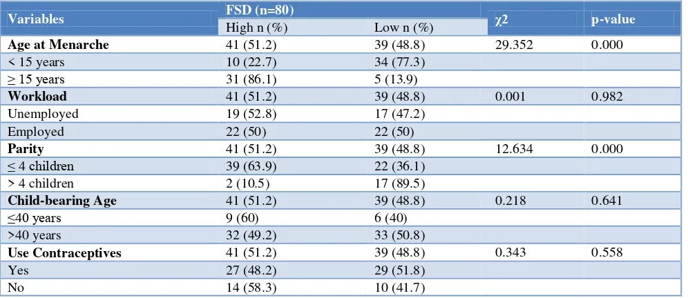
Table 4: Prevalence of FSD by socio-demographic characteristics
Variables FSD (n=80) χ2 p-value
High n (%) Low n (%)
Age at Menarche 41 (51.2) 39 (48.8) 29.352 0.000
< 15 years 10 (22.7) 34 (77.3)
≥ 15 years 31 (86.1) 5 (13.9)
Workload 41 (51.2) 39 (48.8) 0.001 0.982
Unemployed 19 (52.8) 17 (47.2)
Employed 22 (50) 22 (50)
Parity 41 (51.2) 39 (48.8) 12.634 0.000
≤ 4 children 39 (63.9) 22 (36.1)
> 4 children 2 (10.5) 17 (89.5)
Child-bearing Age 41 (51.2) 39 (48.8) 0.218 0.641
≤40 years 9 (60) 6 (40)
>40 years 32 (49.2) 33 (50.8)
Use Contraceptives 41 (51.2) 39 (48.8) 0.343 0.558
Yes 27 (48.2) 29 (51.8)
No 14 (58.3) 10 (41.7)
Table 5: Logistic regression for FSD by selected variables from bivariate analysis.
Variable β S.E. p value Adjusted OR 95% CI for Adjusted OR
Lower Upper
Age at menarche
3.270 0.681 0.000 26.309 6.924 99.969 < 15 years
≥ 15 years Parity
-2.925 0.922 0.002 0.054 0.009 0.327
≤ 4 children
> 4 children
Association of FSD with socio-demographic
characteristics
Among the eight demographic variables tabulated in Table 1, three of them (alcohol consumption, smoking and medical history) were excluded from further analysis because all respondents (100%) showed the same characteristics. The Chi-square test was then used to evaluate the association between the five remaining variables (age at menarche, workload, parity, child-bearing age, and contraceptive) and prevalence of FSD. The results of bivariate (Chi-square test) analysis are presented in Table 4.
Based on the results of Chi-suare test it was found that only two variables of socio-demographic characteristics showing a significant association with the prevalence of FSD, that is age at menarche and parity. The results of
logistic regression analysis for FSD by selected variables from bivariate analysis (age at menarche and parity) are presented in Table 5.
DISCUSSION
reported that predictors of pre-diabetes are male, old-age, high socio-economic status, low education level, hypertension, obesity, central obesity, and smoking.20 Mihardja et al, in contrast, suggested that diabetes mellitus affected more women than men which increased with age and body mass index.21 The occurence of the diabetes was highly correlated with hypertension. Whereas, Widjaja et al, reported that within sex groups, the prevalence of prehypertension was higher in females.22
Regarding the FSD situation of the respondents, except for only one of the 80 participants, 79 of the rest are suffered from sexual disorders (Table 2). These findings actually confirmed the previous assumption that the women who are in perimenopause age generally decreased sexual function.23-25 There are many circumstances that cause perimenopausal women experience sexual dysfunction including physiological changes and the decreased physical activity. Among the physiological changes that are often associated with changes in sexual function in perimenopausal women is hormonal changes.26 The relationship between the decline in sexual function as a result of reduced physical activity in perimenopausal women has also been studied. In women whose average age of 49.8 years, Cabral et al found that physically active women obtained higher score in all FSFI domains (desire, arousal, lubrication, orgasm, satisfaction and pain) and total FSFI score (20.9), indicating better sexual function than their moderately active (18.8) and sedentary (15.6) counterparts.27 The next important finding of this study is respondents who menstruate at the age <15 years tend to show quality of sexual function better than those who menstruate at
age ≥15 years. Seemingly, this phenomenon related to the main estrogenic hormone conditions, 17-β-estradiol. As reported by Emaus et al, in perimenopausal women, early age at menarche result in high levels of 17-β-estradiol throughout the menstrual cycle.28 Estrogenic hormone, as has been widely suggested, is most importanct hormone related to women sexual function in all age levels. In peri-and postmenopausal women, estrogenic hormones therapy proved to alleviate climacteric symptoms and improve well-being.29,30 Estrogen therapy, even proven to improve sexual function in postmenopausal women with vulvovaginal atrophy.31
The last, as indicated in Table 4, respondents with parity
≥4 tend to have better sexual function at their
perimenopause age. This phenomenon, although it still needs to be tested through more in-depth study, can also be associated with the female hormonal circumstances. Women who gave birth reported to have somewhat higher average age of menopause occurrence in comparison to women who did not give birth.32 It seems give-birth associated with the function of ovary. As indicated by Barret et al that the ovarian function between nulliparous and parous women was clearly
different.33 Parity has positive effect on bone mineral density (BMD) in postmenopausal women, whereas the mechanism of bone formation formation itself is maintaining at the cellular level by estrogen.34
In summary, early menarche and high parity contributed to better sexual-quality of life in perimenopausal women. It seems that the low age at menarche and high parity depict the high activity of ovary so that the estrogenic hormones can be maintained.
Funding: This study was funded by Midwifery Academy of Patriot Bangsa Husada, Lampung Tengah, Lampung, Indonesia
Conflict of interest: None declared
Ethical approval: Informed consent used in the study was approved by Ethics Committee for Medical and Health depressive disorders, and hormonal variability. Rev Paul Med. 2001;119(2):78-83.
2. Graziottin A, Leiblum SR. Biological and Psychosocial Pathophysiology of Female Sexual Dysfunction During the Menopausal Transition. J Sex Med. 2005;2(suppl 3):133–45.
3. Genazzani AR, Gambacciani M, Simoncini T. Menopause and aging, quality of life and Sexuality. Position Statement of International Menopause Society Expert Workshop, 1–4 December 2006, Pisa, Italy. Climacteric. 2007;10:88–96.
4. Soules MR, Sherman S, Parrott E, Rebar R, Santoro N, Utian W, et al. Executive summary: Stages of Reproductive Aging Workshop (STRAW). Climacteric: J Int Menopause Society. 2001;4(4):267-72.
5. Ishak IH, Low WY, Othman S. Prevalence, Risk Factors, and Predictors of Female Sexual Dysfunction in a Primary Care Setting: A Survey Finding. J Sexual Med,. 2010;7(9):3080–7.
6. Jaafarpour M, Khani A, Khajavikhan J, Suhrabi Z. Female Sexual Dysfunction: Prevalence and Risk Factors. J Clin Diagn Res. 2013;7(12):2877–80. 7. Aslan E, Fynes M. Female sexual dysfunction. Int
Urogynecol J. 2008;19(2):293–305.
8. Palacios S, Castaño R, Graziottin A. Epidemiology of female sexual dysfunction. Maturitas. 2009;63(2):119-23.
9. Esposito K, Maiorino MI, Bellastella G, Giugliano F, Romano M, Giugliano D. Determinants of female sexual dysfunction in type 2 diabetes. Int J Impotence Res. 2010;22:179–84.
with and without Metabolic Syndrome. J Sexual Med. 2012;9(2):434–41.
11. Kothiyal P and Sharma M. 2013. Post-menopausal quality of life and associated factors- Areview. J Scientific Innovative Res. 2013;2(4):814-23. 12. García-Portilla MP. Depression and perimenopause:
a review. Actas Esp Psiquiatr. 2009;37(4):213-21. 13. Pathak RK, Parashar P. Age at Menopause and
Associated Bio-Social Factors of Health in Punjabi Women. Open Anthropol J. 2010;3:172-80.
14. Brown PE. The Age at Menarche. Brit J Prev Soc Med. 1966;20:9-14.
15. Leal I, Lourenço S, Oliveira RV, Carvalheira A, Maroco J. The impact of childbirth on female sexuality. Psychol Community Health. 2012;1(1):127-39.
16. Lemeshow S, Hosmer Jr DW, Klar J, Lwanga SK. Adequacy of Sample Sizein Health Studies. World Health Organization and John Wiley & Sons, Chichester, England. 1991: 239.
17. Wiegel, M, Meston C, Rosen R. The Female Sexual Function Index (FSFI): Cross-Validation and Development of Clinical Cutoff Scores. J Sex Marital Therapy. 2005;31:1-20.
18. WHO, 2005. Alcohol, Gender and Drinking Problems: Perspectives from Low and Middle Income Countries. Available at: http://www.who.int/substance_abuse/publications/ alcohol gender_drinking_problems.pdf Accessed on 3 March 2017.
19. Reimondos A, Utomo ID, McDonald P, Hull T, Suparno H, Utomo A. Smoking and Young Adults in Indonesia. The 2010 Greater Jakarta Transition to Adulthood Survey. Available at: http://www.who.int/fctc/signatories_parties/en/index .html Accessed on 3 March 2017.
20. Soewondo P, Pramono LA. Prevalence, characteristics, and predictors of pre-diabetes in Indonesia. Med J Indones. 2011;20:283-94.
21. Mihardja L, Soetrisno U, Soegondo S. Prevalence and clinical profile of diabetes mellitus in productive aged urban Indonesians. J Diabetes Invest. 2014;5:507–12.
22. Widjaja FF, Santoso LA, Barus NRV, Pradana GA, Estetika C. Prehypertension and hypertension among young Indonesian adults at a primary health care in a rural area. Med J Indones. 2013;22:39-45. 23. Gonzalez M, Viafara G, Caba F, Molina T, Ortiz C.
Libido and orgasm in middle-aged woman. Maturitas. 2006;53(1):1-10.
24. Kingsberg SA. Hypoactive Sexual Desire Disorder: Understanding the Impact on Midlife Women. Female Patient. 2011;36:1-4.
25. McCool ME, Zuelke A, Theurich MA, Knuettel H, Ricci C, Apfelbacher C. Prevalence of Female Sexual Dysfunction Among Premenopausal Women: A Systematic Review and Meta-Analysis of Observational Studies. Sex Med Rev. 2016;4(3):197–212.
26. Mattar CN, Chong YS, Su LL, Agarwal AA, Wong PC, Choolani M. Care of Women in Menopause: Sexual Function, Dysfunction and Therapeutic Modalities. Ann Acad Med Singapore. 2008;37:215-23.
27. Cabral PUL, Canário ACG, Spyrides MHC, Uchôa SAC, Júnior JE, Giraldo PC, et al. Physical activity and sexual function in middle-aged women. Rev Assoc Med Bras. 2014;60(1):47-52.
28. Emaus A, Espetvedt S, Veierød MB, Barbash RB, Furberg AS, Ellison PT, et al. 17-β-estradiol inrelation to age at menarche at menarche and adult obesity inpremenopausal women. Human Reproduction. 2007;23(4):919-27.
29. Nichols KC, Schenkel L, Benson H. 17 beta-estradiol for postmenopausal estrogen replacement therapy. Obstet Gynecol Surv. 1984;39(4):230-45. 30. Davis SR, Guay AT, Shifren JL, Mazer NA.
Endocrine Aspects of Female Sexual Dysfunction. J Sexual Med. 2004;1(1):82–6.
31. Santoro N, Worsley R, Miller KK, Parish SJ, Davis SR. Role of Estrogens and Estrogen-Like Compounds in Female Sexual Function and Dysfunction. J Sexual Med. 2016;13(3):305–16. 32. Rizvanovic M, Balicc D, Begic Z, Babovic A,
Bogadanovic G, Kameric L. Parity and Menarche as Risk Factors of Time of Menopause Occurrence. Med Arh. 2013;67(5):336-8.
33. Barrett ES, Parlett LE, Windham GC, Swan SH. Differences in ovarian hormones in relation to parity and time since last birth. Fertil Steril. 2014;101(6):1773–80.
34. Bayray A, Enquselassie F. The Effect of Parity on Bone Mineral Density in Postmenopausal Women:A Systematic Review. J Osteopor Phys Act. 2013;1:2. 35. Okman-Kilic T. Estrogen Deficiency and
Osteoporosis. In: Dionyssiotis Y, editors. Advances in Osteoporosis. Publisher: In Tech, Chapters published; 2015: 174.