Volume 2013, Article ID 690918,6pages http://dx.doi.org/10.1155/2013/690918
Research Article
The Effect of Ag Content of the Chitosan-Silver Nanoparticle
Composite Material on the Structure and Antibacterial Activity
Solmaz Akmaz,
1Esra Dilaver Ad
J
güzel,
1Muzaffer Yasar,
1and Oray Erguven
21Department of Chemical Engineering, Istanbul University, Avcilar, 34320 Istanbul, Turkey
2Department of Medical Microbiology, Cerrahpasa Medical Faculty, Istanbul University, Cerrahpasa, 34098 Istanbul, Turkey Correspondence should be addressed to Solmaz Akmaz; [email protected] and Muzaffer Yasar; [email protected]
Received 11 September 2013; Accepted 26 November 2013
Academic Editor: Jie Dai
Copyright © 2013 Solmaz Akmaz et al. This is an open access article distributed under the Creative Commons Attribution License, which permits unrestricted use, distribution, and reproduction in any medium, provided the original work is properly cited.
The aim of this study is to investigate the antibacterial properties and characterization of chitosan-silver nanoparticle composite
materials. Chitosan-silver nanoparticle composite material was synthesized by adding AgNO3and NaOH solutions to chitosan
solution at 95∘C. Different concentrations (0,02 M, 0,04 M, and 0,06 M) of AgNO3 were used for synthesis. Chitosan-silver
nanoparticle composite materials were characterized by Transmission electron microscopy (TEM), X-ray diffraction (XRD),
ultraviolet (UV) spectrophotometer, and Fourier transform infrared (FTIR) spectrometer techniques.Escherichia coli,Acinetobacter
baumannii,Staphylococcus aureus,Enterococcus faecalis,Pseudomonas aeruginosa, andStreptococcus pneumoniaewere used to test the bactericidal efficiency of synthesized chitosan-Ag nanoparticle composite materials. The biological activity was determined by the minimum bacterial concentration (MBC) of the materials. Antibacterial effect of chitosan-silver nanoparticle materials was increased by increasing Ag amount of the composite materials. The presence of small amount of metal nanoparticles in the composite was enough to significantly enhance antibacterial activity as compared with pure chitosan.
1. Introduction
Chitosan obtained from a natural polymer chitin has antibac-terial feature. As a polycationic polymer, chitosan is an envi-ronmental friendly material because of its biodegradability. Nontoxic and antibacterial features of chitosan make it usable for many areas related to human health [1–5]. Due to the functional groups as NH2and OH in the chitosan structure, chitosan is used as an excellent chelating agent [6].
Silver (Ag) ion has been used for a long time as antibac-terial agent due to its strong inhibiting effect on bacteria. Recently, nanoparticle Ag has taken considerable attention to provide maximum bactericidal effect with minimum amount of Ag [7–10]. Generally chemical reducing agents such as NaBH4, ascorbic acid are used for preparation of Ag nanopar-ticle [7–11].
Chitosan is used as metal nanoparticle-chitosan mate-rial in biomedical applications because of its advantages of biodegradability, antibacterial properties, and excellent chelating agent. Both of Ag and chitosan are antibacterial
agents so chitosan-Ag nanoparticle composite material has more antibacterial effect.
Chattopadhyay developed a mild method for synthesizing a chitosan-Ag nanoparticle material in the medium of aqueous sodium hydroxide [14]. They studied the catalytic activity of chitosan-Ag nanoparticles photometrically.
Transmission electron microscopy (TEM), X-ray pow-der diffraction (XRD), UV spectrophotometer, and Fourier transform infrared (FTIR) spectrophotometer techniques were used for characterization of the size and structure of chitosan-Ag nanoparticles [12–16]. The antibacterial effec-tiveness was determined by measuring the minimum bac-terial concentration (MBC) against usuallyEscherichia coli [13,16].
In this study, chitosan-Ag nanoparticle composite was synthesized by mild method in the aqueous sodium hydrox-ide [13,14]. Both of the structural characterization and anti-bacterial effectiveness of chitosan-Ag nanoparticles against three Gram-positive and three Gram-negative bacteria were investigated. The effect of Ag concentration of the composite material on the structure and antibacterial activity was also investigated.
2. Materials and Methods
2.1. Materials. Chitosan (>75 deacetylated), silver nitrate, and glacial acetic acid were purchased from Sigma Aldrich chemical Co. Ltd. Sodium hydroxide was obtained from Merck.
Escherichia coli(E. coliATCC 25922),Acinetobacter bau-mannii(A. baumanniiATCC 19606),Staphylococcus aureus (S. aureus ATCC 25923), Enterococcus faecalis (E. faecalis ATCC 29212),Pseudomonas aeruginosa(P. aeruginosaATCC 27853), andStreptococcus pneumoniae(S. pneumoniaeATCC 49619) were supplied by Microbiologics. Mueller Hinton Broth (Merck microbiology), Chrome agar (Himedia), and Blood agar (%5, Biopen) were used for MBC measurements. Millipore water purification system was used for providing deionized water.
2.2. Preparation of Chitosan-Ag Nanoparticle Composite Mate-rials. Approximately 100 mg of chitosan was placed in a beaker with 50 mL water at 95∘C. Chitosan-Ag nanoparticles were prepared by mixing freshly prepared 1 mL 0.02 M AgNO3 and then 100𝜇L 0.3 M NaOH with homogenizer (18000 rpm, Art Miccra D-1). The color of mixture turned yellow in about one minute after addition of NaOH solution due to the formation of Ag nanoparticles. Then the mixture was stirred for 10 min. Finally, the resulting suspension was filtered and the reddish yellow material was washed until neu-tral with distilled water. Chitosan-Ag nanoparticle material was dried for structural characterization and bacterial tests [13,14].
In order to investigate the effect of Ag content on the antibacterial activity, chitosan-Ag nanoparticle materials with different amount of Ag metal were synthesized by using 0.04 M and 0.06 M AgNO3solution by keeping other conditions unchanged.
The effect of temperature on the formation of Ag nanoparticle was also investigated at 50∘C and 75∘C.
2.3. Characterization of Chitosan-Ag Nanoparticle Compos-ite Materials. Ag nanoparticles of chitosan-Ag nanoparticle composite materials in 0.1% (v/v) acetic acid were investi-gated by HP-8473 UV spectrophotometer. Maximum absorp-tion peak of Ag nanoparticle was observed at 413 nm.
Structure analysis of the samples were carried out by X-ray powder diffraction (XRD) on Rigaku D/Max-2200/PC diffractometer with monochromatic Cu resource (A4 1L-Cu/60 kV, 2.0 kW), the 2-theta ranging from 10∘to 80∘with Cu K𝛼radiation (𝜆= 1.5404 ˚A).
All infrared measurements were performed on a Perkin Elmer Fourier transform infrared (FTIR) spectrophotometer using the attenuated total reflection (ATR) technique and the spectral range is from 650 to 2000 cm−1.
The size of Ag nanoparticles in chitosan-Ag nanopar-ticle materials was determined by transmission electron microscopy (TEM). After dissolving in ethanol with ultra-sonic mixer, samples were dropped onto carbon support film coated with copper TEM grids (200 mesh). Transmission electron microscopy measurements were obtained on JEOL JEM 2100 HRTEM operating at 200 kV (LaB6 filament). Images were taken by Gatan Model 694 Slow Scan CCD Camera.
2.4. Determination of Antibacterial Activity of Chitosan-Ag Nanoparticle Composite Materials. The biological activity was determined by the minimum bacterial concentration (MBC) of the composites. Three Gram-negative bacteria (E. coli(ATCC 25922),A. baumannii(ATCC19606), andP. aeruginosa(ATCC27853)) and three Gram-positive bacteria (S. aureus (ATCC25923), E. faecalis (ATCC29212), and S. pneumoniae (ATCC49619)) were used for measurement of MBC of chitosan-Ag nanoparticle materials. Bacteria except S. pneumoniae were cultured on Chromo agar plates and incubated at 37∘C for 24 hours.S. pneumoniaewas grown on blood agar plate and incubated at 37∘C for 24 hours.
Chitosan-Ag nanoparticle materials synthesized by using 0.02 M, 0.04 M, and 0.06 M AgNO3 solution and chitosan without Ag nanoparticle (pure chitosan) were dissolved in 0.1% (v/v) acetic acid solution.
2.5. Determination of Minimal Bacterial Concentrations (MBC) of Chitosan-Ag Nanoparticle Composite Materials. 1 mL Mueller hinton broth dilution was added to each test tube. Test tubes were plugged by hydrophobic cotton and were sterilized at 121∘C for 20 min.
Each of chitosan-Ag nanoparticle material solutions and pure chitosan solution in 0.1% (v/v) acetic acid was diluted to obtain four different concentrations of the solutions.
Chitosan
300 350 400 450 500
Wavelength (nm)
Figure 1: UV-vis absorption spectra of chitosan-Ag nanoparticle
materials synthesized with 0.02 M, 0.04 M, and 0.06 M AgNO3and
pure chitosan.
inoculated on Chromo agar plates to control the effectiveness of diluted four different concentrations of solutions for each bacterium except S. pneumoniae. The suspensions with S. pneumoniaewere grown on blood agar plate and then all of the plates were incubated at 37∘C for 24 hours. The absence of bacteria on the inoculated agar plates with minimum concentration of solutions indicated minimum bactericidal concentration (MBC).
3. Results and Discussion
3.1. Structural Analysis of Chitosan-Ag Nanoparticle Compos-ite Materials. Ag nanoparticle-containing chitosan compos-ite materials were investigated to understand the interactions between Ag and chitosan and the formation of Ag nanoparti-cles by increasing AgNO3concentration. XRD patterns indi-cated the existence of Ag nanoparticles in chitosan matrix, FTIR spectra provided information of structural change, UV spectra gave Ag concentration in chitosan, and TEM images showed that the size of the Ag nanoparticles well penetrated into chitosan.
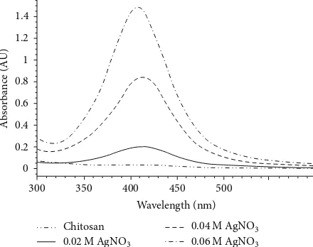
UV-vis absorption spectra of chitosan-Ag nanoparticle materials synthesized by using 0.02 M, 0.04 M, and 0.06 M AgNO3and pure chitosan are shown inFigure 1.
The maximum absorption peak was observed at about 413 nm which is the characteristic absorption peak for Ag nanoparticles. Also UV absorption peak of chitosan-Ag nanoparticles prepared by other researchers was recorded in the range 410–420 nm [8, 13–16]. Figure 1 also displays that the intensity of the absorption peak of Ag nanoparticles increased by increasing the concentration of AgNO3used for preparing chitosan composite material. So this result showed that the amount of Ag nanoparticles of chitosan composite materials increased by increasing AgNO3concentration.
Figure 2illustrates FTIR spectra of chitosan and chitosan-Ag nanoparticle materials synthesized with 0.02 M, 0.04 M, and 0.06 M AgNO3. The broad absorption peak at 3700– 3100 cm−1is the merged characteristic bands for OH and NH2
4000.0 3600 3200 2800 2400 2000 1800 1600 1400 1200 1000
800
Figure 2: FTIR spectra of chitosan-Ag nanoparticle materials
synthesized with 0.02 M, 0.04 M, and 0.06 M AgNO3 and pure
chitosan.
Figure 3: XRD patterns of chitosan-Ag nanoparticle materials
synthesized with 0.02 M, 0.04 M, and 0.06 M AgNO3 and pure
chitosan.
groups [14] and CONH2 absorption band is also observed at near 1657 cm−1 in the FTIR spectrum of chitosan [3]. The shift was observed from 1657 cm−1 in the Ag loaded chitosan spectra. This shift may indicate the binding of Ag nanoparticles to N–H bond of chitosan [3,12,14,16].
XRD patterns of chitosan and Ag-loaded chitosan are shown inFigure 3. XRD pattern of pure chitosan exhibits a strong characteristic peak at about 2𝜃= 20∘for chitosan. XRD patterns of chitosan-Ag nanoparticle materials synthesized with 0.04 M and 0.06 M AgNO3 showed another peak at about 38∘ corresponding to Ag nanoparticle in addition to the chitosan at 20∘[8,11,12]. The peak at 2𝜃= 38∘associated with the crystal face of (111) of Ag [11] became more obvious upon increasing concentration of AgNO3used for preparing chitosan composite material.
Figure 4: TEM image of chitosan-Ag nanoparticle materials
syn-thesized with 0.02 M AgNO3.
Figure 5: TEM image of chitosan-Ag nanoparticle materials
synthe-sized with 0.04 M AgNO3.
matrix with the average diameter of around 3–8 nm. Com-pared with Figures6and4,Figure 5showed that Ag particles synthesized with 0.06 M AgNO3are more densely and more than 50% of the particles are in the range of 5–8 nm in diameter in chitosan matrix.
3.2. Effect of Temperature on the Formation of Chitosan-Ag Nanoparticle Composite Materials. The effect of temperature on the formation Ag nanoparticle was also investigated. The materials were prepared by adding 0.02 M AgNO3 at 50∘C, 75∘C, and 95∘C. The color of material did not turn reddish yellow which indicates the formation of Ag nanoparticle after the addition of NaOH solution at 50∘C.
UV-vis absorption spectra of chitosan-Ag nanoparticle materials synthesized with 0.02 M AgNO3, at 50∘C, 75∘C, and 95∘C are shown inFigure 7.
The Intensity of the absorption peak of Ag nanoparticles at 413 nm increased by increasing temperature keeping other conditions unchanged. This result showed that the formation of Ag nanoparticles of chitosan composite material is related
Figure 6: TEM image of chitosan-Ag nanoparticle materials
synthe-sized with 0.06 M AgNO3.
300 350 400 450 500
Wavelength (nm)
A
b
so
rba
n
ce (A
U)
0.25
0.2 0.15 0.1 0.05 0
95∘C 75∘C
50∘C
Figure 7: UV-vis absorption spectra chitosan-Ag nanoparticle
materials synthesized with 0.02 M AgNO3at 50∘C, 75∘C, and 95∘C.
to the synthetic temperature and the optimum temperature for reduction of Ag ions is about 95∘C.
Table 1: Antibacterial effect of chitosan-Ag nanoparticle composite materials.
Minimum Bactericidal Concentration, MBC (ppm)
Material S. Pneumoniae A. Boumanni E. Coli P. Aeruginosa E. Faecalis S. Aureus
Composite A 100–200 50–100 100–200 50–100 50–100 50–100
Composite B 100–200 50–100 50–100 25–50 50–100 50–100
Composite C 50–100 25–50 50–100 <25 25–50 25–50
Pure chitosan 200–400 >400 >400 >400 200–400 >400
Composite A: Chitosan-Ag nanoparticle material synthesized with 0.02 M AgNO3; Composite B: Chitosan-Ag nanoparticle material synthesized with 0.04 M AgNO3; Composite C: Chitosan-Ag nanoparticle material synthesized with 0.06 M AgNO3.
Figure 8: Antibacterial activity of chitosan-Ag nanoparticle
materi-als synthesized with 0.02 M AgNO3.
Figure 9: Antibacterial activity of chitosan-Ag nanoparticle
mate-rials synthesized with 0.04 M AgNO3.
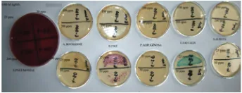
Figure 8 shows antibacterial activity of chitosan-Ag nanoparticle material synthesized with 0.02 M AgNO3. MBC of this chitosan-Ag nanoparticle material is between 100 and 200 ppm against S. pneumoniaeand E. coli, but very little growth appeared on Chromo agar plate with 100 ppm of chitosan-Ag nanoparticle material in suspension towardsE. coli. MBC is between 50 and 100 ppm againstA. baumannii, E. faecalis, P. aeruginosa, andS. aureus.
Figure 9 indicates antibacterial activity of chitosan-Ag nanoparticle material synthesized with 0.04 M AgNO3. MBC of chitosan-Ag nanoparticle material is between 100 and 200 ppm againstS. pneumonia,but a little growth appeared on agar plate with 100 ppm of chitosan-Ag nanoparticle material. MBC is between 50 and 100 ppm against E. coli, A. baumannii, S. aureus,and E. faecalis bacteria, but very little growth appeared on Chromo agar plate with 50 ppm of chitosan-Ag nanoparticle material towardsE. coliandE. faecalis. MBC is between 25 and 50 ppm againstP. aeruginosa. Figure 10 shows antibacterial activity of chitosan-Ag nanoparticle materials synthesized with 0.06 M AgNO3. MBC of chitosan-Ag nanoparticle material is between 50 and 100 ppm againstS. pneumoniaandE. coliand between 25 and 50 ppm against E. faecalis, A. baumannii, and S. aureus.E. coliwas grown a little on Chromo agar plate with 50 ppm of chitosan-Ag nanoparticle material in comparison with 25 ppm of chitosan-Ag nanoparticle material and a little
Figure 10: Antibacterial activity of chitosan-Ag nanoparticle
mate-rials synthesized with 0.06 M AgNO3.
Figure 11: Antibacterial activity of chitosan.
growth appeared on Chromo agar plate with 25 ppm of chi-tosan-Ag nanoparticle materials towardsS. aureus. There was no growth forP. aeruginosaon Chromo agar plate.
Antibacterial effect of chitosan-Ag nanoparticle materials increased with increasing AgNO3concentration.
The antibacterial effect of pure chitosan was also investi-gated and shown inFigure 11. The results showed that MBC is between 200 and 400 ppm of chitosan againstE. faecalisand S. pneumoniaeand MBC is bigger than 400 ppm towardsE. coli, A. baumannii, S. aureus,andP. aeruginosa.
4. Conclusion
Conflict of Interests
None of the authors have any conflict of interests regarding the publication of this paper.
Acknowledgment
This work was supported by Research Fund of the Istanbul University Project no. 5521.
References
[1] E. I. Rabea, M. E.-T. Badawy, C. V. Stevens, G. Smagghe, and W. Steurbaut, “Chitosan as antimicrobial agent: applications and
mode of action,”Biomacromolecules, vol. 4, no. 6, pp. 1457–1465,
2003.
[2] I. M. Helander, E.-L. Nurmiaho-Lassila, R. Ahvenainen, J. Rhoades, and S. Roller, “Chitosan disrupts the barrier prop-erties of the outer membrane of Gram-negative bacteria,”
International Journal of Food Microbiology, vol. 71, no. 2-3, pp. 235–244, 2001.
[3] L. Qi, Z. Xu, X. Jiang, C. Hu, and X. Zou, “Preparation and
antibacterial activity of chitosan nanoparticles,”Carbohydrate
Research, vol. 339, pp. 2693–2700, 2004.
[4] E. Renbutsu, M. Hirose, Y. Omura et al., “Preparation and biocompatibility of novel UV-curable chitosan derivatives,”
Biomacromolecules, vol. 6, no. 5, pp. 2385–2388, 2005. [5] X. Zhang, X. Geng, H. Jiang, J. Li, and J. Huang, “Synthesis
and characteristics of chitin and chitosan with the (2-hydroxy-3-trimethylammonium)propyl functionality, and evaluation of
their antioxidant activity in vitro,”Carbohydrate Polymers, vol.
89, pp. 486–491, 2012.
[6] A. J. Varma, S. V. Deshpande, and J. F. Kennedy, “Metal
com-plexation by chitosan and its derivatives: a review,”
Carbohy-drate Polymers, vol. 55, no. 1, pp. 77–93, 2004.
[7] A. Dror-Ehre, H. Mamane, T. Belenkova, G. Markovich, and A. Adin, “Silver nanoparticle-E. coli colloidal interaction in water
and effect on E. coli survival,”Journal of Colloid and Interface
Science, vol. 339, no. 2, pp. 521–526, 2009.
[8] P. Chen, L. Song, Y. Liu, and Y. Fang, “Synthesis of silver
nano-particles by𝛾-ray irradiation in acetic water solution containing
chitosan,”Radiation Physics and Chemistry, vol. 76, pp. 1165–
1168, 2007.
[9] J. S. Kim, E. Kuk, K. N. Yu et al., “Antimicrobial effects of silver
nanoparticles,” Nanomedicine: Nanotechnology, Biology, and
Medicine, vol. 3, no. 1, pp. 95–101, 2007.
[10] X. L. Cao, C. Cheng, Y. L. Ma, and C. S. Zhao, “Preparation of silver nanoparticles with antimicrobial activities and the
researches of their biocompatibilities,” Journal of Materials
Science, vol. 21, pp. 2861–2868, 2010.
[11] J. P. Ruparelia, A. K. Chatterjee, S. P. Duttagupta, and S. Mukherji, “Strain specificity in antimicrobial activity of silver
and copper nanoparticles,”Acta Biomaterialia, vol. 4, no. 3, pp.
707–716, 2008.
[12] S. W. Ali, S. Rajendran, and M. Joshi, “Synthesis and character-ization of chitosan and silver loaded chitosan nanoparticles for
bioactive polyester,”Carbohydrate Polymers, vol. 83, no. 2, pp.
438–446, 2011.
[13] P. Sanpui, A. Murugadoss, P. V. D. Prasad, S. S. Ghosh, and A. Chattopadhyay, “The antibacterial properties of a novel
chitosan-Ag-nanoparticle composite,”International Journal of
Food Microbiology, vol. 124, no. 2, pp. 142–146, 2008.
[14] A. Murugadoss and A. Chattopadhyay, “A “green” chitosan-silver nanoparticle composite as a heterogeneous as well as
micro-heterogeneous catalyst,”Nanotechnology, vol. 19, no. 1,
Article ID 015603, 2008.
[15] Y.-K. Twu, Y.-W. Chen, and C.-M. Shih, “Preparation of silver
nanoparticles using chitosan suspensions,”Powder Technology,
vol. 185, no. 3, pp. 251–257, 2008.
[16] D. Wei, W. Sun, W. Qian, Y. Ye, and X. Ma, “The synthesis of chitosan-based silver nanoparticles and their antibacterial
activity,”Carbohydrate Research, vol. 344, pp. 2375–2382, 2009.
[17] J.-W. Rhim, S.-I. Hong, H.-M. Park, and P. K. W. Ng, “Prepa-ration and characterization of chitosan-based nanocomposite
films with antimicrobial activity,”Journal of Agricultural and
Food Chemistry, vol. 54, no. 16, pp. 5814–5822, 2006.
[18] R. Tankhiwale and S. K. Bajpai, “Silver-nanoparticle-loaded
chitosan lactate films with fair antibacterial properties,”Journal
of Applied Polymer Science, vol. 115, no. 3, pp. 1894–1900, 2010. [19] X. Chen and H. J. Schluesener, “Nanosilver: a nanoproduct in
medical application,”Toxicology Letters, vol. 176, no. 1, pp. 1–12,
2008.
[20] N. Liu, X.-G. Chen, H.-J. Park et al., “Effect of MW and
concen-tration of chitosan on antibacterial activity ofEscherichia coli,”
Carbohydrate Polymers, vol. 64, pp. 60–65, 2006.
[21] N. P. Panyala, E. M. Pena-Mendez, and J. Havel, “Silver or silver nanoparticles: a hazardous threat to the environment and
human health?”Journal of Applied Biomedicine, vol. 6, no. 3, pp.
117–129, 2008.
[22] R. Das, S. S. Nath, D. Chakdar, G. Gope, and R. Bhattacharjee, “Preparation of silver nanoparticles and their characterization,”
Journal of Nanotechnology, vol. 5, pp. 1–6, 2009.
[23] K.-H. Cho, J.-E. Park, T. Osaka, and S.-G. Park, “The study of antimicrobial activity and preservative effects of nanosilver
ingredient,”Electrochimica Acta, vol. 51, no. 5, pp. 956–960,
2005.
[24] I. Sondi and B. Salopek-Sondi, “Silver nanoparticles as antimi-crobial agent: a case study on E. coli as a model for
Gram-negative bacteria,”Journal of Colloid and Interface Science, vol.