arXiv:physics/0612183v1 [physics.bio-ph] 19 Dec 2006
Internal protein dynamics shifts the distance to the mechanical transition state
Daniel K. West
School of Physics & Astronomy and
School of Biochemistry & Microbiology, University of Leeds, Leeds LS2 9JT, United Kingdom
Emanuele Paci and Peter D. Olmsted∗
School of Physics & Astronomy and
Astbury Centre for Structural Biology, University of Leeds, Leeds LS2 9JT, United Kingdom
Mechanical unfolding of polyproteins by force spectroscopy provides valuable insight into their free energy landscapes. Most phenomenological models of the unfolding process are two-state and/or one dimensional, with the details of the protein and its dynamics often subsumed into a zero-force unfolding rate and a single distancex1D
u to the transition state. We consider the entire phase space
of a model protein under a constant force, and show that the distance x1D
u contains a sizeable
contribution from exploring the full multidimensional energy landscape. Proteins with more degrees of freedom are expected to have larger values for x1D
u . We show that externally attached flexible
linkers also contribute to the measured unfolding characteristics.
Atomic force microscopy or optical tweezers are now routinely used to study the mechanical properties of pro-teins [1, 2]. An important issues is the unfolding behavior of folded domains, including the strength, and the depen-dence on fold topology [3] and secondary structure [3, 4]. The simplest description of unfolding treats the unfolding domain as moving in a one dimensional potential G(x), where the reaction coordinatexis assumed to be directly coupled to the applied force. In perhaps the simplest ap-proximation, the rate of unfolding ku(F) of a two-state
(native and denatured) protein under force can then be calculated using Bell’s formula based on Kramers’ rela-tion for escape from a well [1, 5]:
ku(F)≃ku0exp
F x1D
u
kBT
, (1)
wherekBis Boltzmann’s constant,Tis the absolute
tem-perature,x1D
u is the transition state displacement along
the force projection, and k0
u is the unfolding rate
con-stant at zero force. This result can be used to calculate the distributions of unfolding times (for applied force) or unfolding forces (for applied pulling speed), and has been applied to many different proteins [6, 7, 8, 9, 10]. This simple result has been corrected to use the entire shape of G(x) rather than just the barrier height and displacementx1D
u and incorporate the cantilever
compli-ance [11, 12], to relax the diffusive limit to faster speeds [13], and to allow for multiple pathways or states [14, 15]. There has been extensive work on the utility of a thexu
and the simple 1D picture, linear force dependence, etc [16, 17][More work to do here!!!]
However, the assumption of a one dimensional reac-tion coordinate grossly simplifies physical reality. The unfolding rate depends dramatically on pulling direction [18, 19], and hence on the multidimensional nature of the free energy landscape. Moreover, the one dimensional parameters have no satisfactory physical interpretation:
x1D
u is defined along the pulling direction, while the
ac-tual unfolding takes place along an unknown reaction co-ordinate(s), presumably involving a few key amino acids. Whilex1D
u ≃0.2 nm is the right order of magnitude for
hydrogen bonds, the explicit connection with molecular configurations remains unclear. In this Letter we explore the role of the multidimensional energy landscape in de-termining the effective 1D unfolding parameters. The key physical ingredient is that an applied force perturbs fluc-tuations transverse to the forcing direction, because of the highly anharmonic nature of angular bonds. Specif-ically, force restricts a protein’s conformational search among the dihedral states of the polypeptide backbone. We calculate this using molecular dynamics (MD) sim-ulations of a simple off-lattice G¯o model [20, 21] for a
u
X X
x
U TS
N
Free Energy G
k0 u
2
topologically simple protein, and show that this restric-tion leads to a sizeable contriburestric-tion tox1D
u .
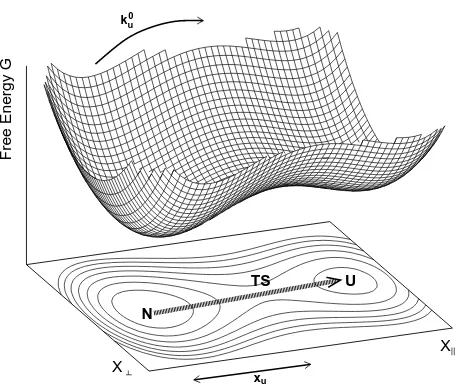
Escape from a multidimensional energy landscape—
The rate of escape from anN-dimensional energy land-scape under an applied forceF (Fig. 1) is given by [22]
ku(F) = Γ(F) exp
where xu is the distance to the transition state, γ is a
friction coefficient, and ∆GT S−N is the height of the
free energy barrier relative to the native basin. G′′RC T S
is the curvature in the unstable direction at the transi-tion state, G′′k
T S are the N −1 stable curvatures at the
transition state, and G′′k
N (F) are theN positive
curva-tures about the native basin under an applied force. If the transition state is sharp i.e., |G′′RC
C | ≫ F/xu, the
curvaturesG′′T S at the transition state, as well asxu, are
approximately independent of force. A more physical representation of the attempt frequency Γ(F) follows by relating a curvature G′′k to the associated fluctuations
byhδx2
T S is the width of the transition state along the
unfolding reaction coordinate,VT Sis the volume of phase
space available for fluctuations at the transition state and
VN(F) is the corresponding volume in the native basin.
In one dimension the weak force dependence of the prefactor Γ(F) can be safely ignored [23]. A weak per-turbation of the native basin volumeVN(F) leads to
ku(F)≃k0uexp the volume of the native basin with force will shiftx1D
u
in the equivalent one dimensional model,
x1Du =xu+λkBT ≡xu+δxu, (6)
whereδxuis adynamical, or entropic, contribution to the
transition state displacement. If the volumes associated with different degrees of freedom randomly increased or decreased with an applied force, there would be little effect. However, we expect the volume of most perturbed degrees of freedom to decrease under an applied force, so that δxu is proportional to the number of perturbed
degrees of freedom. For a force-independent volume we recover Eq. 1, withδxu= 0.
Calculation of phase space volumes—Phase space
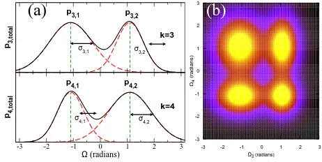
fluc-tuation volumes were calculated from MD simulation tra-jectories. MD was performed for protein L (PDB refer-ence: 1HZ6[24]) using the CαG¯o model of Refs. [20, 25]. The simulation protocol is described in detail in [3]. A real protein does not fluctuate about a single well-defined average structure; because dihedral angles typically ac-cess discrete values, the acac-cessible states are fluctua-tions about many well defined structures, or nodes in phase space. Fig. 2 shows the phase space explored by a tetramer with 2 unimodal and 2 bimodal dihedral distri-butions.
The total unfolding rate ktot
u (F) is the weighted sum
probability of nodeβ. The quantityVeff
N (F) is the
effec-tivephase space volume of the native basin.
Ω3 (radians)
FIG. 2: The phase space of an oligomer with 4 dihedral an-gles: two of these are unimodal (k = 1,2, not shown) and two are bimodal (k = 3,4). (a) The probability distribu-tion funcdistribu-tionpk,total for each bi-modal dihedral angle (black solid line) can be resolved into separate distributionspk,n(red
dashed lines) about well defined averages (green vertical dot-ted lines). There are four possible structures corresponding to fluctuations around{Ω¯3,1, ¯Ω4,1},{Ω¯3,1, ¯Ω4,2},{Ω¯3,2, ¯Ω4,1}and
{Ω¯3,2, ¯Ω4,2}(b) The phase space projected onto{Ω3,Ω4}.
The occupation probabilityPβ(F) of each node is
Pβ(F) =
3
where N is the number of atoms. Each term in the product is the normalised probability that, in node β, a given dihedral angle Ωk participates in itsnth dihedral
state (peak) (Fig. 2a). For large numbers of nodesM a mean field approach, in which all nodes are assumed to be equally populated, works well when states are sufficiently uncorrelated in time, as in this case [26].
The volumeVNβ(F) of fluctuations about each nodeβ
is given by VNβ(F) =
√
detCβ, where C
ijβ = hδriδrjiβ
is the covariance matrix for fluctuations δri in eachCα
position ri. Here the angle brackets denote an average
within nodeβ. We calculateCβ by transforming
coordi-nates to bond lengths, bond angles, and dihedrals angles. We ignore correlations between bond and dihedral angles, which is an excellent approximation here [26]. Hence, the effective volume of phase space is given by
1
Veff
N (F) ≃
1
Vθ(F) M
X
β=1
Pβ(F)
VΩβ(F)
. (10)
whereVθ(F) and VΩβ(F) are the volumes of phase space
explored by the bond and dihedral angles respectively.
50 100 150 200 250 300
10-52 10-51
VN eff (F) (Å
2N-5
)
0 100 200 300 400 500
force (pN) 10-4
100 104 108
average unfolding time (ps)
0 linkers 16 linkers 32 linkers
FIG. 3: (a) Effective phase space volumeVeff
N (F) for protein L
(◦). Dashed line is a fit to lnVeff
N (F)≃lnV
eff
N (0)−δxuF/kBT,
which yields the dynamic contribution to the transition state placementδxu= 0.054±0.008 nm (Eq. 6). (b) Average
un-folding times for protein L using MD atT = 300 K, withnl
attached glycine linkers (◦:nl= 0, x1Du = 0.191±0.004 nm,:
nl= 16, x
1D
u = 0.241±0.004 nm,⋄: nl= 32, x
1D
u = 0.267±
0.004 nm). The linear fits yield logτ = A−x1D
u F/(kBT).
Error bars are of order the symbol size.
Results—Fig. 3a shows the phase space volume as a
function of force calculated from MD simulations of pro-tein L. The dynamic contribution to the transition state placement is δxu = 0.054±0.008 nm. The reduction
of phase space volume comes from (1) the narrowing of the dihedral distributions, and (2) the reduction in the
number of multi-modal dihedral peaks. The latter ef-fect dominates, since the loss of a single dihedral peak immediately removes many nodes of phase space. Sim-ulations of the unfolding of the same protein L domain (Fig. 3b) yield an effective 1D transition displacement
x1D
u = 0.191±0.004 nm, from measuring an exponential
dependence of the unfolding time τu on applied force,
τu ∼ e−F x
1D
u /(kBT), as predicted from a single reaction
coordinate description, Eq. (1). Hence we conclude that the bare transition state position wasxu= 0.137 nm, and
the large shift ofδxu= 0.054±0.008 nm is between 34%
and 45%.
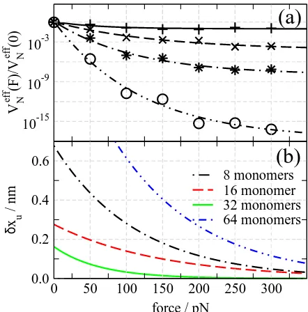
Linker Effects— For convenience, protein domains
are often pulled with long linkers, or unfolded protein strands. The linkers also fluctuate about discrete di-hedral states when stretched. The “lumpiness” of this phase space is irrelevant for weakly stretched strands, but dominates the response for strongly stretched strands. Since force is coupled to the folded domain through the linkers, the total available phase space is the product of protein and linker phase spaces, and the measuredx1Du
depends on the restriction of the linkers’ phase space.
10-15
10-9
10-3
V N
eff
(F)/V
N
eff
(0)
0 50 100 150 200 250 300
force / pN 0.0
0.2 0.4 0.6
δ
x u
/ nm
8 monomers 16 monomer 32 monomers 64 monomers
(a)
(b)
FIG. 4: (a) Volume of phase space calculated using the mean field method and (b) the dynamic contribution to the distance to the transition stateδxu=−kBT ∂[lnVNeff]/∂F, for strands
of flexible glycine linkers. A constant force was applied for a total time of 1µsat T=300K.
To test this, homogeneous linkers strands were con-structed from a dihedral potential based on glycine. Fig. 4 shows the normalised effective phase space volume
Veff
N (F)/VNeff(0) and the correspondingδxu as a function
of force for different number of atomsnlper linker. The
depen-4
dence of the volume of the transition state VT S (Eq. 4).
Although we cannot easily characterize the (unstable) transition state, we can compare the shiftsδxumeasured
directly from the unfolding times (Fig. 3) with predic-tions from the phase space volumes (Fig. 4). The dif-ference δxu(nl = 32)−δxu(nl = 16) ≃ 0.03−0.05 nm
from the calculation of phase space volumes (at forces of order 200−300 pN) agrees with the difference xu(nl=
32)−xu(nl= 16) = 0.026 nm measured from pulling
sim-ulations. This gives us confidence that for this model of protein L the transition state is sharp and its volume does not change appreciably under an applied force.
Discussion— We have shown that an externally
ap-plied force restricts a fluctuating protein’s accessible phase space volume, which increases the transition state displacementx1D
u in the equivalent one dimensional
two-state model. This contribution can be appreciable be-cause of the many degrees of freedom in a protein. Larger proteins have a potentially larger x1D
u , depending on
which degrees of freedom couple to the applied force; this will depend critically on the topology of the fold and the direction in which it is pulled. Most importantly, we predict that x1D
u should be greater for proteins
un-folded through longer attached linker strands. This may have biological significance;e.g. the long unfolded PEVK regions in titin [27] may play help modify the unfold-ing characteristics of titin. Finally, we note that many experiments have unfolded concatamers of multiple do-mains, for convenience of attachment and to generate larger statistics [9, 10, 15, 18, 19, 28, 29, 30, 31]. We sur-mise that in all of these cases the dynamic contribution to x1D
u was significant, and also included a contribution
from already unfolded domains, which act as “linkers” for the last few domains to unfold in a given pull.
Acknowledgements— DKW acknowledges the
Well-come Trust for a PhD studentship. We thank D. J. Brockwell, J. Clarke, T. McLeish, and S. Radford for helpful discussions.
∗ Electronic address: [email protected]
[1] M. Rief, M. Gautel, F. Oesterhelt, J. M. Fernandez, and H. E. Gaub, Science276, 1109 (1997).
[2] M. Rief, J. M. Fernandez, and H. E. Gaub, Phys. Rev. Lett.81, 4764 (1998).
[3] D. K. West, D. J. Brockwell, P. D. Olmsted, S. E. Rad-ford, and E. Paci, Biophys. J.90, 287 (2006).
[4] V. Ortiz, S. O. Nielsen, M. L. Klein, and D. E. Discher, J. Mol. Biol.349, 638 (2005).
[5] G. I. Bell, Science200, 618 (1978).
[6] J. M. Fernandez and H. Li, Science303, 1674 (2004). [7] M. Schlierf, H. Li, and J. M. Fernandez, Proc. Natl. Acad.
Sci. USA101, 7299 (2004).
[8] A. F. Oberhauser, P. K. Hansma, M. Carrion-Vazquez, and J. M. Fernandez, Proc. Natl. Acad. Sci. USA98, 468 (2001).
[9] D. J. Brockwell, G. S. Beddard, E. Paci, D. K. West, P. D. Olmsted, D. A. Smith, and S. E. Radford, Biophys. J.89, 506 (2005).
[10] D. J. Brockwell, G. S. Beddard, J. Clarkson, R. C. Zi-nober, A. W. Blake, J. Trinick, P. D. Olmsted, D. A. Smith, and S. E. Radford, Biophys. J.83, 458 (2002). [11] M. Schlierf and M. Rief, Biophys. J.90, L33 (2006). [12] G. Hummer and A. Szabo, Biophys. J.85, 5 (2003). [13] O. K. Dudko, G. Hummer, and A. Szabo, Phys. Rev.
Lett.96, 108101 (2006).
[14] D. Bartolo, I. Der´enyi, and A. Ajdari, Phys. Rev. E65, 051910 (2002).
[15] P. M. Williams, S. B. Fowler, R. B. Best, J. Toca-Herrera, K. A. Scott, A. Steward, and J. Clarke, Nature422, 446 (2003).
[16] P. C. Li and D. E. Makarov, J. Chem. Phys.121, 4826 (2004).
[17] S. Kirmizialtin, L. Huang, and D. E. Makarov, J Chem Phys122, 234915 (2005).
[18] M. Carrion-Vazquez, H. Li, H. Lu, P. E. Marszalek, A. F. Oberhauser, and J. M. Fernandez, Nature Struct. Biol. 10, 738 (2003).
[19] D. J. Brockwell, E. Paci, R. C. Zinober, G. S. Beddard, P. D. Olmsted, D. A. Smith, R. N. Perham, and S. E. Radford, Nature Struct. Biol.10, 731 (2003).
[20] J. Karanicolas and C. L. Brooks III, Prot. Sci.11, 2351 (2002).
[21] D. K. West, P. D. Olmsted, and E. Paci, J. Chem. Phys. 124, 154909 (2006).
[22] P. Hanggi, P. Talkner, and M. Borkovec, Rev. Mod. Phys. 62, 251 (1990).
[23] E. Evans and K. Ritchie, Biophys. J.72, 1541 (1997). [24] J. W. O’Neill, D. E. Kim, D. Baker, and K. Y. Zhang,
Acta Crystallogr. D Biol. Crystallogr.57, 480 (2001). [25] J. Karanicolas and C. L. Brooks III, J. Mol. Biol.334,
309 (2003).
[26] D. K. West, E. Paci, and P. D. Olmsted, Phys. Rev. E. in preparation, (2006).
[27] W. A. Linke, M. Ivemeyer, P. Mundel, M. R. Stockmeier, and B. Kolmerer, Proc. Natl. Acad. Sci. USA95, 8052 (1998).
[28] M. Rief, J. Pascual, M. Saraste, and H. E. Gaub, J. Mol. Biol.286, 553 (1999).
[29] R. B. Best, B. Li, A. Steward, V. Daggett, and J. Clarke, Biophys. J.81, 2344 (2001).
[30] M. Carrion-Vasquez, A. F. Oberhauser, S. B. Fowler, P. E. Marszalek, S. E. Broedel, J. Clarke, and J. M. Fer-nandez, Proc. Natl. Acad. Sci. USA96, 3694 (1999). [31] R. B. Best, S. Fowler, J. L. Toca-Herrera, A. Steward, E.