How to cite:
Shofiyah F, Muid M, Sujuti H (2017) Differences in the Levels of Interleukin-1β (IL-1β) and Interleukin-1 Receptor Antagonist (IL-1ra) in Children with Status Epilepticus and Febrile Seizure. J. Trop. Life. Science 7 (3): 213 – 217.
*Corresponding author: Fita Shofiyah
Department of Biomedical Sciences, Faculty of Medicine. Brawijaya University
Jalan Veteran, Malang, Indonesia 65145 E-mail: [email protected]
VOL. 7, NO. 3, pp. 213 – 217, September 2017 Submitted January 2016; Revised January 2017; Accepted February 2017
Differences in the Levels of Interleukin-1
β(IL-1
β) and Interleukin-1 Receptor Antagonist (IL-1ra)
in Children with Status Epilepticus and Febrile Seizure
Fita Shofiyah 1*, Masdar Muid 2, Hidayat Sujuti 3
1 Department of Biomedical Sciences, Faculty of Medicine, Brawijaya University, Malang, Indonesia 2 Department of Pediatric, Saiful Anwar Public Hospital, Malang, Indonesia
3 Biochemistry Laboratory, Faculty of Medicine, Brawijaya University, Malang, Indonesia
ABSTRACT
Proinflammatory cytokines are elevated in status epilepticus and febrile seizure and associated with tissue damage. This study aimed to investigate the differences in interleukin 1 beta (IL-1β) and interleukin 1 receptor agonist (IL-1ra) levels in status epilepticus as compared with febrile seizure and febrile. This cross-sectional study was designed to include 45 subjects divided equally into three groups (status epilepticus, febrile seizure, and febrile). Both IL-1β and IL-1ra were measured by using an ELISA method. Results showed that IL-1β levels were significantly higher in the status epilepticus group as compared with the febrile seizure and febrile groups (p < 0.05). IL-1ra levels in the status epilepticus group were significantly lower compared with the febrile seizure group (p = 0.04). Consistently, the IL-1β/IL-1ra ratio in the status epilepticus group was substantially higher as compared with the febrile seizure group (p = 0.01). We concluded that IL-1β and the IL-1β/IL-1ra ratio were significantly higher in status epilepticus. IL-1ra levels were considerably greater in the febrile seizure group
Keywords: Febrile seizure, IL-1β, IL-1ra, status epilepticus
INTRODUCTION
Status epilepticus is defined as a seizure that lasts for more than 30 minutes. Status epilepticus in children is an emergency condition that can be life-threatening. The incidence rate of status epilepticus is 10 – 58 per 100 000 people per year in the US population. The an-nual incidence rate of status epilepticus in children is 20 per 100,000 people in developing countries [1, 2].
A seizure is a clinical manifestation of neurologic dysfunction and is usually found in the emergency room. In the pediatric population, seizures are mostly caused by febrile seizure [3]. A febrile seizure is defined as a seizure associated with fever and age without intra-cranial infection or impairment of the central nervous system [4]. The peak incidence of febrile seizure occurs at 18 months of age (age range 3 months to 5 years) [4, 5]. The incidence of febrile seizure in US and Europe ranges from 2% to 5%, whereas in the Asianpopulation,
it is twice that of the US and Europe (Japan 8.3%–9.9%, India 10.1%, and Guam 14%) [8]. The mortality rate of febrile seizure is 0.64% – 0.75%, and most patients re-cover without neurologic sequelae [4, 6].
Recently, abnormalities in proinflammatory cyto-kine expression were found in patients with seizures [7, 8]. An imbalance of pro- and anti-inflammatory cyto-kines could lead to progressive damage of the brain pa-renchyma and could be clinically related to complica-tions [9]. Both febrile seizure and status epilepticus are distinguished on the basis of clinical manifestation. However, the prognosis in status epilepticus is worse than that in febrile seizure [10].
ated with seizure and febrile conditions [9, 11]. Another research demonstrated that IL-1β and IL-1ra levels were elevated in patients with febrile seizures compared with controls [7]. This study aimed to investigate the differ-ences in IL-1β and IL-1ra levels in status epilepticus as compared with febrile seizure.
MATERIALS AND METHODS
This was a cross-sectional study designed to compare IL-1β and IL-1ra levels in patients with status epilepticus and febrile seizure. This study was conducted in the emergency ward of the Pediatric Department, Dr. Saiful Anwar Hospital Malang, and the Biochemistry Laboratory, Faculty of Medicine, Brawijaya University Malang. This study was approved by the Ethical were as follows: having status epilepticus, febrile seizure,
or febrile and permission from the subject’s parents to
join this study (informed consent). Criteria for inclusion in the status epilepticus group were seizures that lasted for more than 30 minutes or 2 minutes or more seizures with decreased consciousness between seizures, and age > 1 month or < 18 years. Criteria for inclusion in the febrile seizure group were simple or complex febrile seizures. The criterion for inclusion in the febrile patient groups was body temperature ≥ 38.5ºC. Criteria for exclusion from the febrile seizure group were severe clinical conditions and previous chronic disease.
Blood sampling
Blood sampling was performed in the emergency room and pediatric ward of dr. Saiful Anwar Public Hospital Malang. Blood sampling was performed by a well-trained nurse. A 2-mL venous blood sample was mixed with EDTA as an anticoagulant, stored in a cooler box, and maintained at 4ºC. The mixture of venous blood and EDTA was immediately transported to the Biomedical Laboratory, Medical Faculty of Brawijaya University. In the laboratory, specimens were centrifu-ged at 1000 rpm for 30 minutes. Plasma (supernatant) was separated from blood cells (pellet) and stored for a later experiment.
Measurement of IL-1β and IL-1ra
Both IL-1β and IL-1ra levels were measured by using
an ELISA (enzyme-linked immunoassay) method. After all serum samples from 24 patients had been collected, measurement of IL-1β and IL-1ra was performed. The extracellular antigen of IL-1β and IL-1ra were dissolved in coating buffer and 50 µL was dispensed into an ELISA microplate (1 : 50) and incubated at 4°C overnight. The antigen suspension was washed twice with buffer solution (PBS-T) for 5 min each and incubated with 1% PBS-T for 45 minutes. After this process, the antigen was washed twice for 5 minutes each. The next step was incubation with anti-IL-1β and anti-IL-1ra primary antibody for 60 minutes. The suspension was removed, and the plate was washed twice with buffer solution for 5 minutes each. The next step was incubation with secondary antibody (anti-mouse) for 60 minutes. The suspension was removed, and the plate was washed twice with phosphate buffer solution for 5 minutes each, followed by addition of streptavidin-horseradish peroxidase (SA-HRP) and then incubated for 60 minutes. The reaction was washed twice with buffer solution for 5 min each. Tetramethylbenzidine (TMB) substrate was added to the microplate and then incubated for 30 minutes. The reaction was stopped by addition of 1 N HCl for 15 minutes. The absorbance was read at a wavelength of 492 nm in an ELISA microplate reader [16].
Statistical analysis
Levels of IL-1β and IL-1ra were analyzed for distribution and homogeneity. Differences of IL-1β and IL-1ra levels in the status epilepticus group, febrile seizure group, and the febrile group was analyzed by using ANOVA (confidence interval 95%). Data were analyzed by using software SPSS (statistical product and service solution) for Windowsversion 16.0.
RESULTS AND DISCUSSION
The normal mature erythrocyte of E. cyanophlyctis
is an oblong-oval shape with a centric nucleus (Figure 1a). In the present study, we observed five majors nu-clear Baseline characteristics
Figure 1. The level of IL-1β (pg/mL) in each group
Figure 2. The level of IL-1ra (ng/mL) in each group
of 15 subjects in the status epilepticus group died, but no one died in the febrile seizure group.
This was a cross-sectional study design including 45 subjects divided equally into three groups: status epilep-ticus, febrile seizure, and febrile groups. In the seizure group, 12 of 30 subjects were male, whereas, in the fe-brile seizure group, 8 of 15 subjects were female. A pre-vious study showed similar characteristics [7]. The aver-age aver-age in this study was 29.4 months. In the febrile seizure group, the average age of subjects was 24.2 months. These data were in accordance with a previous study [7]. Febrile seizures often occur in children aged 17 – 23 months. In early life, neuronal cells in the brain tissue are in a developmental window; therefore, the fe-brile condition as a traumatic insult could provoke a sei-zure [7, 10].
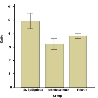
Figure 3. The level of IL-1β/ IL-1ra (ng/mL) in each group
Levels of IL-1β and IL-1ra
Results showed that IL-1β levels were significantly higher in the status epilepticus group than in the febrile seizure and febrile groups (one-way ANOVA, p < 0.05) (Figure 1). IL-1ra levels in the status epilepticus group were significantly lower than in the febrile seizure group (one-way ANOVA, p < 0.05) (Figure 2). Consistently, the IL-1β/IL-1ra ratio in the status epilepticus group was significantly higher than in the febrile seizure group (one-way ANOVA, p < 0.05) (Figure 3).
Zhang and colleagues also showed that HFMD cases with clinical meningoencephalitis caused by viral infec-tion (enterovirus and coxsackie virus) showed higher levels of IL-1β than those without meningoencephalitis [13]. A previous study demonstrated that bacterial me-ningoencephalitis (meningococcus or pneumococcus) caused an elevation of IL-1β [8]. IL-1β might be associ-ated with tissue damage, which is the underlying cause of morbidity and mortality in meningoencephalitis [14]. This study also showed that IL-1ra levels were ele-vated in all groups (reference value 0.2 ng/mL). How-ever, IL-1ra levels in the febrile seizure group were sig-nificantly higher than in the status epilepticus group. Other types of seizure, such as epileptic seizure, have shown an elevation of IL-1ra levels [8]. However, Virta and colleagues showed different results regarding IL-1ra levels. They demonstrated that both IL-1ra levels and the IL-1ra/IL-1β ratio were significantly higher in the febrile seizure group than in the control group [15]. Theoreti-cally, elevated IL-1ra in febrile seizure might be caused by compensatory mechanisms of body systems that act to stop IL-1β action and thus prevent prolonged and re-current seizure. Scheld and colleagues showed elevated IL-1ra levels in the meningoencephalitis group [14]. Griffiths and colleagues also demonstrated that IL-1ra levels were lower in HFMD with viral encephalitis (en-terovirus) as compared with HFMD without encephali-tis [11]. Later data indicated that IL-1ra might have the beneficial effect of decreasing the severity of disease as-sociated with overexpression of IL-1β [11]. Consistent with previous results, the IL-1β/IL-1ra ratio was signifi-cantly higher in the status epilepticus group than in the febrile seizure group. An elevated IL-1β/IL-1ra ratio was also shown in other types of seizure, such as epileptic seizure [8], febrile seizure [7], and viral meningoenceph-alitis [11].
The IL-1β/IL-1ra ratio in the status epilepticus group was significantly higher than in the febrile seizure and febrile groups. The lowest IL-1β/IL-1ra ratio was shown in the febrile seizure group. These data were sim-ilar to a previous study by Virta, which demonstrated that the IL-1β/ILra ratio was decreased in a febrile sei-zure [15]. Another also demonstrated that the IL-1β/ILra ratio was decreased in the febrile group com-pared with the febrile seizure group [7].
CONCLUSION
We concluded that IL-1β and the IL-1β/IL-1ra ratio were significantly higher in the status epilepticus group compared with the febrile seizure and febrile groups.
IL-1ra levels were substantially greater in the febrile seizure group compared with the status epilepticus group.
ACKNOWLEDGMENT
We would like to thank the Department of Child Health, Faculty of Medicine, University of Brawijaya/dr. Saiful Anwar general Hospital, Malang, Indonesia for providing the grant to accomplish this research. We also thank Prof. ER. Dr. Kusworini, Sp.PK (K); dr. Satrio, Sp.A (K) for her excellent guidance to accomplish this research. We also thank Mrs. Fitri from Biochemistry Laboratory and Mrs. Hajeng from Clinical Pathology Laboratory for her excellent assistance in ELISA and blood sampling.
REFERENCES
1. Rijkers K, Majoie HJ, Hoogland G et al. (2009) The role of interleukin-1 in seizure and epilepsy: A critical review. Ex-perimental Neurology 216 (2): 258-271. 258-71. doi: 10.1016/j.expneurol.2008.12.014.
2. Yu N, Di Q, Hu Y et al. (2012) A meta-analysis of pro-in-flammatory cytokines in the plasma of epileptic patients with recent seizure. Neuroscience Letters 514 (1): 110-115. doi: 10.1016/j.neulet.2012.02.070.
3. Riviello JJ, Ashwal S, Hirtz D et al. (2006) Practice parame-ter: diagnostic assessment of the child with status epilepticus (an evidence-based review): report of the Quality Standards Subcommittee of the American Academy of Neurology and the Practice Committee of the Child Neurology Society. Neurology 67 (9): 1542-1550.
4. Pusponegoro HD (2004) Kejang demam patofisiologi dan penatalaksanaannya. In: Kustiowati E, ed. Kumpulan maka-lah pertemuan nasional – I epilepsi. Semarang, Penerbit UNDIP.
5. Johnston MV (2007) Seizures in childhood. In: Behrman RE, Kliegman RM, Jenson HB, eds. Nelson textbook of pediat-rics. 18th ed. WB Saunders Co., Philadelphia, pp 2457-2471. 6. Johnson EA, Kan RK (2010) The acute phase response and soman induced status epilepticus: temporal, regional and cel-lular changes in rat brain cytokine concentrations. Journal of Neuroinflammation 7 (40): 1-9. doi: 10.1186/1742-2094-7-40.
7. Fernandes D, Pereira J, Silvestre J, Bento L (2014) Acute bac-terial meningitis in the intensive care unit and risk factors for clinical outcomes: Retrospective study. Journal of Critical Care 29 (3): 347-350. doi: 10.1016/j.jcrc.2013.12.
9. Choi J, Koh S (2008) Role of brain inflammation in epilep-togenesis. Yonsei Medical Journal 49 (1): 1-18. doi: 10.3349/ymj.2008.49.1.1.
10. Barichello T, Generoso J, Collodel A et al. (2012) Pathophys-iology of acute meningitis caused by Streptococcus pneu-moniae and adjunctive therapy approaches Arquivos de Neuro-Psiquiatria 70 (5): 366-372. 10.1590/S0004-282X2012000500011.
11. Griffiths M, Mong HO, Wong SC et al. (2012) In Enterovi-rus 71 encephalitis with cardio-respiratory compromise, ele-vated Interleukin -1β, interleukin 1 receptor antagonist, and granulocyte colony stimulating factor levels are marker of poor prognosis. The Journal of Infectius Diseases 206 (6): 881-892. doi: 10.1093/infdis/jis446.
12. Dube CM, Vezzani A, Behrens M (2005) Interleukin-1 beta contributes to the generation of experimental febrile sei-zures. Annals of Neurology 57 (1): 152– 155. doi: 10.1002/ana.20358.
13. Zhang S, Xu M, Jun Li, Ding S (2015) Immunologic charac-terization of Cytokine responses to enterovirus 71 and Cox-sackievirus A16 infection in children. Medicine (Baltimore) 94 (27): e1137. doi: 10.1097/MD.0000000000001137. 14. Scheld WM, Koedel U, Nathan B, Pfister HW (2002)
Path-ophysiology of bacterial meningitis: mechanism(s) of neu-ronal injury. The Journal of Infectious Diseases 186 (Suppl 2): S225-S233. doi: 10.1086/344939
15. Virta M, Hurme M, Helminen M (2002) Increased plasma level of pro and antiinflamatory cytokines in patients with febrile seizures. Epilepsia 43 (8): 920-923.