Available online at http://journal.walisongo.ac.id/index.php/jnsmr
Preparation and Microstructure of Ag Doped on ZnO
Sheilla Rully Anggita1, and Heri Sutanto 2
1 Department of Physic Education, Faculty of Science and Technology UIN Walisongo, Indonesia 2 Department of Physic, Faculty of Science and Matematika, Diponegoro University, Indonesia
Abstracts
Corresponding author: [email protected] Recived: 06 November 2016, Revised : 10 December 2016 Accepted: 27 December 2016.
Various concentration of silver (Ag) dopant on ZnO (ZnO:Ag) 2-8 mol% with sol-gel method were deposited on glass substrates that has been heated to a temperature of 250°C for 1h using a thermal spray coating technique and then annealed on 400C for 1h. The obtained samples were characterized by Scanning Electron Microscopy (SEM). The effect of various Ag dopant on ZnO on the microstructure are investigated. The results showed that the microstructure of ZnO:Ag are the surface of ZnO make ganglia phase with Ag grains covered the surface. The addition concentration of Ag promotes the growth of ganglia phase and grain size. The surface of ZnO:Ag mol% the growth grain Ag is more dominant than the growth of ZnO. The dopant Ag with a concentration of 4-8mol% ganglia structure begin to form with the presence of grains. Obtained the surface of ZnO:Ag 4 mol% begins to growth ganglia phase with the largest grain diameter is 304.8 nm. ©2016 JNSMR UIN Walisongo. All rights reserved.
Keywords: Thermal Spray; Coating; ZnO; Ag dopant; Microstructure
1.
Introduction
Zinc Oxide (ZnO) is one of semiconductor that acts as a photocatalyst. The photocatalytic activity of ZnO has been explored and reported. ZnO has the ability to degrade pollutants more effective, economic, and environment-friendly[1]. ZnO is used as a photocatalyst material due to low corrosion, relatively easy synthesis, and has a high photocatalytic activity[2][3]. Several studies with several methods have been performed to enhance the
photocatalytic activity of ZnO. ZnO doped with various transition metal dopants has been investigated. By increasing the concentration of doped transition metal, the energy level is changed which is able to improve the physical and optical properties [4]. In previous research utilizing silver (Ag) doped on ZnO (ZnO:Ag) is able to increase photocatalytic activity[3][4].
Copyright @2016, JNSMR, ISSN: 2460-4453
produced various microstructures. Some of them are microrods [3], nanocrystalline [4][5][6], nanorods [7], grains [8] and ganglia-like phase structure [9]. Ag doped ZnO microstructure photocatalyst with high activity has been intensively researched [1-9]. Meanwhile, the higher surface area is also an important factor for enhancement of photocatalytic perfomance of Ag doped ZnO catalyst [1]. Ganglia phase structure ZnO:Ag were found that Ag grain were agglutinated to the ZnO ganglia phase. Ganglia phase structure with grains of ZnO: Ag becomes more effective than pure ZnO. The addition of Ag on ZnO give good impact to the photocatalytic activity because of the surface of ganglia structure with grains have higher surface area than the surface of pure ZnO [9]. From the method that has been done by some previous researchers in forming the ganglia phase structure of ZnO:Ag, in the present study, various concentration of dopant Ag on ZnO using sol-gel method and deposited with thermal spray-coating technique on their microstructure are investigated. acetate dehydrate was dissolve in 2-propanol and MEA 0,3M was added drop wise to zinc acetate solution, then stirred by a magnetic stirrer at a temperature of 70° C for 30 minute to get ZnO sol-gel [10]. Similarly for synthesis of sol-gel ZnO:Ag 0,3M zinc acetate solution with amount of silver nitrate with concentration range from 2-8 mol% was added droped wise to ZnO solution.
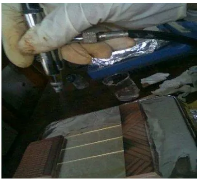
Furthermore to get thin film, sol-gel ZnO and ZnO:Ag were deposited by spray thermal coating (shown at Figure1) at the substrate temperature of 250°C onto glass substrate for 1 hour. Deposition of ZnO and ZnO:Ag film was carried out at atmospheric pressure in a flowing non-sealed system. The post deposition
annealing of ZnO and ZnO:Ag film was annealed at the temperature of 400ºC for 1 hour with the heating and cooling rates of 5 ºC/minute.
Figure 1. Thermal spray coating technique
The synthesized of ZnO and ZnO:Ag microstructure were observed by Scanning Electron Microscopy (SEM). SEM data obtained was used to analyze the surface of ZnO and ZnO:Ag. In-house image analysis technique was used to determined the grain size and it’s gel method has been successfully deposited on a glass substrate measuring 2.54 cm x 7.62 cm x 0.1 cm using thermal spray coating technique. After the coating ZnO: Ag deposited on a glass substrate, and then characterized using Scanning Electron Microscopy (SEM) to determine the morphology of ZnO layer and ZnO: Ag.
more tightly, allowing the substrate color is getting darker.
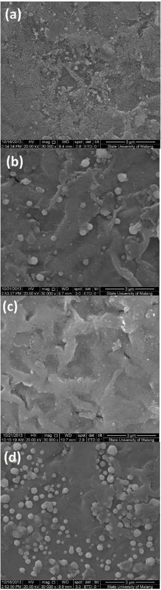
Figure 2. Thin Film of (a) ZnO and ZnO:Ag (b) 2 mol%, (c) 4 mol%, (d) 6 mol% (e) 8 mol%
Fig. 3 (a) shows the SEM image of the sample ZnO thin film with a magnification of 10,000 times. Seen in the picture surface morphology of ZnO thin film with a thermal spray coating method has ganglia-like shape surface [11]. Previous research reports morphology of ZnO was deposited on a glass substrate by using a dip-coating has similar structure such as ganglia [12] as Figure. 3 (b).
The microstructure of Ag doped ZnO as Ag concentration are raised from 2 to 8 mol% are shown in Fig. 4. Microstructure of ZnO: Ag is the ganglia phase with grains agglutinated to the surface. Grain size was more varied due to the influence of the addition of Ag dopant on ZnO. It is estimated ganglia is as ZnO and grains is as Ag.
The grains of Ag have various size for the various concentration. Figure 4 (a) ZnO: Ag 2% shows the growth of Ag grains more dominant than the formation of ganglia phase of ZnO. Thin film of ZnO: Ag 2% have surface with uniform distribution of the the spherical grains about 76,5 nm is observed. Small amount of Ag suggest that no chemical reaction between Ag and ZnO. In previous research the volume of ZnO unit cell decrease as a small amount of Ag is added. The size of Ag+ ion (0.122 nm) is
larger than Zn2+ ions (0.088 nm) and therefore
level of substitution of Zn by Ag is expected to be quite low[6] [8]. In other words, small
amount of Ag mol% no chemical reaction so the growth of Ag more dominant than the growth of ZnO.
(a)
(b)
Figure 3. (a) SEM image of Pure ZnO using thermal spray coating technique[11] and (b) Pure ZnO using dip-coating technique[12]
Fig. 4 (b), (c) and (d) the addition of Ag
with a concentration of mol% ganglia
structure begin to form with the presence of grains. The addition of Ag dopant influence on the formation of ZnO. Increased concentrations of Ag is added the growth of ZnO was increase. The addition of Ag noticeably reduces the scattering of grain sizes [8]. In other words, Ag
concentration mol% the growth of ZnO
Copyright @2016, JNSMR, ISSN: 2460-4453
Figure 4. SEM Image of thin film ZnO:Ag (a) 2 mol%, (b) 4 mol%, (c) 6 mol%, (d) 8 mol%
The addition of Ag dopant on ZnO was also made into variant grain sizes as shown in Table 1. Thin film of ZnO: Ag 4% indicates that most large grain size in comparison with a thin layer of ZnO: Ag else that the average diameter of 304.8 nm. In Figure 4 (a) shows a thin layer of ZnO: Ag 2% have a surface with uniform grain and has a diameter size of the smallest grain that is 76.5 nm.
Table 1. Diameter size of grains ZnO and ZnO:Ag
Sample diameter size (nm)
ZnO -
ZnO:Ag 2% 76.5
ZnO:Ag 4% 304.8
ZnO:Ag 6% 173.4
ZnO:Ag 8% 252.5
4.
Conclusion
Acknowledgment
The authors wish to thank DPPM DIKTI for supporting this work.
References
[1] K. B. Dermenci, B. Ebin, and S. Gürmen, Production of Spherical Ag / ZnO Nanocomposite Particles for
Photocatalytic Applications, pp. –
572, 2012.
[2] X. Yin, W. Que, D. Fei, F. Shen, and Q. Guo,
Ag nanoparticle/ZnO nanorods
nanocomposites derived by a seed-mediated method and their
photocatalytic properties, J. Alloys
Compd., vol. 524, pp. 13–21, 2012.
[3] Z. G. Jia, K. K. Peng, Y. H. Li, and R. S. Zhu,
Preparation and photocatalytic
performance of porous ZnO microrods
loaded with Ag, Trans. Nonferrous Met.
Soc. China (English Ed., vol. 22, no. 4, pp. 873–878, 2012.
[4] R. Chauhan, A. Kumar, R. P. Chaudhary, and T. Education, Synthesis and characterization of silver doped ZnO nanoparticles, Arch. Appl. Sci. Res., vol. 2, no. 5, pp. 378–385, 2010.
[5] C. Karunakaran, V. Rajeswari, and P.
Gomathisankar, Optical, electrical,
photocatalytic, and bactericidal properties of microwave synthesized nanocrystalline Ag-ZnO and ZnO, Solid State Sci., vol. 13, no. 5, pp. 923–928, 2011.
[6] P. Amornpitoksuk, S. Suwanboon, S. Sangkanu, A. Sukhoom, N. Muensit, and J. Baltrusaitis, Synthesis, characterization, structure and electrical properties of ZnO, J. Eur. Ceram. Soc., vol. 27, no. 16, pp. 4521–4527, 2007.
[9] D. Lapisan, T. Zno, A. Dan, S. Rully, and H. Sutanto, Aplikasinya Untuk Degradasi
Polutan, vol. , no. , pp. 85–90, 2014.
[10] D. (. S. Sistesya, S)FAT OPT)S LAP)SAN Zno:Ag YANG DIDEPOSISI DI ATAS SUBSTRAT KACA MENGGUNAKAN METODE CHEMICAL SOLUTION DEPOSITION (CSD) DAN APLIKASINYA PADA DEGRADASI ZAT WARNA
MET(YLENE BLUE, Youngster Phys. J.,
vol. 1, no. 4, 2013.
[11] M. Pusvitasari, Deposisi Lapisan Tipis Zinc Oxide (ZnO) di Atas Substrat Kaca dengan Metode Sol-Gel untuk Aplikasi
Degadrasi Warna, Youngster Phys. J., vol.
1, no. 1, 2012.
![Figure 3. (a) SEM image of Pure ZnO using thermal spray coating technique[11] and (b) Pure ZnO using dip-coating technique[12]](https://thumb-ap.123doks.com/thumbv2/123dok/1896126.1586532/3.612.331.522.129.432/figure-using-thermal-spray-coating-technique-coating-technique.webp)