Corresponding author: [email protected]
Correlation of methylation of toll-like
receptor 4 (TLR4) and interleukin-6 (IL6)
promoter with insulin resistance in obese
adolescents
Ratih Feraritra Danu Atmaja1, Dian Caturini Sulistyonigrum2, Emy Huriyati2, Ahmad Hamim
Sadewa1, Rina Susilowati3*
1Department of Biochemistry, 2Department of Nutrition and Health, 3Department of Histology and Cell Biology, Faculty of Medicine, Universitas Gadjah Mada, Yogyakarta, Indonesia
DOI: http://dx.doi.org/10.19106/JMedSci004801201602
ABSTRACT
Chronic inlammation can lead to insulin resistance in obesity. Increased mRNA expression of inlammatory markers such as toll-like receptor 4 (TLR4) and interleukin 6 (IL6) were reported both in the tissue and the peripheral blood of obese adolescent and correlated with insulin resistance. DNA methylation surrounding TSS region is known to regulate the level of a gene expression. The aim of the study was to investigate the methylation status of TLR4 and IL6 promoter in peripheral blood of obese adolescent and its correlation to insulin resistance. This was a cross sectional study with observational analytic approached. Fifty adolescents with obesity from Yogyakarta Special Region, aged 15-18 years, z-score value >2 SD, no experience of any acute infections within 2 weeks and signed in informed consent, were selected in this study. Subjects with diabetic mellitus, incomplete data and DNA concentration of <20 µg/mL were excluded. DNA samples were extracted from peripheral blood of the subjects. The bisulite converted DNA was analyzed for methylation level with direct sequencing. Correlation analysis using Spearman test was performed with signiicance value was set at p<0.05. No methylation in TLR4 promoter was detected in all subjects. The methylation level of IL6 was transformed to categorical variable. Four (8%) subjects with insulin resistance and 2 (4%) without insulin resistance had methylation on a CpG site. Methylation of IL6 was not statisticaly different between insulin resistance and non-resistance adolescent (p=0.635). The signiicant correlation was not found between methylation level in IL6 promoter with insulin resistance (HOMA-IR) (r=0.051). Cell speciic of methylation mechanism, characteristic of subjects and types of samples were suspected causing those results. In conclusion, no correlation between methylation levels of TLR4 promoter and IL6 with insulin resistance in obese adolescent was observed in this study.
ABSTRAK
Inlamasi kronis dapat menyebabkan resistensi insulin pada obesitas. Peningkatan ekspresi mRNA pada penanda inlamasi seperti toll-like receptor 4 (TLR4) dan interleukin 6 (IL6) dilaporkan terjadi di jaringan dan darah tepi pada remaja obes dan berhubungan dengan resistensi insulin. Metilasi DNA di sekitar regio Transcriptional Start Site (TSS) berperan pada pengaturan ekspresi gen. Penelitian ini bertujuan untuk mengkaji status promoter TLR4 dan IL6 darah tepi remaja obes dan hubungannya dengan resistensi insulin. Rancangan studi potong lintang dengan pendekatan analitik observasional digunakan pada penelitian ini. Penelitian melibatkan 50 remaja obes di Daerah Istimewa Yogyakarta (DIY), berumur 15-18 tahun, mengalami obesitas dengan skor z >2 SD, tidak memiliki riwayat infeksi akut selama 2 minggu terakhir dan menandatangani surat pernyataan persetujuan keikut sertaan dalam penelitian. Kriteria eksklusi adalah subjek dengan diabetes mellitus, data tidak lengkap, dan konsentrasi DNA <20 µg/mL. Sampel DNA diekstraksi dari darah tepi untuk selanjutnya dikonversi dengan bisulit, dan dianalisis untuk mengetahui tingkat metilasi dengan sekuensing DNA secara langsung. Analisis statistik dilakukan menggunakan uji korelasi Spearman dengan nilai kemaknaan p<0,05. Hasil penelitian menunjukkan bahwa metilasi pada promoter TLR4 tidak terdeteksi pada seluruh subjek. Tingkat metilasi IL6 diubah menjadi variabel kategorik. Empat (8%) subjek dengan resitensi insulin dan 2 (4%) subjek sensitif insulin menunjukkan metilasi pada sisi CpG. Metilasi pada IL6 secara statistik tidak menunjukkan adanya perbedaan bermakna antara remaja dengan resistensi insulin dan sensitif resistensi (p=0.635). Tidak ada hubungan antara tingkat metilasi promoter IL6 dengan resistensi insulin (HOMA-IR) (r=0.051). Mekanisme metilasi spesiik sel, karakteristik subjek dan tipe-tipe sampel diduga berpengaruh terhadap hasil yang diperoleh pada penelitian ini. Kesimpulan dari penelitian ini adalah tidak adanya hubungan antara tingkat metilasi promoter TLR4 dan IL6 dengan resistensi insulin pada remaja obes.
INTRODUCTION
Indonesia has been included in top 10 countries with the highest prevalence of obesity in the world.1 The prevalence of obese
adolescent in Indonesia increased from 1.4%
in 2007 to 7.3% in 2013.2 Around 5.7 %
adolescents aged 16-18 years is overweight, while 1.6 % is included in the category
of obese.2 Yogyakarta Special Region is a
province in Indonesia that has the highest prevalence of adolescent obesity in 2010 and as one of the cities where the prevalence of obese adolescent prevalence above the national average in 2013.2,3
Obesity at a young age will give long-term health impacts, for example the tendency of being obese in adulthood, increasing the risk of developing degenerative diseases
such as type 2 diabetes mellitus (T2DM), hypertension, ischemia and stroke, until a decline in life expectancy (life expectance).4-6
Insulin resistance (IR) is one of major factors which cause T2DM manifestation. The level of IR by homeostatic model assessment-insulin resistance (HOMA-IR) is known having linear correlation with body mass index (BMI) and fat deposition percentage in adolescent.7
Inlammation is the most studied pathway
to explain individuals with obesity may develop to insulin resistance. This pathway
includes inlammatory activation through
toll-like receptor 4 (TLR4) and interleukin
6 (IL6). The pro-inlammatory enzymes
of biochemical parameters. The remaining precipitate including buffycoat were stored in 4oC until used. In this study, 50 obese subjects
followed by the data such as fasting glucose blood (mg/dL), insulin level (mg/dL) and HOMA-IR were chosen by random sampling
with sex stratiication from the previous study.
This study was approved by the Medical and Health Research Ethics Committee of the Faculty of Medicine, Universitas Gadjah Mada, Yogyakarta.
Obese adolescent were deined according
to Indonesia Basic Health Research (2013)
and WHO (2007) as those having z-score of > 2. Insulin resistance were deined as HOMA-IR > 3.2.17 Fasting blood glucose ≥ 126 mg/
dl was deined as T2DM.2 Value of
HOMA-IR > 3.2 was deined as insulin resistance. Methylation level of each subject was deined
as an average of methylation level of all CpG in target region based on chromatogram from sequencing results. Methylation level of each CpG was calculated by Lewin’s equations.18
DNA extraction and bisulite treatment
Genomic DNA was extracted by DNA
genomic Promega Wizard® Genomic DNA Puriication Kit. Four hundred nanogram of genomic DNA was used for bisulite treatment
according to the procedure of EZ - methylation
Zymo Gold Kit with a total elution volume of
10 mL and stored at -20°C.
Ampliication of target region
Region target of IL6 at -290 to +10
was ampliied using primer forward
AGTTATTAATAAAAGAAAAA-3’ and reverse 5’- ACAAAATAAACCTCAA-ACATCTC-3’. While primer for TLR4
ampliication were forward 5’-GTTTTTTT-AGTTATTGGTTTGTAGG-3’ and reverse 5’- AAACAAACATCATCCTAACATCATC-3’ which is targeting region at -94 to +202.
Cycling condition for ampliication of IL6
The increase of expressing gene of TLR4 and IL6 has been reported in obese and T2DM.11,12
This increase mainly occur in insulin targeting cells such as adipocytes, hepatocytes, and skeletal muscle cells.13-15 One of mechanisms
which regulate the level of gene expression is DNA methylation. Although the status and
level of methylation is speciic to each type
of cell but the increase of mRNA TLR4 and IL6 has been reported in polymorphnuclear cell of individuals with obesity and positively correlates with insulin resistance.11,16 It has
been suggested that there is DNA methylation regulation which cause such increase. How-ever, there was no report about the status of promoter methylation of IL6 and TLR4 with IR in obese adolescent’s peripheral blood. This study aims to investigate how the level of the status of promoter methylation of IL6 and TLR4 in obese adolescent and its correlation with insulin resistance.
MATERIALS AND METHODS
Subjects
Samples of this study was a part of samples from the study conducted by Huriaty in 2014 (unpublished). In the previous study, 4000 adolescents from 10 senior high schools in Yogyakarta Special Region followed screening tests such as height (cm) and weight (kg) for grouping by nutritional status. Following the exclusion of subjects with the history of T2DM and kidney diseases, as many as 263 adolescents, aged 15-18 years, was not
having any infection in 2 weeks (conirmed
resistance. The data were then grouped based on the status of insulin resistance by gender
stratiication to analyse the mean difference
between groups. were 95oC for 5 minutes, 50 cycle of 95oC
for 1 minute, 55oC for 1 minute dan 72oC for
1 minute 50 and inal extension for 5 minute
at 72oC. Similar cycling condition was used
for TLR4 ampliication except for annealing
temperature was 60oC for 1 minute. The PCR
product was stored at 4oC until being used.
Primer used in this study was designed using Meth Primer.19
Direct sequencing
Thirthy microliter of PCR product and 10 µl sent to 1st base to be sequenced by Sanger
method. Sequencing of IL6 used forward
primer whereas TLR4 used reverse primer.
PCR clean-up and sequencing protocol was done according to daily protocol of 1st base.
Statistical analysis
Data were presented as mean ± standard deviation (SD) or as median (minimum-maximum) depending on the types of data. The mean difference between groups was tested using unpaired t-test. Correlation analysis between methylation of TLR4 and IL6 promoter was performed using Spearman test. The differences between groups were
considered statistically signiicant if a p value
< 0.05.
RESULTS
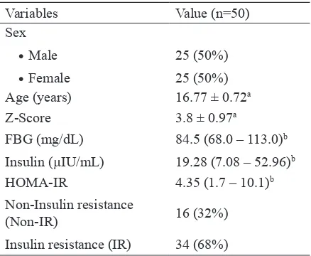
The chracteristics of subject are presented in TABLE 1. Fifty 50 adolescents with obesity consisting of 25 (50%) male and 25 (50%) female from Yogyakarta Special Region were involved in this study. The mean age of subjects was 16.77±0.72 years. Z -score value mean obtained based on the WHO criteria was 3.8±0.97. Median level of fasting blood glucose, insulin and HOMA-IR values, were 84.5 (68.0-13.0), 19.28 (7.08-52.96) and 4.35 (1.7-10.1), respectively. A total of 68 % of the total subjects in this study had insulin
TABLE 1. Subject characteristics
Variables Value (n=50)
Insulin (µIU/mL) 19.28 (7.08 – 52.96)b
HOMA-IR 4.35 (1.7 – 10.1)b
Non-Insulin resistance
(Non-IR) 16 (32%)
Insulin resistance (IR) 34 (68%)
a Presented in mean ± standard deviation (SD) b Presented in median value (minimum-maximum)
Mean difference of age and z-score was
performed using unpaired t-test whereas FBG level, insulin level, and HOMA-IR was performed using Mann Whitney test. The age of subjects was similar across groups. Z-score
values and FBG level was signiicantly
different within female between the insulin resistance and non-insulin resistance groups (p<0.05) but not in boys. Levels of insulin and
HOMA-IR values were signiicantly higher in
insulin resistance group in both female and male (p < 0.001) (TABLE 2).
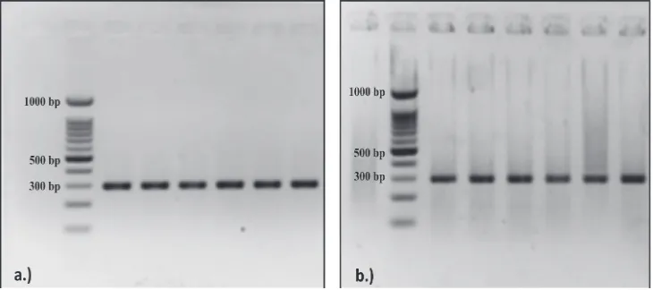
Ampliication of bisulite coverted DNA
FIGURE 1a and 1b show that the
ampliication of bisulite -modiied DNA (Bis
- DNA) of gene promoter target region of IL6 and TLR4 was successfully performed. It was
characterized by an intense single band on
TABLE 2. Mean difference between groups
Category
Female Male
IR (n=18)
Non-IR (n=7)
IR (n=16)
Non-IR (n=9)
Agea 16.78 ± 0.7 16.68 ± 0.7 16.65 ± 0.6 17.02 ± 0.9
Z-scorea 4.2 ± 0.9 3.16 ± 0.6* 3.74 ± 1.0 3.51 ± 1.0
FBGb (mg/dL) 86.5 (73-113)
77* (68-90)
87.5 (74-111)
76 (69-101)
Insulinb (mg/dL) 25.53 (16.85-40.19)
14.94** (9.62-18.41)
22.18 (16.21-52.96)
13.33** (7.08-16.99)
HOMA-IRb 5.15
(3.8 - 9.2)
2.9** (1.7-3.1)
4.9 (3.4-10.1)
2.5** (1.7 - 3.1)
a Presented in mean ± standard deviation (SD) b Presented in median value (minimum-maximum) * Signiicance p<0.05
**Signiicance p<0.001
FIGURE 1. Bisulite-modiied DNA (Bis-DNA) amplicon visualization of the target region
of IL6 and TLR4 gene promoter on 2% agarose gel.
Sequencing was performed to conirm that the region ampliied by the primer was a target
region. The nucleotide sequences obtained from sequencing was aligned to compare with DNA sequences from NCBI using Multalin. The results show that the alignment of the
ampliied region is equal to the target region
with nucleotide similarities 90 %.
Direct Bisulite Sequencing PCR TLR4
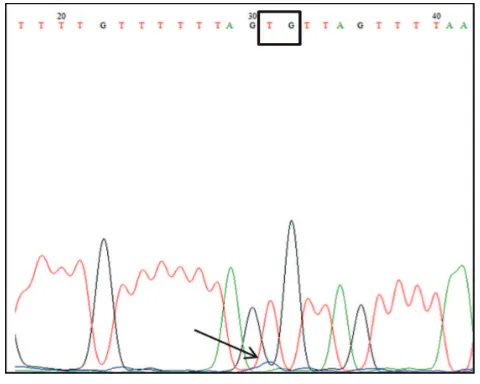
The results of TLR4 promoter region -94 to +202 sequencing showed that the entire nucleotide C of predicted CpG changed into nucleotides T except on CpG1. Cytosine of CpG1 (based on reference sequence) was read as guanine in chromatogram. It indicates that the entire targeted CpG in the target region was unmethylated. The percentage of methylation also could not be calculated because no overlapping found between nucleotides C and T in the predicted CpG.
Targeted IL6 promoters in this study were around -290 to +10 nucleotides of TSS that includes 5 CpGs (FIGURE 3). Several overlapping peak between nucleotide C peak and nucleotide T was found in predicted CpG site of IL6 electrogram. Peak ratio between them was calculated using Lewin’s equation to get methylation level of each CpG.
FIGURE 2. Electrogram of target region of TLR4 gene promoter. Region target arround at -94 to +202 of the TSS that include 7 CpG . There was no
methylation found in all CpG of region target which is characterized by
the entire nucleotide C in CpG turned into a nucleotide T.
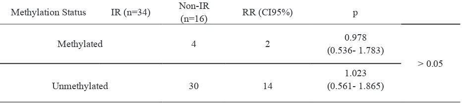
Only 6 from 50 subjects showed methylation in the promoter IL6 (FIGURE 4) . The level of methylation in IL6 target region obtained in the study were very low, ranging from 0 to 12.91%. The analysis was performed
TABLE 3. Fisher test of methylation of IL6 between IR and non-IR group
Methylation Status IR (n=34) Non-IR
(n=16) RR (CI95%) p
Methylated 4 2 0.978
(0.536- 1.783)
> 0.05
Unmethylated 30 14
1.023 (0.561- 1.865)
Among methylated group, 4 (8% ) subjects was insulin resistance while 2 (4%) subjects was non-insulin resistance. Relative risk of methylated group being insulin resistance was lower (RR=0.978) compared with unmethylated group (RR=1.023). However the risk was not statistically different between
groups ( p > 0.05).
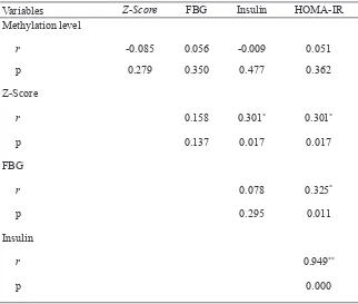
Correlation analysis by Spearman test showed that there was no statistically
using different proportions of methylation level data that has been transformed into a categorical variable. Subjects who showed any methylation minimal at one CpG would
be categorized as methylated group (TABLE
3).
FIGURE 4. Distribution of methylation level. Only 6 out of 50 subjects showed any methylation with low level of methylation.
signiicant correlation found between
methylation level and HOMA-IR neither
with z-score, FBG level and insulin level. Signiicant positive correlation was shown between z-score and insulin level as well as z-score and HOMA-IR but z-score and FBG level was not correlated signiicantly
correlation between FBG level and HOMA -
IR was statistically signiicant. Strong positive
correlation was shown between insulin level
and HOMA-IR. Signiicant correlation was
found between insulin level and HOMA-IR (r=0.949) (TABLE.4).
Variables Z-Score FBG Insulin HOMA-IR
Methylation level
r -0.085 0.056 -0.009 0.051
p 0.279 0.350 0.477 0.362
Z-Score
r 0.158 0.301* 0.301*
p 0.137 0.017 0.017
FBG
r 0.078 0.325*
p 0.295 0.011
Insulin
r 0.949**
p 0.000
TABLE 4. Correlation between variables
DISCUSSION
Our data show that a total of 68 % of the subjects in this study had insulin resistance
(TABLE 1). These data conirm the results
of the previous research that reported that more than a half of the population of children and adolescents with obesity have insulin resistance.20 The median value of insulin levels
in this study was 19.28 μIU/mL with the range
of insulin levels obtained was between 7.08
to 52.96 μIU/mL (TABLE 1). Although the
value limit of normal insulin levels in previous studies showed varying results, insulin levels in the present study were higher than the cut-off range of hyperinsulinemia in adolescents aged 14-19 years which was 18.15 to 23.73
μIU/mL.21 A study by Rodriguez-Moran et
al.,22 used 16 μIU/mL for hyperinsulinemia
cut-off category in adolescents.22 A study
of cardiovascular disease in children and adolescents by the American Heart Association (AHA) established that insulin levels by 15-20
μIU/mL as a borderline high of insulin normal
levels.23
A signiicant difference occurred between
the value of HOMA-IR in the insulin resistance group and the non-insulin resistance both group. High value of HOMA-IR in this study
was probably to be more inluenced by insulin
compared to insulin and HOMA-IR (TABLE 4). Hyperinsulinemia in adolescents has been reported to be a physiological condition which is affected by growth hormone and
an increasing body mass during puberty.24
Insulin secretion will return to normal level by the end of puberty.25 However, the
condition of hyperinsulinemia accompanied by obesity during adolescence may increase the risk of insulin resistance which is known as a major factor related to cardiovascular
disease and type 2 diabetes.23 Low-grade
chronic inlammation and imbalance levels of
adipokines in obesity conditions may interfere insulin signal transduction which decreases glucose uptake by cells. The decrease of glucose uptake by cell results in increasing pancreas secretion of insulin as an effort to increase glucose uptake in order to keep blood glucose at normal level.
The degree of insulin resistance has been reported to be linear to BMI and body fat percentage.7 Insulin resistance in obesity is
associated with metalamation or inlammatory
response that occurs as a result of excessive
nutrients consumption.26 Free fatty acids,
which is reported to be increasing in condition of obesity, are one of inducing factor for
inlammatory response through TLR-4.13,14,27,28
Proinlammatory enzymes such as JNK and IĸKβ which are active in downstream
TLR-4 signaling can lead to impaired insulin signaling through phosphorylation at IRS 1 serine residues.8 Cytokine IL-6, one of the
proinlammatory cytokine which is secreted
through TLR-4 signals, can also cause the IRS as an adapter protein loses its’ function. SOCS3 which is active in IL-6 signal transduction, is able to decrease the phosphorylation of IRS 1 and even increase the ubiquitilation of IRS 1 and subsequently affect the insulin signal transduction.9,10 These cellular mechanisms
not only cause chronic inlammatory
conditions continue to occur, but also interfere insulin signaling, and thus the glucose uptake will be impaired. A decrease in glucose uptake will lead to the increase of insulin secretion induction.
Although all subjects were obese, mean difference test results showed that the
z-score value affects the incidence of insulin
resistance in adolescent girls but not in boys (p <0.05) (TABLE 2). Level of FBG was
also signiicant in girls group only. Insulin
resistance in boy adolescent may occur by hormonal effect on the balance of adipokines. Adiponectin level was reported to decrease in obese condition. During puberty in male adolescents, adiponectin levels decrease with increasing testosterone and negatively
correlate with HOMA-IR.29
Promoter methylation levels of TLR4 and IL6 in this study were detected using
direct-BSP methods and were analyzed
with Lewin’s equation. The present study targetted ±300 bp promoter region of the TSS. Differences of distance between the methylated region with the transcription start site (TSS) order is known to be affecting the expression level. Methylation around TSS such as exon one, and downstream promoter
signiicantly showed lower genes expression
than methylation on the upstream promoter or intragenic region and last exons.30 A study
about pattern distribution and methylation level has shown that gene with highest expression has lower methylation on region
around 500bp from TSS.31 Target region in
this study has transcription factor binding site as predicted by PROMO software.32 Each
transcription factor was reported to have a role in the regulation of the transcription of the gene.
predicted CpG. As far as our knowledge, no studies have reported the methylation level of TLR4 on peripheral blood cells adolescents with obesity and its correlation with insulin resistance. Studies on TLR-4 methylation has been performed in intestinal epithelial cells and gingival tissue. A study by Takahashi et al.,33 using cell intestinal epithelial cell line
reported that methylation at -69 to +277 serve to maintain homeostasis condition of normal
lora in the digestive tract by emphasizing the
expression of TLR-4.33 The different results
between our study and theirs may occur because of the characteristic of methylation
mechanisms itself which are speciic to
each cell.34 Research on TLR4 promoter
methylation at CpG -646 and -822 positions in the gingival tissues showed that almost the whole subjects in the regions were not methylated and were not associated with
TLR-4 mRNA transcripts.35 Because all subjects
were not methylated in the region target of the TLR4 promoter, it can be concluded that there was no correlation between the degree of methylation in the TLR4 target region with insulin resistance in adolescents with obesity.
Methylation of CpG on the promoter region has been known to affect the effectiveness of transcription initiation complex formation. However, it was reported that methylation can also occur in untranslated regions, exons, the non-coding region, and repetitive sequence although its mechanism
to inluence the transcription process is not
widely known.36,37 Therefore it is possible
that epigenetic regulation does not occur in the target region but occurs in other regions. Remely et al.,38 reported that the gene TLR4
exon 1 methylation level of intestinal cells found in individuals with obesity is lower
than the control group.38 Methylation in
exon 1 of a gene affects the transcriptional silencing occurence more than the promoter
methylation in the upstream portion.30
However, the mechanisms of how gene’s body
methylation inluence the transcription process
effectiveness is still unknown. Methylation in gene’s body is suspected to play a role in the splicing process.39
Targeted IL6 promoters in this study are inside -290 to +10 nucleotides of TSS that includes 5 CpGs (FIGURE 3). The level of methylation in IL6 target region obtained in the present study are very low, ranging from 0 to 12.91% and only 6 of the 50 subjects showed methylation in IL6 target region (FIGURE 4) Therefore, the data is transformed into categorical form with minimum requirements 1 of CpGs which shows any methylation is
categorized as methylated. Fisher test results
in TABLE 3 shows that the relative risk of individuals with methylation on IL6 promoter suffering insulin resistance is smaller (RR=0.978) compared with individuals who were methylated (RR=1.023). However, there
is no signiicant difference between the insulin
resistance with no insulin resistance groups (TABLE 3).
There was no statistically signiicant
correlation between the levels of IL6 promoter methylation with other variables such as
z-score, GDP, levels of insulin and
HOMA-IR value. Zhang et al.,40 study showed similar
results which shows no correlation between
IL6 promoter methylation with BMI.40
Different results were reported by Na et al.,41
which signiicantly had more IL6 promoter
hypermethylation in peripheral blood cells of women with obesity than overweight group or normal BMI group. The occuring hypermethylation is suspected as a negative feedback to suppress the IL-6 levels on the condition of obesity.41
Promoter DNA methylation may
inluence the transcription process by either
factors to the binding site or together with
MBD, histone deacetylase enzyme and
histone methyltransferase enzyme causing modiications of histones. The histone modiication can cause chromatin structure
to become more compact and therefore the transcription factor binding sites become inaccessible and the transcription process may also be disrupted. DNA methylation mechanism will lead to repression of transcription thus decreasing the expression of a gene or vice versa.42,43
Methylation around TSS such as exon-1 and downstream promoter (250bp) results in lower gene expression than methylation on upstream promoter, intragenic region or last exon. Nonetheless, Nile et al.,44 showed that
methylation on -1099 signiicantly represses
the expression of mRNA of TLR4.44 This may
occur if the methylated CpG is precisely on
TFBS and affects binding afinity between
transcription factor and TFBS.45
There are several possibilities of why methylation in the TLR4 promoter target region was not found and methylation in the IL6 target region was in low levels in this study. First, every subject in this study was an individual with obesity. Obesity is known as a
condition due to chronic inlammation which is characterized by increased expression
of TLR4 and IL6 not only on the insulin target cells, but also on the peripheral blood cells.11,12,28 Thus, low levels of methylation of
IL6 in the target region or even the absence of methylation at TLR4 CpG target allegedly is indeed an up-regulation form of TLR-4 and IL-6 on the obesity condition through DNA methylation.
Toll-like receptor 4 in circulation is mainly expressed by monocytes which are known to secrete IL-6 mostly through the activation of TLR-4 in circulation on the obesity condition46
However, the composition of monocytes is only about 2-8% of total leukocytes in the
circulation.47 The level of obtained methylation
is also suspected to be very low considering that this study used a sample buffycoat and not
speciic mononuclear cells. Other possibilities
for improvement of IL6 mRNA and TLR4 were widely reported increases in peripheral blood cells of individuals with obesity are not regulated by other DNA methylation but by other mechanisms such as mRNA stability, the effectiveness of the enhancer and suppressor,
polymorphism inluences or the existence of
miRNA. The weakness of this study is not to measure the mRNA of TLR-4 or IL-6, therefore it is not known whether there is a relationship between the methylation status in the studied region with the produced mRNA transcripts. Besides, this study did not observe the levels of free fatty acids which act as an
initiator of inlammation through TLR4.
The characteristics of subjects in this study had been designed to prevent bias in the obtained methylation results by determining short age limit which is 15-18 years old and
making gender stratiication. Selection of stratiication by gender was based on research
which showed differences in methylation levels between men and women; not only in the X chromosome, but also in autosomes.48-50
The inluence of age on methylation occurs
in almost all tissues and healthy cells (non-cancer) in humans and can be used to predict a person’s age in forensic or can be used as a routine examination for predicting the risk of age-related diseases.51-53
ACKNOWLEDGEMENTS
We would like to thank the children and their parents who have participated in this study.
REFERENCES
1. The American Thoracic Society. Candida
2. Olsen I. Attenuation of Candida albicans
vir-ulence with focus on disruption of its vacuole functions. J Oral Microbiol 2014; 6:23898. http://dx.doi.org/10.3402/jom.v6.23898
3. Elis D. Candida albicans, Mycology online, University of Adelaide. http://www.mycol -ogy.adelaide.edu.au/Fungal_Descriptions/ Yeasts/Candida/Candida_albicans.html
4. Fisher JF, Kavanagh K, Sobel JD, Kauffman
CA, Newman CA. Candida urinary tract in-fection: pathogenesis. Clin Infect Dis 2011; 52 Suppl 6:437-51.
http://dx.doi.org/ 10.1093/cid/cir110
5. Pfaller MA, Jones RN, Messer SA, Edmond
MB, Wenzel RP. National surveillance of nosocomial blood stream infection due to
candida albicans: frequency of occurrence
and antifungal susceptibility in the SCOPE Program. Diag Microbiol Infect Dis 1998;
31(1):327-32. http://dx.doi.org/10.1016/
S0732-8893(97)00240-X
6. Akbari F, Kjellerup BV. Elimination of
bloodstream infections associated with can-dida albicans bioilm in intravascular cathe -ters. Pathogens 2015; 4(3):457-69.
http://dx.doi.org/10.3390/pathogens4030457
7. Cleveland AA, Harrison LH, Farley MM,
Hollick R, Stein B, Chiller TM, et al. Declin-ing incidence of candidemia and the shiftDeclin-ing epidemiology of candida resistance in two US metropolitan areas, 2008–2013: results from population-based surveillance. PLoS ONE 2015; 10(3):e0120452. http://dx.doi. org/10.1371/journal.pone.0120452
8. Vermitsky JP, Self MJ, Chadwick SG, Trama
JP, Adelson ME, Mordechai E, et al. Survey of vaginal-lora Candida species isolates from women of different age groups by use of species-speciic PCR detection. J Clin Mi -crobiol 2008; 46(4):1501-3.
http://dx.doi.org/10.1128/JCM.02485-07
9. De Bernardis F, Arancia S, Sandini S,
Gra-ziani S, Norelli S. Studies of immune re -sponses in Candida vaginitis. Pathogens 2015; 4(4):697-707
http://dx.doi.org/10.3390/pathogens4040697
10. Sobel JD. Vulvovaginal candidosis. Lancet
2007; 369(9577):1961-71.
h t t p : / / d x . d o i . o r g / 1 0 . 1 0 1 6 / S 0 1 4 0 -6736(07)60917-9
11. Van Schalkwyk J, Yudin MH. Vulvovaginitis:
screening for and management of trichomo-niasis, vulvovaginal candidiasis, and bacte-rial vaginosis. J Obstet Gynaecol Can 2015;
37(3):266-76. http://dx.doi.org/10.1016/
S1701-2163(15)30316-9
12. Williams D, Lewis M. Pathogenesis and
treatment of oral candidosis. J Oral Microbiol 2011; 3:5771. http://dx.doi.org/10.3402/jom. v3i0.5771
13. Zomorodian K, Haghighi NN, Rajaee N,
Pakshir K, Tarazooie B, Vojdani M, et al. As-sessment of Candida species colonization and denture-related stomatitis in complete den-ture wearers. Med Mycol 2011; 49(2):208-11. http://dx.doi.org/10.3109/13693786.2010.50 7605
14. Kelly BP. Supericial Fungal Infections. Pe
-diatr Rev 2012; 33(4):22-37. http://dx.doi. org/10.1542/pir.33-4-e22
15. Rex JH, Walsh TJ, Sobel JD, Filler SG,
Pappas PG, Dismukes WE, et al. Practice guidelines for the treatment of candidiasis. Infectious Diseases Society of America. Clin Infect Dis 2000; 30(4):662-78. http://dx.doi. org/10.1086/313749
16. Machado AG, Komiyama EY, Santos SS,
Jorge AO, Brighenti FL, Kago-Ito CY. In vi -tro adherence of Candida albicans isolated from patients with chronic periodontitis. J Appl Oral Sci 2010; 19(4):384-7. http://dx. doi.org/10.1590/S1678-77572011005000014
17. Schmid J, Hunter PR, White GC, Nand AK,
Cannon RD. Physiological traits associated with success of Candida albicans strains as commensal colonizers and pathogens. J Clin Microbiol 1995; 33(11):2920-6.
18. Calderone R, Suzuki S, Cannon R, Cho T,
h t t p : / / d x . d o i . o rg / 1 0 . 1 0 8 0 / m m y. 3 8 . s1.125.137
19. Cannon RD, Chafin WL. Oral colonization
by Candida albicans. Crit Rev Oral Biol Med 1999; 10(3):359-83. http://dx.doi.org/10.117 7/10454411990100030701
20. Tsuchimori N, Sharkey LL, Fonzi WA, French
SW, Edwards JE Jr, Filler SG. Reduced viru -lence of HWP1-deicient mutants of Candida albicans and their interactions with host cells. Infect Immun 2000; 68(4):1997-2002. http://dx.doi.org/10.1128/IAI.68.4.1997-2002.2000
21. Yang YL. Virulence factors of Candida
species. J Microbiol Immunol Ifect 2003; 36(4):223-8.
22. Staniszewska M, Bondaryk M, Siennicka K,
Piłat J, Schaller M, Kurzatkowski W. Role of Aspartic Proteinases in Candida albicans Vir-ulence. Part I. Substrate Speciicity of Aspar -tic Proteinases and Candida albicans Patho-genesis. Post Mikrobiol 2012; 51(2):127-35.
23. Naglik JR, Challacombe SJ, Hube B. Candi
-da albicans secreted aspartyl proteinases in virulence and pathogenesis. Microbiol Mol Biol Rev 2003; 67(3):400-28.
http://dx.doi.org/10.1128/MMBR.67.3.400-428.2003
24. Schaller M, Schackert C, Korting HC, Janus
-chke E, Hube B. Invasion of Candida albi-cans correlates with expression of secreted aspartic proteinases during experimental in-fections of human epidermis. J Invest Derma -tol 2000; 114(4):712-7.
h t t p : / / d x . d o i . o r g / 1 0 . 1 0 4 6 / j . 1 5 2 3 -1747.2000.00935.x
25. Mardegan RC, Foglio MA, Gonçalves RB,
Höling JF. Candida albicans proteinases. Braz J Oral Sci 2006; 5(16):944-52.
26. Borst A, Fluit AC. High levels of
hydrolyt-ic enzymes secreted by Candida albhydrolyt-icans isolates involved in respiratory infections. J Med Microbiol 2003; 52(Pt 11):971-4. http:// dx.doi.org/10.1099/jmm.0.05228-0
27. Monroy-Pérez E, Paniagua-Contreras G, Va
-ca-Paniagua F, Negrete-Abascal E, Vaca S.
SAP expression in candida albicans strains isolated from mexican patients with vaginal candidosis. Int J Clin Med 2013; 4(1):25-31. http://dx.doi.org/10.4236/ijcm.2013.41006
28. Staniszewska M, Bondaryk M, Siennicka K,
Piłat J, Schaller M, Kurzatkowski W. Role of Aspartic Proteinases in Candida albicans Virulence. PART II: Expression of SAP 1-10 Aspartic Proteinase during Candida albicans Infection in vivo. Post Mikrobiol 2012; 51 (2): 137–142
29. Ghannoum MA. Potential role of
phospholi-pases in virulence and fungal pathogenesis. Clin Microbiol Rev 2000; 13(1):122-43. http://dx.doi.org/10.1128/CMR.13.1.122-143.2000
30. Williams DW, Jordan RP, Wei XQ, Alves CT,
Wise MP, Wilson MJ, et al. Interactions of Candida albicans with host epithelial surfac-es. J Oral Microbiol 2013; 5:22434. http:// dx.doi.org/10.3402/jom.v5i0.22434
31. Samaranayake YH, Dassanayake RS, Cheung
BP, Jayatilake JA, Yeung KW, Yau JY, et al. Differential phospholipase gene expression by Candida albicans in artiicial media and cultured human oral epithelium. APMIS 2006; 114(12):857-66.
http://dx.doi.org/10.1111/j.1600-0463.2006. apm_479.x
32. Ibrahim AS, Mirbod F, Filler SG, Banno Y,
Cole GT, Kitajima Y, et al. Evidence impli-cating phospholipase as a virulence factor of Candida albicans. Infect Immun 1995;
63(5):1993-8. http://dx.doi.org/10.1111/
j.1567-1364.2009.00570.x
33. Almeida RS, Wilson D, Hube B. Candida
al-bicans iron acquisition within the host. FEMS Yeast Res 2009; 9(7):1000-12.
http://dx.doi.org/
10.1111/j.1567-1364.2009.00570.x
34. Rossoni RD, Barbosa JO, Vilela SF, Jorge
AO, Junqueira JC. Comparison of the hemo
35. Rosa EA, Rached RN, Ignacio SA, Rosa RT,
da Silva WJ, Yau JY, et al. Phenotypic eval-uation of the effect of anaerobiosis on some virulence attributes of Candida albicans. J Med Microbiol 2008; 57(Pt 10):1277-81. http://dx.doi.org/10.1099/jmm.0.2008/ 001107-0
36. Wibawa T, Praseno, Aman AT. Virulence
Candida albicans isolated from HIV Infect-ed and non infectInfect-ed individuals. SpringerPlus 2015; 4:408.
http://dx.doi.org/10.1186/s40064-015-1215-0
37. Molero G, Díez-Orejas R, Navarro-García
F, Monteoliva L, Pla J, Gil C, et al. Candida albicans: genetics, dimorphism and pathoge -nicity. Internatl Microbiol 1998; 1(2):95-106.
38. Mayer FL, Wilson D, Hube B. Candida
al-bicans pathogenicity mechanisms. Virulence 2013; 4(2):119-28. http://dx.doi.org/10.4161/ viru.22913
39. Pukkila-Worley R, Peleg AY, Tampakakis
E, Mylonakis E. Candida albicans hyphal formation and virulence assessed using a Caenorhabditis elegans infection model. Eu-karyot Cell 2009; 8(11):1750-8. http://dx.doi. org/10.1128/EC.00163-09
40. Saville SP, Lazzell AL, Monteagudo C, Lo
-pez-Ribot JL. Engineered control of cell morphology in vivo reveals distinct roles for yeast and ilamentous forms of Candida al -bicansduring infection. Eukaryot Cell 2003; 2(5):1053-60.
http://dx.doi.org/10.1128/EC.2.5.1053-1060.2003
41. Vautier S, Drummond RA, Chen K, Murray
GI, Kadosh D, Brown AJ, et al. Candida albi-cans colonization and dissemination from the murine gastrointestinal tract: the inluence of morphology and Th17 immunity. Cell Micro-biol 2015; 17(4):445-50.
http://dx.doi.org/10.1111/cmi.12388
42. Kumamoto CA, Vinces MD. Contributions
of hyphae and hypha-co-regulated genes to Candida albicans virulence. Cel
Mi-crobiol 2005; 7(11):1546-54 http://dx.doi. org/10.1111/j.1462-5822.2005.00616.x
43. Han TL, Cannon RD, Villas-Bôas SG. The
metabolic basis of Candida albicans morpho-genesis and quorum sensing. Fungal Genet Biol 2011; 48(8):747-63.
http://dx.xoi.org/10.1016/j.fgb.2011.04.002
44. Baillie GS, Douglas LJ. Role of dimorphism
in the development of Candida albicans bio-ilms. J Med Microbiol 1999; 48(7):671-9. http://dx.doi.org/10.1099/00222615-48-7-671
45. Jacobsen ID, Wilson D, Wächtler B, Brunke
S, Naglik JR, Hube B. Candida albicans di -morphism as a therapeutic target. Expert Rev Anti Infect Ther 2012; 10(1):85-93.
http://dx.doi.org/10.1586/eri.11.152
46. Xie J, Tao L, Nobile CJ, Tong Y, Guan G, Sun
Y, et al. White-opaque switching in natural MTLa/a isolates of Candida albicans: evolu -tionary implications for roles in host adapta-tion, pathogenesis, and sex. PLoS Biol 2013; 11(3):e1001525.
h t t p : / / d x . d o i . o rg / 1 0 . 1 3 7 1 / j o u r n a l . pbio.1001525
47. Huang G. Regulation of phenotypic
transi-tions in the fungal pathogen Candida albi-cans. Virulence 2012; 3(3):251-61. http:// dx.doi.org/10.4161/viru.20010
48. Sasse C, Hasenberg M, Weyler M, Gunzer M,
Morschhäusera J. White-opaque switching of Candida albicans allows immune evasion in an environment-dependent fashion. Eu-karyot Cell 2013; 12(1):50-8. http://dx.doi. org/10.1128/EC.00266-12
49. Anderson J, Cundiff L, Schnars B, Gao MX,
Mackenzie I, Soll DR. Hypha formation in the white-opaque transition of Candida albi-cans. Infect. Immun 1989; 57(2):458-67.
50. Morschhäuser J. Regulation of white-opaque
switching in Candida albicans. Med Micro-biol Immunol 2010; 199(3):165-72. http:// dx.doi.org/10.1007/s00430-010-0147-0
51. Wibawa T. Candida albicans bioilm: forma
52. Ramage G, Rajendran R, Sherry L,
Wil-liams C. Fungal bioilm resistance. Int J Mi -crobiol 2012; 2012:528521. http://dx.doi. org/10.1155/2012/528521
53. Pierce CG, Chaturvedi AK, Lazzell AL,
Powell AT, Saville SP, McHardy SF, et al. A novel small molecule inhibitor of Candida albicans bioilm formation, ilamentation and virulence with low potential for the develop-ment of resistance. NPJ Bioilms Micro
-biomes 2015; 1pii:15012. http://dx.doi. org.10.1038/npjbioilms
54. Wibawa T, Nurrokhman, Baly I, Daeli
PR, Kartasasmita G, Wijayanti N. Cyclo
-sporine a decreases the luconazole mini -mum inhibitory concentration of Candida
albicans clinical isolates but not bioilm
formation and cell growth. Trop Biomed
2015; 32(1): 76-182.
55. Finkel JS, Mitchell AP. Genetic control of Candida albicans bioilm development. Nat Rev Microbiol 2011; 9(2):109-18. http://dx.doi.org/10.1038/nrmicro2475 56. Naglik JR. Candida Immunity. New J Sci
2014; 2014(ID 390241):1-27.
http://dx.doi.org/10.1155/2014/390241
57. Netea MG, Sutmuller R, Hermann C,Van
der Graaf CA, Van der Meer JW, van Krieken JH, et al. Toll-like receptor 2 suppresses immunity against Candi-da albicans through induction of IL-10
and regulatory T cells. J Immunol 2004; 172(6):3712-8.