46
DAFTAR PUSTAKA
1. Baratawidjaja K, Rengganis I. Imunologi Dasar, Edisi Kedelapan. Jakarta:
Balai Penerbit Fakultas Kedokteran Indonesia; 2009.
2. Munandar Riza S. Pengaruh Pemberian Simunox Dosis bertingkat
terhadap kadar Reactive Oxygent Intermediate (ROI) pada mencit swiss.
Semarang: Fakultas Kedokteran Universitas Diponegoro; 2010.
3. Davidson College biology department. Innate Immune Response
[homepage on the internet]. c2007 [cited 2007 by Dr.Sophia Sarafova].
Available from: http://www.bio.davidson.edu/people/sosarafova/assets/
bio307/emrivard/Innate%20Immune%20Response.html
4. Tolak Angin- Tolak Angin Anak. Obat Herbal Terstandard Tolak Angin.
Available from: http://xa.yimg.com/kq/groups/78262509/931225350
/name/TA-TAA-SARI.pdf
5. Panda H. Hand Book on Herbal Medicines. Gramedia; 2004.
6. Sheng zhang, Wang zhao-yu, Wang tie-shang, Li miao-xia. Composition
and Antimicrobial Activities of Essential Oil of Fructus Amomi. Natural
Product Research & Development. Vol. 23 Issue 3, pp 464 – 72; 2011.
7. World Health Organization. Caryophilli flos. 2004. Available from:
http://apps.who.int/medicinedocs/en/d/Js4927e/7.html
8. Akinboro A dkk. Mutagenic and antimutagenic assessment of methanol
leaf extract of Myristica fragrans (Houtt.) using in vitro and in vivo
9. Abbas A, Lichtman A, Pillai S. Cellular and Molecular Immunology,
SixthEdition. Philadelphia: Elsevier-Saunders; 2007.
10. Hunt Richard, dkk. Microbiology and immunology online. University of
South Carolina; 2004.
11. Goodbourn S. Interferons: cell signalling, immune modulation, antiviral
response and virus countermeasures. Gen Virol. vol. 81 no. 10 pp 2341-64;
2000.
12. Yanto B. Peranan Sitokin Terhadap Reaksi Radang di Rongga Mulut.
Medan: Universitas Sumatera Utara; 2000.
13. Surati S. Pengaruh Ekstrak Daun Salam (Sygyzium Polyanthum) Terhadap
Aktivitas Makrofag Pada Mencit BALB /c yang diinfeksi Salmonella
Typhimurium. Masters thesis, Diponegoro University; 2012.
14. Chomarat P, dkk. IL-6 switches the differentiation of monocytes from
dendritic cells to macrophages. Nature Immunology , No. 1 , pp. 510-14;
2000.
15. Nilsson Anna. Mechanisms involved in macrophage phagocytosis of
apoptotic cells. University of Umea, sweden; 2009.
16. David M. Mosser and Justin P. Edwards. "Exploring the full spectrum of
macrophage activation". Nature Reviews Immunology 8 (12). doi:10.1038/nri2448. ; pp. 958–69; 2008.
17. Wikipedia free ensiklopedia. Macrophage [last modified: 29 january 2014].
18. Munandar Riza S.Pengaruh pemberian simunox dosis bertingkat terhadap
kadar Reactive Oxygent Intermediate (ROI) pada mencit swiss. Semarang:
Fakultas Kedokteran Universitas Diponegoro; 2010
19. Nathan C, Shiloh MU. Reactive oxygen and nitrogen intermediates in
the relationship between mammalian hosts and microbial pathogens.
Proc Natl Acad Sci USA; 97: pp. 8841-48; 2000.
20. Feng, L. Involvement of reactive oxygen intermediates in
cyclooxygenase-2 expression induced by interleukin-1, tumor necrosis factor-alpha, and
lipopolysaccharide. J Clin Invest; 95(4): pp. 1669–75; 1995.
21. Dharmana E, Susilaningsih N , Wijayahadi N. Pengaruh Pemberian Tolak
Angin Anak terhadap Proliferasi limfosit, Produksi IFN-γ, Fungsi
fagositosis Makrofag dan Produksi ROI. Semarang: Fakultas Kedokteran
Universitas Diponegoro; 2009.
22. Gupta SC, Hevia D, Patchva S, Park B, Koh W, Aggarwal BB. Upsides
and downsides of reactive oxygen species for cancer: the roles of reactive
oxygen species in tumorigenesis, prevention, and therapy. Antioxid. Redox
Signal. 16 (11): pp 1295–1322.; 2012.
23. Alam Ikbal G. Leukosit. Prodi keperawatan FPOK. Available from:
file.upi.edu/Direktori/FPOK/Jur._Pend._Kesehatan_&_Rekreasi/Prosi._Ke
perwatan/197610152008011-Ikbal_Gentar_Alam/Leukosit.pdf
24. Hughes R, Andrew PW, Kilvington S. Enhanced killing of
Acanthamoeba cysts with a plant peroxidase-hydrogen peroxide-halide
25. Desjardins, michel. Dkk. Phagocytosis: latex leads the way . Current
Opinion in Cell Biology Volume 15, Issue 4, pp 498–503; 2003.
26. Zulkalli, M.D. dkk. Oryza sativa L. husk as heavy metal adsorbent:
Optimization with lead as model solution. Bioresource Technology
Volume 97, Issue 1, pp. 21–5; 2006.
27. Tando, Wita. Efek Daun Cengkeh (Caryophylli Folium) Sebagai
Antelmintik Terhadap Ascaris suum In Vitro. Universitas Kristen
Maranatha; 2004.
28. Sunarti, siti. Jungrahab (Baeckea frutescensL.).Satu-satunya Tumbuhan
Obat Dari marga baeckeadi Indonesia dan Koleksinya di Herbarium
Bogoriense; 2011.
29. Baliga, M. S.; Rao, S. Radioprotective potential of mint: A brief review. J
Cancer Res Ther. 6 (3): pp 255–62; 2010.
30. Moss, Mark; Hewitt, Steven; Moss, Lucy; Wesnes, Kieth. "Modulation of
cognitive performance and mood by aromas of peppermint and
ylang-ylang". The International journal of neuroscience118(1): pp.59–77; 2008.
31. Meika D. Pengaruh Pemberian Simunox Dosis Bertingkat terhadap Fungsi
Fagositosis Makrofag pada Mencit Swiss. 2010
32. Khaerunnisa, ST. Application of Bioactive Compound From Root of
Alang-Alang as antioxidant. Department of Chemistry, Faculty of Science
and Technology, Airlangga University; 2009.
33. Meniran Tanaman Pemacu Kekebalan Tubuh [cited 2014 July 07].
34. Schuhmacer A. Virucidal effect of peppermint oil on the enveloped
viruses herpes simplex virus type 1 and type 2 in vitro. Phytomedicine. pp
504–10; 2003
35. Imai H. Inhibition by the essential oils of peppermint and spearmint of the
growth of pathogenic bacteria. Microbios. pp 31-9; 2001.
36. Sunarti S. Jungrahab : The Only Medicinal Plant of Baeckeae in Indonesia
and Its Collection at Herbarium Bogoriense. LIPI Bogor; 2011.
37. Wikipedia free ensiklopedia. ROS [last modified: 9 june 2014]. Available
from: http://en.wikipedia.org/wiki/Reactive_oxygen_species.
38. Devasagayam, Tilak JC, Boloor KK, Sane Ketaki S, Ghaskadbi Saroj S,
Lele RD. Free Radicals and Antioxidants in Human Health: Current Status
and Future Prospects. Journal of Association of Physicians of India (JAPI)
METODE PEMERIKSAAN LABORATORIUM LAMPIRAN 1.
PROSEDUR ISOLASI MAKROFAG PERITONEAL MENCIT
(dikutip dari Lewis JG, 1995)
Untuk mendapatkan 1-3 x 10⁶ sel/ml
Alat :
1. Gunting dan pinset
2. Semprit 10 ml steril dengan jarum ukuran 18 atau 20 gauge 3. Tabung sentrifus 50 ml steril
4. Pipet Pasteur steril 5. Tabung berlapis silikon 6. Hemacytometer
Laminar flow hood 7. Refrigerated centrifuge
Bahan / reagen :
1. Sodium pentobarbital, 50 mg/ml 2. Ethanol, 70% (v/v)
3. Free Hank’s balanced salt solution (CMF-HBSS), mengandung Ca2+ dan Mg2+ (GIBCO)
4. Asam acetat 3 % (v/v) + crystal violet 1 mg/100 ml 5. Roswell Park Memorial Institute (RPMI)-1640 (GIBCO) 6. Fetal Bovine Serum, FBS
7. Glutamin (GIBCO)
8. Penicillin-Streptomycin (GIBCO)
Prosedur :
1. Mencit dibunuh dengan dislokasi cervix atau inhalasi dengan sodium pentobarbital atau CO2, dibaringkan telentang dan seluruh permukaan
ventral disiram ethanol 70%
2. Buat irisan kecil pada kulit menggunakan gunting pada medial abdomen. Robek kulit menggunakan 2 pinset kearah kepala dan ekor mencit, sehingga kulit terkelupas, dan tampak peritoneum. Basahi peritoneum dengan ethanol 70% untuk menyingkirkan bulu- bulu yang rontok
3. Injeksikan 5 ml RPMI dalam rongga peritoneum menggunakan semprit 10ml dengan jarum no. 18 atau 20 (lebih baik pakai no 26). Injeksi dilakukan di bagian perut bawah dimana lemak berada di sekitar kandung kemih. Lokasi ini dipilih karena setelah penyuntikan jarum akan ditarik dan abdomen/peritoneum dipijat, lemak akan melekat pada lubang jarum sehingga cairan tidak keluar selama pemijatan. Peritoneum dipijat pelan kemudian disemprot lagi dengan ethanol 70%
4. Sedot kembali cairan dalam rongga peritoneum sampai habis (dengan jarum 18/ 20 ujung jarum menghadap ke atas/ventral), bila masih ada sisa cairan dihisap menggunakan pipet Pasteur steril. Masukkan cairan ke dalam tabung sentrifuse steril
5. Cairan disentrifus 800 xg pada suhu 20o C selama 5 menit. Bila cairan terkontaminasi darah, maka cuci sel- sel tersebut 2 kali menggunakan RPMI
6. Tambahkan medium komplit yang terdiri dari RPMI 1640 mengandung penicillin (50 U/ml), streptomycin (50µg/ml), glutamine (20mM) dan 10% FBS
7. Setelah itu cairan peritonium diambil sebagian dimasukkan ke dalam tolliven blue. Hitung sel- sel dengan Hemacytometer
8. Kultur sel dalam medium komplit dengan kepadatan 5 x 10⁵ sel/ml selama 2 jam dalam CO2 inkubator pada suhu 37o C
9. Cuci sel- sel tersebut dengan RPMI sebanyak 3 kali, kemudian tambahkan 1 ml medium komplit untuk selanjutnya dikultur dalam CO2 inkubator
pada suhu 37o C selama 24 jam
Modifikasi dari Lewis JG. Isolation of Alveolar Macrophages, Peritoneal Macrophages, and Kupffer cells. In : Methods in Immunotoxicology vol 2, editor : Burleson GR, Dean JH, Munson AE. New York: A John Wilye Liss & sons Inc Publ, 1995;15-26
LAMPIRAN.2
PROSEDUR PEMERIKSAAN FAGOSITOSIS MAKROFAG DENGAN LATEX BEADS
Bahan dan Alat : 1. Makrofag
2. Latex beads 3 um (Sigma.Cat.L30) 3. PBS 4. RPMI 5. Microplate 24 well 6. Coverslip 7. Object glass 8. Inkubator CO2
9. Mikroskop cahaya + kamera foto
Prosedur Pemeriksaan :
1. Suspensi makrofag yang telah dihitung dikultur pada microplate 24 well yang telah diberi coverslips bulat, setiap sumuran 200µl (5 x 10⁵ sel), inkubasikan dalam incubator CO2 5%, 37oC selama 30 menit
2. Tambahkan medium komplet 1 ml/sumuran, inkubasikan selama 2 jam 3. Sel dicuci dengan RPMI 2 kali, kemudian tambahkan medium komplet 1
ml/sumuran, inkubasikan sampai 24 jam (tergantung keperluan)
4. Makrofag peritoneum yang dikultur sehari sebelumnya, dicuci 2 kali dengan RPMI
5. Latex beads diresuspensikan sehingga mendapat konsentrasi 2,5 x 10⁷/ml 6. Tambahkan suspense latex 200 µl/sumuran, inkubasi selama 60 menit
pada suhu 37oC, CO2
7. Cuci 3 kali dengan PBS untuk menghilangkan partikel yang tak difagosit 8. Keringkan pada suhu ruang, fiksasi dengan metanol absolute 30 detik 9. Setelah kering, coverslips dipulas dengan Giemsa 20% selama 30 menit 10. Cuci dengan aquadest, angkat dari sumuran kultur dan keringkan pada
suhu kamar
11. Setelah kering dimounting pada object glass
12. Presentase sel yang memfagosit partikel latex dihitung dari 300 sel yang diperiksa dengan mikroskop cahaya 400x, dengan replikasi penghitungan 2 kali
Modifikasi dari Phagocytosis of Sheep Red Blood Cells by Macrophages :Neldon DL, Lange RW, Rosenthal GJ, Comment CE, Burleson R. Macrophage nonspecific
Phagocytosis Assays. In : Methods in immunotoxicology vol 2, editor: Burleson GR, Dean JH, Munson AE. New York : A John Wilye Liss & sons Inc Publ, 1995;39-57 Index fagositosis = (presentase makrofag yang memfagosit) x (rata- rata jumlah partikel yang difagosit oleh sel yang positif)
Coligan JE, Kruisbeek AM, Margulies DH, Shevach EM, Strober W (editor). Current Protocols in Immunology vol 2. New York: John Wilye & Sons Inc, 1991;14.6.3
LAMPIRAN 3.
PROSEDUR PEMERIKSAAN ROI dengan cara reduksi NBT
(Metode Leijh dkk, 1986)
Prinsip :
Makrofag distimulasi dengan PMA sehingga mensekresi Anion Superoksid (O2-) yang akan mengoksidasi NBT formazan (tdk larut), dg pewarnaan Neutral Red tampak butir2 biru.
Bahan /Alat :
- PMA ( Phorbol 12-Myristate 13-Acetate)
- NBT (Nitro Blue Tetrazolium), dilarutkan dg aquabides 1mg/ml - Makrofag peritoneal
- Neutral Red sol 2% - Plate 24 well
- Coverslip bulat diameter 12 mm - Object glass
- RPMI, PBS Cara kerja :
- Suspensi makrofag yang telah dihitung dikultur pada microplate 24 well yang telah diberi coverslips bulat, setiap sumuran 200 l (5 x 10⁵ sel), inkubasikan dalam inkubator CO2 5%, 37oC selama 30 menit,
- Tambahkan medium komplet 1 ml/sumuran, inkubasikan selama 2 jam.
- Sel dicuci dengan RPMI 2 x , kemudian tambahkan medium komplet 1 ml/ sumuran, inkubasikan sampai 24 jam.
- Makrofag peritoneum yang dikultur sehari sebelumnya, dicuci 2 x dengan RPMI.
- Tambahkan 500 l larutan NBT yang mengandung PMA 125 ng/ml. Pada sumuran kontrol hanya diberi NBT saja (tanpa PMA)
- inkubasikan dalam inkubator CO2 5%, 37oC selama 60 menit.
- Sel dicuci dengan PBS 3x, dikeringkan pada suhu kamar - Fiksasi dengan metanol absolut selama 30 detik
- Setelah kering diwarnai dg 2% larutan Neutral Red selama 15 menit, cuci dg aquadest, keringkan pada suhu kamar.
- Sel dg reduksi NBT (biru) dihitung persentasinya dr 200 sel, dapat juga dengan scoring 1-4 atau 1-5.
Dengan adanya anion superoksida (O2-) pada kultur makrofag yang
diinduksi Phorbol 15-Myristate 13-Acetate (PMA), NBT akan teroksidasi membentuk presipitat formazan yang tidak terlarut (biru). Hasilnya dibaca dengan mikroskop cahaya, dengan : presipitat 25-50%
Derajat 1 : presipitat <25 % Derajat 2 : presipitat 25-50%
Derajat 3 : presipitat >50-75 %
Derajat 4 : presipitat >75%
Masing-masing sediaan dibaca pada 50 sel makrofag dan dihitung berdasarkan jumlah sel yang ada dikalikan derajat masing-masing, kemudian dibagi dengan total jumlah sel yang ada.
LAMPIRAN 4. Indeks Fagositosis Makrofag
Hasil output data program SPSS for windows 16.00
Tabel uji Deskriptif
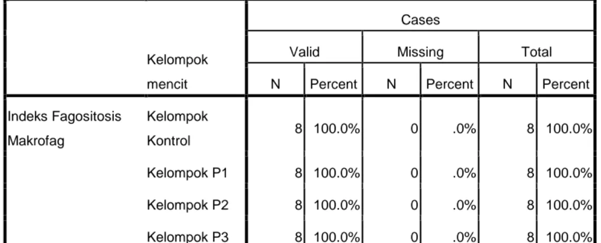
Case Processing Summary
Kelompok mencit
Cases
Valid Missing Total
N Percent N Percent N Percent
Indeks Fagositosis Makrofag Kelompok Kontrol 8 100.0% 0 .0% 8 100.0% Kelompok P1 8 100.0% 0 .0% 8 100.0% Kelompok P2 8 100.0% 0 .0% 8 100.0% Kelompok P3 8 100.0% 0 .0% 8 100.0% Descriptive Statistics
Kelompok mencit N Minimum
Maximu m Mean Std. Deviation Kelompok Kontrol Indeks Fagositosis Makrofag 8 .1800 .2833 .227500 .0332552 Valid N (listwise) 8
Kelompok P1 Indeks Fagositosis
Makrofag 8 .2667 .3500 .295012 .0285043
Valid N (listwise) 8
Kelompok P2 Indeks Fagositosis
Makrofag 8 .2400 .4400 .336250 .0622128
Valid N (listwise) 8
Kelompok P3 Indeks Fagositosis
Makrofag 8 .2300 .3567 .296250 .0483252
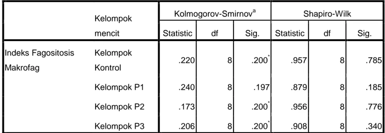
Tabel Uji Normalitas
Tabel Uji Lavenne
Tabel Uji one way ANOVA
Tests of Normality Kelompok
mencit
Kolmogorov-Smirnova Shapiro-Wilk
Statistic df Sig. Statistic df Sig.
Indeks Fagositosis Makrofag Kelompok Kontrol .220 8 .200 * .957 8 .785 Kelompok P1 .240 8 .197 .879 8 .185 Kelompok P2 .173 8 .200* .956 8 .776 Kelompok P3 .206 8 .200* .908 8 .340
a. Lilliefors Significance Correction
*. This is a lower bound of the true significance.
Test of Homogeneity of Variances Indeks Fagositosis Makrofag
Levene Statistic df1 df2 Sig.
1.725 3 28 .185
ANOVA Indeks Fagositosis Makrofag
Sum of Squares df Mean Square F Sig.
Between Groups .049 3 .016 8.013 .001
Within Groups .057 28 .002
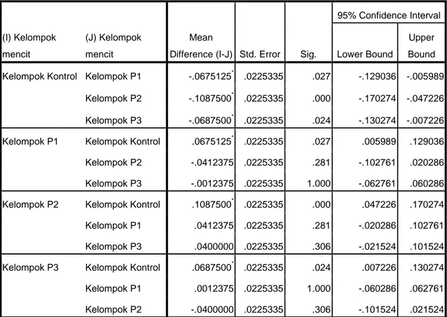
Tabel uji post-hoc
Multiple Comparisons Indeks Fagositosis Makrofag
Tukey HSD (I) Kelompok mencit (J) Kelompok mencit Mean
Difference (I-J) Std. Error Sig.
95% Confidence Interval
Lower Bound
Upper Bound
Kelompok Kontrol Kelompok P1 -.0675125* .0225335 .027 -.129036 -.005989
Kelompok P2 -.1087500* .0225335 .000 -.170274 -.047226
Kelompok P3 -.0687500* .0225335 .024 -.130274 -.007226
Kelompok P1 Kelompok Kontrol .0675125* .0225335 .027 .005989 .129036
Kelompok P2 -.0412375 .0225335 .281 -.102761 .020286
Kelompok P3 -.0012375 .0225335 1.000 -.062761 .060286
Kelompok P2 Kelompok Kontrol .1087500* .0225335 .000 .047226 .170274
Kelompok P1 .0412375 .0225335 .281 -.020286 .102761
Kelompok P3 .0400000 .0225335 .306 -.021524 .101524
Kelompok P3 Kelompok Kontrol .0687500* .0225335 .024 .007226 .130274
Kelompok P1 .0012375 .0225335 1.000 -.060286 .062761
Kelompok P2 -.0400000 .0225335 .306 -.101524 .021524
*. The mean difference is significant at the 0.05 level.
Case Processing Summary
N %
Cases Valid 8 100.0
Excludeda 0 .0
Total 8 100.0
a. Listwise deletion based on all variables in the procedure.
Reliability Statistics P2 Cronbach's Alpha N of Items
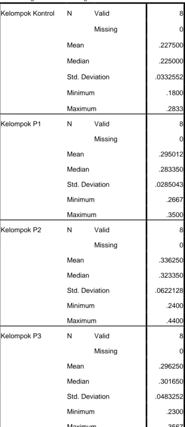
Tabel Deskriptif tambahan nilai median
Statistics Indeks Fagositosis Makrofag
Kelompok Kontrol N Valid 8
Missing 0 Mean .227500 Median .225000 Std. Deviation .0332552 Minimum .1800 Maximum .2833 Kelompok P1 N Valid 8 Missing 0 Mean .295012 Median .283350 Std. Deviation .0285043 Minimum .2667 Maximum .3500 Kelompok P2 N Valid 8 Missing 0 Mean .336250 Median .323350 Std. Deviation .0622128 Minimum .2400 Maximum .4400 Kelompok P3 N Valid 8 Missing 0 Mean .296250 Median .301650 Std. Deviation .0483252 Minimum .2300 Maximum .3567
LAMPIRAN 5. Produktivitas ROI
Hasil output data program SPSS for windows 16.00
Tabel uji Deskriptif
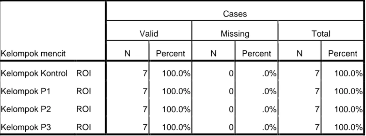
Case Processing Summary
Kelompok mencit
Cases
Valid Missing Total
N Percent N Percent N Percent
Kelompok Kontrol ROI 7 100.0% 0 .0% 7 100.0%
Kelompok P1 ROI 7 100.0% 0 .0% 7 100.0%
Kelompok P2 ROI 7 100.0% 0 .0% 7 100.0%
Kelompok P3 ROI 7 100.0% 0 .0% 7 100.0%
Case Summaries ROI
Kelompok mencit N Mean Median Minimum Maximum Std. Deviation
Kelompok Kontrol 7 1.7114 1.6600 1.46 2.32 .28068 Kelompok P1 7 2.4343 2.4200 2.00 3.08 .32408 Kelompok P2 7 2.3971 2.3600 2.14 2.68 .19746 Kelompok P3 7 2.4657 2.5000 2.20 2.90 .24812 Total 28 2.2521 2.3000 1.46 3.08 .40604 Tests of Normality Kelompok mencit Kolmogorov-Smirnova Shapiro-Wilk
Statistic df Sig. Statistic df Sig.
Kelompok Kontrol ROI .373 7 .004 .735 7 .009
Kelompok P1 ROI .375 7 .004 .812 7 .054
Kelompok P2 ROI .152 7 .200* .960 7 .815
Kelompok P3 ROI .201 7 .200* .917 7 .446
a. Lilliefors Significance Correction
Uji Kruskal-Wallis
Ranks
Kelompok mencit N Mean Rank
ROI Kelompok Kontrol 7 5.14
Kelompok P1 7 17.43 Kelompok P2 7 17.00 Kelompok P3 7 18.43 Total 28 Uji Mann-Whitney Kelompok K- Kelompok P1 Test Statisticsb ROI Mann-Whitney U 2.000 Wilcoxon W 30.000 Z -2.907
Asymp. Sig. (2-tailed) .004
Exact Sig. [2*(1-tailed Sig.)] .002a a. Not corrected for ties.
b. Grouping Variable: Kelompok mencit
Test Statisticsa,b ROI
Chi-Square 12.241
df 3
Asymp. Sig. .007
a. Kruskal Wallis Test b. Grouping Variable: Kelompok mencit
Ranks
Kelompok mencit N Mean Rank Sum of Ranks
ROI Kelompok Kontrol 7 4.29 30.00
Kelompok P1 7 10.71 75.00
Uji Mann-Whitney Kelompok K- Kelompok P2 Uji Mann-Whitney Kelompok K- Kelompok P3 Uji Mann-Whitney Kelompok P1- Kelompok P2 Ranks Kelompok mencit N Mean Rank Sum of Ranks ROI Kelompok P1 7 7.64 53.50 Kelompok P2 7 7.36 51.50 Total 14 Ranks Kelompok mencit N Mean Rank Sum of Ranks
ROI Kelompok Kontrol 7 4.43 31.00
Kelompok P2 7 10.57 74.00 Total 14 Test Statisticsb ROI Mann-Whitney U 3.000 Wilcoxon W 31.000 Z -2.747
Asymp. Sig. (2-tailed) .006
Exact Sig. [2*(1-tailed Sig.)] .004a a. Not corrected for ties.
b. Grouping Variable: Kelompok mencit
Test Statisticsb
ROI
Mann-Whitney U 3.000
Wilcoxon W 31.000
Z -2.747
Asymp. Sig. (2-tailed) .006
Exact Sig. [2*(1-tailed Sig.)] .004a a. Not corrected for ties.
b. Grouping Variable: Kelompok mencit Ranks Kelompok mencit N Mean Rank Sum of Ranks
ROI Kelompok Kontrol 7 4.43 31.00
Kelompok P3 7 10.57 74.00 Total 14 Test Statisticsb ROI Mann-Whitney U 23.500 Wilcoxon W 51.500 Z -.129
Asymp. Sig. (2-tailed) .897
Exact Sig. [2*(1-tailed Sig.)] .902a a. Not corrected for ties.
Uji Mann-Whitney Kelompok P1- Kelompok P3 Uji Mann-Whitney Kelompok P2- Kelompok P3 Ranks Kelompok
mencit N Mean Rank
Sum of Ranks ROI Kelompok P2 7 7.07 49.50 Kelompok P3 7 7.93 55.50 Total 14 Test Statisticsb ROI Mann-Whitney U 21.500 Wilcoxon W 49.500 Z -.388
Asymp. Sig. (2-tailed) .698
Exact Sig. [2*(1-tailed Sig.)] .710a a. Not corrected for ties.
b. Grouping Variable: Kelompok mencit Ranks
Kelompok
mencit N Mean Rank
Sum of Ranks ROI Kelompok P1 7 7.07 49.50 Kelompok P3 7 7.93 55.50 Total 14 Test Statisticsb ROI Mann-Whitney U 21.500 Wilcoxon W 49.500 Z -.385
Asymp. Sig. (2-tailed) .701
Exact Sig. [2*(1-tailed Sig.)] .710a a. Not corrected for ties.
b. Grouping Variable: Kelompok mencit Case Processing Summary
N %
Cases Valid 7 100.0
Excludeda 0 .0
Total 7 100.0
a. Listwise deletion based on all variables in the procedure.
Reliability Statistics Cronbach's
Alpha N of Items
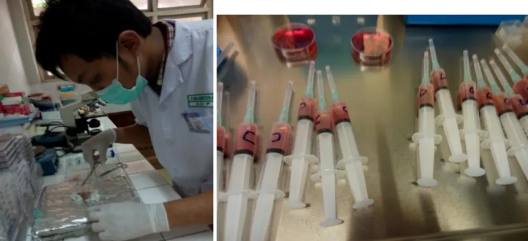
LAMPIRAN 6. Foto Penelitian
Proses Pembedahan mencit Pengumpulan cairan peritoneum
Pengelompokan Mencit Microwell 24 plate
Terlihat aktivitas fagositosis makrofag terhadap lateks
Curriculum Vitae
Nama : Akhsananta Lian Ferdiansyah
NIM : 22010110120004
Tempat tanggal lahir : Mojokerto, 30 Juni 1993
Jenis Kelamin : Laki-laki
Alamat : Jalan Gondang Timur IV no 30 Tembalang Semarang
Nomor telfon : 085731283002
Email : [email protected]
Riwayat Pendidikan
1. SD : SDN Bubutan IV Surabaya (2004) 2. SMP : SMPN 3 Surabaya (2007)
3. SMA : SMAN 6 Surabaya (2010)
4. PTN : Fakultas Kedokteran Universitas Diponegoro-2010
Riwayat organisasi