Detection and molecular characterization of canine adenovirus
type 2 (CAV-2) in dogs with respiratory tract symptoms in
shelters in Turkey
Mehmet Ozkan Timurkan1*, Hakan Aydin1, and Feray Alkan2 1Department of Virology, Faculty of Veterinary Medicine, Atatürk University, Erzurum,Turkey
2Department of Virology, Faculty of Veterinary Medicine, Ankara University, Ankara,Turkey ________________________________________________________________________________________ TIMURKAN, M. O., H. AYDIN, F. ALKAN: Detection and molecular characterization of canine adenovirus type 2 (CAV-2) in dogs with respiratory tract symptoms in shelters in Turkey. Vet. arhiv 88, 467-479, 2018.
ABSTRACT
Canine adenoviruses are agents responsible for two different infections in Canidae. While canine adenovirus type 1 (CAV-1) causes contagious hepatitis (HCC) in dogs, canine adenovirus type 2 (CAV-2) is responsible for infectious laryngotracheitis (ILT). CAV-2, especially in the respiratory tract, leads to an infection that can result in death in young and cohabitant animals. In public housing such as shelters, in addition to opportunistic infections, a disorder defined as canine infectious respiratory disease (CIRD) may also occur frequently. In this study, 155 nasal swabs were collected from dogs in two shelters where cases of respiratory system infections were closely monitored. These samples were tested for CAV-2 using polymerase chain reaction (PCR) with primers designed for the CAV E3 (Early) gene. Positive amplicons were subjected to DNA sequencing. CAV-2 nucleic acids were present in 2.5% (4/155) of the test samples. The phylogenetic assessment of the amplicon sequences revealed a 97.7%-98.9% similarity in the local viruses. The partial sequence analyses of the E3 gene of CAV-2 showed that Turkish and Chinese strains have differences in 9 amino acids. These differences redounded on phylogenetic analyses, and the virus which was considered as a single group, is now subdivided into two subgroups. One subgroup comprises American-European isolates and the other one consists of Turkish and Chinese isolates, so this subdivision can be classified into at least two subgroups, designated China-Turkey and America-Europe. To our knowledge, this is the first study that has examined the possible role of CAV-2 in respiratory system infections in dogs in Turkey, to provide novel and updated information regarding CAV-2.
Key words: canine adenovirus 2; molecular characterization; PCR; Turkey
________________________________________________________________________________________
*Corresponding author:
Mehmet Ozkan Timurkan, Department of Virology, Faculty of Veterinary Medicine, Ataturk University, 25240,
Introduction
Dog adenoviruses, canine adenovirus type 1 (CAV-1) and canine adenovirus type 2 (CAV-2), are double-stranded deoxyribonucleic acid (DNA) icosahedral viruses
belonging to the genus Mastadenovirus, of the family Adenoviridae (DUARTE et al.,
2014; WALKER et al., 2016). These viruses are genetically and antigenically related. CAV-1 targets the digestive tract tissue, and causes hepatitis contagiousa canis infection by damaging the liver. CAV-2 targets lung tissue where it causes infectious laryngotracheitis
(HU et al., 2001).
CAV-2 has an affinity for the epithelium of the upper airway, the nasal cavity, and especially the throat, larynx, and trachea tissue. However, one study reportedly found it in the intestinal system (MACARTNEY et al., 1988), while another found that it had
crossed the blood-brain barrier and reached the brain (BENETKA et al., 2006). This virus
was first detected in dogs in a Canadian shelter in 1961 that had symptoms of upper respiratory tract infection (DITCHFIELD et al., 1962). In subsequent years, the presence
of CAV-2 was reported in many countries such as Italy, Brazil, and India (BALBONI et al., 2013; PARTHIBAN et al., 2009; SILVA et al., 2014) and was also detected in Turkey,
based on serological data alone (BULUT et al., 2013; CAN SAHNA and ASLAN, 2015). Different methods can be used to diagnose adenoviral infections. Although various
methods, such as virus isolation, haemaglutination, and neutralization, can be utilised for the diagnosis of adenoviruses, factors such as time, labour, and the systems affected by
the infections (HU et al., 2001) have recently increased the preference for polymerase
chain reaction (PCR)-based methods (SILVA et al., 2014; BOOMKENS et al., 2005;
DECARO et al., 2007). The CAV-2 genome consists of gene regions that encode various proteins with specific functions. The most important ones are E1A, E1B (19K and 55K), E3, E4, pIVa2, pIIIa, pVI, pVII, pVIII, Pol, pTP, and DBP. E3 encodes several proteins
and is the only genome region that encodes the membrane proteins that modulate the host
response to infection (BURGERT and BLUSCH, 2000; LICHTENSTEIN et al., 2004).
In this study, swab samples collected from dogs in shelters were tested using PCR, targeting the E3 gene-region to obtain information about the possible role of CAV-2 in respiratory system infections, and molecular characterization of local CAV-2 strains.
Materials and methods
Samples. In this study, a total of 155 nasal swabs were collected from 155 dogs
belonging to 2 shelters. Dogs from both the shelters were found to have severe clinical
signs of respiratory system infection. The samples were collected from animals that
exhibited clinical signs, such as cough and nasal discharge.
The nasal swabs were resuspended in 1 mL sterile phosphate buffer solution (PBS) and stored at -20 °C until the time the nucleic acid isolation was to be performed. Materials
were also tested for canine respiratory coronavirus, canine influenza virus, and distemper
virus, possible aetiological agents of respiratory system infections in dogs.
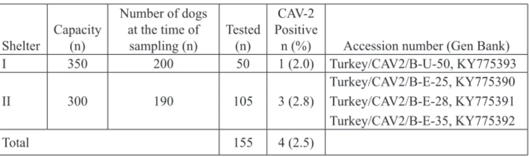
Table 1. Distribution of diagnostic materials and results
Shelter Capacity (n) Number of dogs at the time of sampling (n) Tested (n) CAV-2 Positive
n (%) Accession number (Gen Bank)
I 350 200 50 1 (2.0) Turkey/CAV2/B-U-50, KY775393 II 300 190 105 3 (2.8) Turkey/CAV2/B-E-25, KY775390 Turkey/CAV2/B-E-28, KY775391 Turkey/CAV2/B-E-35, KY775392 Total 155 4 (2.5)
Extraction and Polymerase Chain Reaction (PCR). Nucleic Acid Extraction Kits (Cat No:GF-RD-100, Vivantis, Malaysia) were used for viral nucleic acid extraction from the swab supernatants. Extraction was carried out according to the manufacturer’s
instructions. Primers and methods suitable for both CAV-1 and CAV-2 were used for
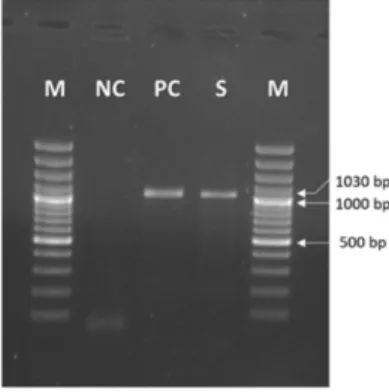
the PCR, as recommended by HU et al., (2001) and CHATURVEDI et al. (2008). Consequently, the formation of PCR products in the expected sizes (508 bp and 1030 bp for CAV-1 and CAV-2, respectively) was analysed using DNA gel electrophoresis.
Sequencing and Phylogenetic Analyses. DNA sequencing of the amplicons of expected
size (1030 bp) for CAV-2 was conducted via service procurement (Pendik Veterinary Control Institute, Istanbul, Turkey).The raw sequencing data were aligned using BioEdit
version 7.0.5 (HALL, 1999) with the Clustal W algorithm. The phylogenetic map of the
aligned sequences was established using MEGA version 6.0 (TAMURA et al., 2013) (Fig. 2). Nucleotide sequence accession numbers, the partial genome sequences of CAV-2 were obtained from Gen Bank under the accession nos.; U77082 (Toronto), KU725673 (SV521), KU315335 (1.18), KU315336 (3.55), KU725672 (SV435), KU315333 (1.6), KU725674 (SV675), JX416842 (113-3f-c04), KF676978 (60-2011-OFS), JX416841 (113-3F-c01), GQ915311 (HB1), EF090910 (India-CAV1), KU755720 (010515), KU755729 (16606), KT853097 (13-0067), M60937 (Gloxo-Vaccine), JN418926 (M1), NC002513 (BoAdV2), GU226970 (BatAdV-TJM), and JN252129 (BatAdV2-PPV1). This molecular study was approved by the Ethics Sub-Committee of the Ataturk University, Faculty of Veterinary
Medicine (date/protocol number: 16.02.2017/2017-008) in Erzurum, Turkey.
Results
The PCR results for CAV-2, based on the E3 gene from the nasal swab samples that
included CAV-2, revealed DNA products of the expected sizes (1030 bp) (Fig. 1). All
According to the PCR and sequencing analyses results, CAV-2 nucleic acids were
detected in 4 (2.5%) of the 155 tested swab samples. The positivity rates of the samples obtained from the shelters were determined as 2.0% and 2.8% (Table 1). The partial
nucleotide sequences of the CAV-2 E3 genes in our samples were deposited in The Gen Bank (Table 1). The percentages of nucleic acid identified in the sequences of our CAV-2
E3 genes were compared and the data are shown in Table 2.
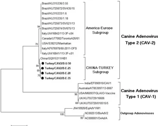
In the phylogenetic analyses of the results obtained from the sequencing reactions,
CAV-2, regarded as a single group in previous studies, was found to constitute 2
different clusters (subgroups) when compared to the data obtained from the Gene Bank.
Chinese and Turkish strains detected in this study belonged to the same subgroup.
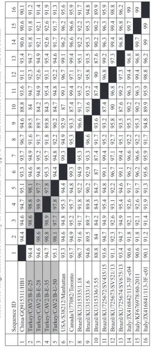
Moreover, when the nucleotide similarities of the strains were examined (Table 2), it
was determined that the strains obtained in this study were 97.7%-98.9% similar to each other and 84.2%-99.7% similar to the reference strains.
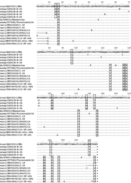
The observations made during the examination of the 792 nucleotide sequence
(almost 256 amino acids) in the partial E3 gene at the level of amino acids in different
situations were noted (Fig. 3). Due to the genetic proximity of Chinese and Turkish isolates, the Chinese isolate (Accession number: GQ915311, HB1) was taken as the
reference isolate, in phylogenetic analyses and amino acid analyses We observed the
consistent appearance of the amino acid change at positions 126 (A→T), 136 (M→K), 138 (N→D), 172 (T→V), 186 (E→D), 216 (V→M), 222 (N→D), 227 (V→I), and 254 (S→P) which are predominant mutations in CAV-2 in the Italian, Brazilian, and
American strains. However, although it was in the same subgroup of Chinese and Turkish strains, amino acid changes were observed at the following seven amino acid
positions: 15 (P→D-N), 97 (W→L), 152 (K→N), 187 (D→N), 184 (I→T), 221 (P→L), and 250 (N→T).
Fig. 1. PCR for canine adenovirus partial E3 gene. M: Marker (100-3000 bp, Thermo Fisher, Germany), NC: Negative control (Nuclease-Free Water), PC: Positive control of Canine
Fig. 2. Phylogenetic tree based on a 792 nucleotide sequence of 20 strains of adenoviruses (CAV-1, CAV-2, EqAdV1, BoAdV2, and BatAdV) generated by using the maximum likelihood algorithm, test of phylogeny is the bootstrap method with 1000 replicates, using the MEGA 6.0 software program. Equine, Bovine and Bat adenoviruses were used as an out group. The round
Fig. 3. Alignment of the deduced amino acid sequences of the partial-length E3 gene (256 amino acids) amplified using PCR from the clinical samples. The accession numbers of the sequences
are described in the “Material and methods” section. Amino acid changes are shown in the
Table 2.
Average nucleotide identity of canine adenovirus type 2 with the reference strain and with our strains
Sequence ID 1 2 3 4 5 6 7 8 9 10 11 12 13 14 15 16 1 China/GQ91531 1/HB1 94.4 94.6 94.7 95.1 93.3 93.7 96.7 94.6 88.8 93.6 91.1 93.4 90.6 90.6 90.1 2 Turkey/CA V2/B-E-25 94.4 98.6 98.8 98.1 94.6 94.9 91.6 89.5 84 94.7 92.3 94.7 91.8 91.8 91.2 3 Turkey/CA V2/B-E-28 94.6 98.6 98.9 97.7 94.8 95.2 91.7 89.7 84.2 94.9 92.6 94.9 92.1 92.1 91.4 4 Turkey/CA V2/B-E-35 94.7 98.8 98.9 97.8 95.3 95.7 91.8 89.8 84.3 95.4 93.1 95.4 92.6 92.6 91.9 5 Turkey/CA V2/B-U-50 95.1 98.1 97.7 97.8 94.4 94.8 92.2 90.2 84.7 94.8 92.2 94.6 91.7 91.7 91.3 6 USA/S38212/Manhattan 93.3 94.6 94.8 95.3 94.4 99.3 94.9 92.9 87 99.1 96.7 99.1 96.2 96.2 95.6 7 Canada/U77082/T oronto 93.7 94.9 95.2 95.7 94.8 99.3 95.3 93.3 87.4 99.4 97.1 99.4 96.6 96.6 95.9 8 Brazil/KU315335/1.18 96.7 91.6 91.7 91.8 92.2 94.9 95.3 96.6 91.7 95.2 92.7 95.1 92.2 92.2 91.7 9 Brazil/KU315333/1.6 94.6 89.5 89.7 89.8 90.2 92.9 93.3 96.6 93.6 93.2 95.8 93.5 95.3 95.3 94.8 10 Brazil/KU315336/3.55 88.8 84 84.2 84.3 84.7 87 87.4 91.7 93.6 87.4 90 87.6 90.2 90.2 89.9 11 Brazil/KU725672/SV435/15 93.6 94.7 94.9 95.4 94.8 99.1 99.4 95.2 93.2 87.4 96.8 99.2 96.3 96.3 95.9 12 Brazil/KU725673/SV521/13 91.1 92.3 92.6 93.1 92.2 96.7 97.1 92.7 95.8 90 96.8 97.3 99.4 99.4 98.8 13 Brazil/KU725674/SV675/13 93.4 94.7 94.9 95.4 94.6 99.1 99.4 95.1 93.5 87.6 99.2 97.3 96.8 96.8 96.2 14 Italy/JX416842/1 13-3F-c04 90.6 91.8 92.1 92.6 91.7 96.2 96.6 92.2 95.3 90.2 96.3 99.4 96.8 99.7 99 15 Italy/KF676978/60-201 1 90.6 91.8 92.1 92.6 91.7 96.2 96.6 92.2 95.3 90.2 96.3 99.4 96.8 99.7 99 16 Italy/JX416841/1 13-3F-c01 90.1 91.2 91.4 91.9 91.3 95.6 95.9 91.7 94.8 89.9 95.9 98.8 96.2 99 99
Discussion and conclusion
Animal care conditions and length of stay in shelters are governed by legislation in Turkey. These shelters are founded by government or civil organisations, and primary care
for newly arriving animals is performed within these shelters, such as initial examination
and vaccination. However, there is risk of severe respiratory and alimentary infections
for reasons such as reactivation of latent viruses and the inability to quarantine incoming
animals due to limited space and caring for more than one species (especially cats and
dogs) in close proximity. Also, similar to dogs living in shelters, it is known that there is a risk of infections for dogs with owners due to their exposure to several factors during
their daily walks. These infections often involve the digestive or respiratory system. The
development of canine infectious respiratory disease (CIRD), previously known as Kennel Cough, caused by viral agents such as CAV-2, parainfluenza virus, canine respiratory
coronavirus, and canine herpes virus, as well as bacterial agents such as Bordotella spp., Mycoplasma spp., and Streptococcus spp. or often with the participation of a few of them
(BUONAVOGLIA and MARTELLA, 2007; DECARO et al., 2016; LAVAN and KNESL,
2015).
Although CAV-1 and CAV-2 are associated with infections involving different clinical manifestations, they are antigenically related. Therefore, some enzyme-linked immunosorbent assay (ELISA)-based serologic tests detect antibodies to both these agents. In fact, some reports on CAV in Turkey (CAN SAHNA and ASLAN, 2015;
BULUT et al., 2013) have reported information on CAV seropositivity. However, to the
best of our knowledge, no studies have reported CAV-2 seroprevalence, virus isolation, or molecular epidemiology.
In this study, among all the dogs with respiratory system infection, 2% and 2.8% of
them in shelters 1 and 2, respectively, showed CAV-2 positivity. SCHULZ et al. (2014) have reported that in addition to CAV-2, canine parainfluenza virus, canine influenza
virus, canine respiratory coronavirus, as well as B. bronchiseptica were detected in varying rates among the shelter and the owned dogs, with or without acute respiratory
system infection. Further, the presence of B. bronchiseptica was significantly higher
in dogs with clinical respiratory infection than in the healthy looking dogs (45.6% and 78.7%, respectively); there was a high prevalence of multiple infections. In this study, we did not collect data pertaining to the presence of viruses and bacterial agents, other
than the distemper virus, canine respiratory coronavirus, and canine influenza virus. Therefore, it was not possible to make a definitive assessment regarding the individual
or combined effect of CAV-2 or some other factors on the clinical status of the shelter dogs. However, we believe that CAV-2 played an individual or contributory role in the development of clinical disorders in some of the dogs in these sheltering units. However,
data regarding age, prior possession, vaccination information, and time of admission to the shelter.
Studies on CAV-2 are limited in the literature. Different clinical manifestations and different species have been reported in previous studies (DECARO et al., 2016; SILVA et al., 2014). In a study conducted by WHETSTONE (1988) in the United States of America (USA), monoclonal antibodies were used to distinguish the CAV types, where immunofluorescence and RFLP were applied comparatively. DECARO et al., (2016) questioned the presence of Bordetella bronchiseptica and some Mycoplasma species,
as well as several viruses (canine respiratory coronavirus, canine parainfluenza, canine
herpesvirus-1, etc.), including CAV-2 because the highest positivity rate was found for
canine parainfluenza in their study population of dogs with CIRD. In a study conducted in Japan (MOCHIZUKI et al., 2008), although canine parainfluenza virus was the most frequently detected factor in dogs with CIRD that were taken to different animal hospitals,
canine respiratory coronavirus, canine adenovirus-2, and canine distemper virus were also detected.
The Gene Bank has nucleotide entries for Brazil and China isolates of CAV-2 (Brazil; KU725674, China; GQ153311). It was determined that our isolates had 84.2%-99.7%
nucleotide similarities with the reference strains (Table 2). The similarities among the
strains in the study were 97.7%-98.9%. In the phylogenetic analyses (Fig. 2) of the strains isolated in our study and the reference strains obtained from the Gene Bank, Chinese isolates (GQ153311) appeared to have formed a subgroup with the strains isolated in this study. Other reference strains (USA, Canada, Brazil, and Italy) have been observed to form another subgroup, distinct from the subgroup including our viruses. Furthermore, some fixed substitutions found at 126, 136, 138, 172, 186, 216, 222, 227, and 254 could
be the result of the differences in the CAV-2 subgroups. These differences could also
account for the substitutions identified for positions 15, 97, 152, 187, 184, 221, and 250 within the same subgroup, although this was a unique feature of the Turkish strains. The
Turkish and Chinese strains of the 7 different amino acids were different from each other. This is a potentially interesting feature of the new subgroup. However, analyses of more
and longer sequences are warranted to confirm this. In addition, another region needed to be amplified to obtain a phylogenetic tree confirming it. The gene regions (E1, E2, E3 and
E4) of CAV-2 are responsible for efficient replication of the virus, viral gene expression
and regulation of host-cell functions (MORRISON et al., 1997). Nine different amino
acids have been identified on the molecular level (amino acid differences) between the
China-Turkey isolates subgroup and the America-Europe subgroup strains. These amino acid differences suggest that the China-Turkey isolates group created a new subgroup. However, this must be analysed in different gene regions of the CAV-2 genome.
In conclusion, in this study, we explored CAV-2 nucleic acid detection and the
shelters in Turkey. Additionally, our results revealed the possibility of a new subgroup of CAV within type 2 (CAV-2). The molecular characterization of the viruses from known dogs in different housing conditions (owned and kept inside a house, kept in front of the door, idling around, etc.) with varying vaccination history and age, would provide new information regarding the epidemiology of CAV-2 infections. Otherwise, knowledge of the possibility of different subgroups of CAV type 2 is also needed to enhance
surveillance of circulating strains. Further, the study of other causative agents of CIRD
in dogs detected with CAV-2 will also contribute to the knowledge on this subject and commercial vaccine use.
References
BALBONI, A., R. VERIN, F. MORANDI, A. POLI, S. PROSPERI, M. BATTILANI (2013):
Molecular epidemiology of canine adenovirus type 1 and type 2 in free-ranging red foxes
(Vulpes vulpes) in Italy. Vet. Microbiol. 162, 551-557.
DOI: 10.1016/j.vetmic.2012.11.015
BENETKA, V., H. WEISSENBOCK, I. KUDIELKA, C. PALLAN, G. ROTHMULLER, K.
MOSTL (2006): Canine adenovirus type 2 infection in four puppies with neurological signs. Vet. Rec. 158, 91-94.
DOI: 10.1136/vr.158.3.91
BOOMKENS, S. Y., E. SLUMP, H. F. EGBERINK, J. ROTHUIZEN, L. C. PENNING (2005): PCR screening for candidate etiological agents of canine hepatitis. Vet. Microbiol. 108, 49-55.
DOI: 10.1016/j.vetmic.2005.03.003
BULUT, O., O. YAPICI, O. AVCI, A. SIMSEK, K. ATLI, I. DIK, S.YAVRU, S. HASIRCIOGLU,
M. KALE, N. MAMAK (2013): The serological and virological investigation of canine
adenovirus infection on the dogs. Sci. World J. 24, ID 587024. DOI: 10.1155/2013/587024
BURGERT, H. G., J. H. BLUSCH (2000): Immunomodulatory functions encoded by the E3 transcription unit of adenoviruses. Virus Genes. 21, 13-25
DOI: 10.1023/A:1008135928310
BUONAVOGLIA, C., V. MARTELLA (2007): Canine respiratory viruses. Vet. Res. 38, 355-373.
DOI: 10.1051/vetres:2006058
CAN SAHNA, K., O. ASLAN (2015): Investigation of Canine Adenovirus (CAV) antigen in urine
and plasma samples of dog by ELISA. Harran Univ. Vet. Fak. Derg. 4, 45-47 (in Turkish). CHATURVEDI, U., A. K. TIWARI, B. RATTA, P. V. RAVINDRA, Y. S. RAJAWAT, S. K.
PALIA, A. RAI (2008): Detection of canine adenoviral infections in urine and faeces by the
polymerase chain reaction. J. Virol. Methods 149, 260-263.
DECARO, N., M. CAMPOLO, G. ELIA, D. BUONAVOGLIA, M. L. COLAIANNI, A. LORUSSO, V. MARI, C. BUONAVOGLIA (2007): Infectious canine hepatitis: an “old” disease reemerging in Italy. Res. Vet. Sci. 83, 269-273.
DOI: 10.1016/j.rvsc.2006.11.009
DECARO, N., V. MARIA, V. LAROCCA, M. LOSURDO, G. LANAVE, M. S. LUCENTE, M. CORRENTE, C. CATELLA, S. BO, G. ELIA, G. TORRE, E. GRANDOLFO, V. MARTELLA, C. BUONAVOGLIA (2016): Molecular surveillance of traditional and emerging pathogens associated with canine infectious respiratory disease. Vet. Microbiol. 192, 21-25.
DOI: 10.1016/j.vetmic.2016.06.009
DITCHFIELD, J., L. W. MACPHERSON, A. ZBITNEW (1962): Association of a canine adenovirus (Toronto A 26/61) with an outbreak of laryngotracheitis (“kennel cough”) A preliminary report. Can. Vet. J. 3, 238-247.
DUARTE, M. D., A. M. HENRIQUES, C. LIMA, C. OCHOA, F. MENDES, M. MONTEIRO, F. RAMOS, T. LUIS, R. NEVES, M. FEVEREIRO (2014): Fatal canine adenovirus type 1 acute infection in a Yorkshire Terrier puppy in Portugal: a case report. Vet. Med-Czech 59, 210-220 DOI: 10.17221/7482-VETMED
HALL, T. A. (1999): BioEdit: A user-friendly biological sequence alignment editor and analysis
program for Windows 95/98/NT. Nucl. Acids Symp. Ser. 41, 95-98.
HU, R. L., G. HUANG, W. QIU, Z. H. ZHONG, X. Z. XIA, Z. YIN (2001): Detection and
differentiation of CAV-1 and CAV-2 by polymerase chain reaction. Vet. Res. Commun. 25, 77-84.
DOI: 10.1023/A:1006417203856
LAVAN, R., O. KNESL (2015): Prevalence of canine infectious respiratory pathogens in
symptomatic dogs presented at US animal shelters. J. Small Anim. Pract. 56, 572-576. DOI: 10.1111/jsap.12389
LICHTENSTEIN, D. L., K. TOTH, K. DORONIN, A. E. TOLLEFSON, W. S. WOLD (2004):
Functions and mechanisms of action of the adenovirus E3 proteins. Int. Rev. Immunol. 23,
75-111.
DOI: 10.1080/08830180490265556
MACARTNEY, L., H. M. CAVANAGH, N. SPIBEY (1988): Isolation of canine adenovirus-2 from the faeces of dogs with enteric disease and its unambiguous typing by restriction endonuclease mapping. Res. Vet. Sci. 44, 9-14.
MOCHIZUKI, M., A. YACHI, T. OHSHIMA, A. OHUCHI, T. ISHIDA (2008): Etiologic study of upper respiratory infections of household dogs. J. Vet. Med. Sci. 70, 563-569.
DOI: 10.1292/jvms.70.563
MORRISON, M. D., ONIONS, D. E., NICOLSON, L. (1997): Complete DNA sequence of canine adenovirus type 1. J. Gen. Virol. 78, 873-878.
PARTHIBAN, M., K. KUMANAN, K. SUNDER, S. S. KUMAR, D. KATHIRESAN (2009):
Molecular detection of canine adenovirus using polymerase chain reaction and sequencing.
Tamilnadu J. Vet. Animal Sci. 5, 140-142.
SILVA, A. P., L. BODNAR, S. A. HEADLEY, A. F. ALFIERI, A. A. ALFIERI (2014): Molecular
detection of canine distemper virus (CDV), canine adenovirus A type 1 and 2 (CAdV-1 and
CAdV-2), and canine parvovirus type 2 (CPV-2) in the urine of naturally infected dogs. Semin. Ciênc. Agrár. 35, 3231-3236.
DOI: 10.5433/1679-0359.2014v35n6p3231
SCHULZ, B. S., S. KURZ, K. WEBER, H. J. BALJER, K. HARTMANN (2014): Detection
of respiratory viruses and Bordetella bronchiseptica in dogs with acute respiratory tract infections. Vet. J. 201, 365-369.
DOI: 10.1016/j.tvjl.2014.04.019
TAMURA, K., G. STECHER, D. PETERSON, A. FILIPSKI, S. KUMAR (2013): MEGA6: Molecular Evolutionary Genetics Analysis version 6.0. Mol. Biol. Evol. 30, 2725-2729. DOI: 10.1093/molbev/mst197
WHETSTONE, C. A. (1988): Monoclonal antibodies to canine adenoviruses 1 and 2 that are
type-specific by virus neutralization and immunofluorescence. Vet. Microbiol. 16, 1-8. DOI: 10.1016/0378-1135(88)90121-6
WALKER, D., S. A FEE, G. HARTLEY, J. LEARMOUNT, M. J. H. O’HAGAN, A. L. MEREDITH, B. M. De C. BRONSVOORT, T. PORPHYRE, C. P. SHARP, A. W.
PHILBEY (2016): Serological and molecular epidemiology of canine adenovirus type 1 in red foxes (Vulpes vulpes) in the United Kingdom. Sci. Rep. 31, ID 36051.
DOI: 10.1038/srep36051
Received: 6 August 2017 Accepted: 12 March 2018
_____________________________________________________________________________________ TIMURKAN, M. O., H. AYDIN, F. ALKAN: Otkrivanje i molekularna
karakterizacija psećeg adenovirusa tipa 2 (CAV-2) kod pasa sa simptomima dišnog
sustava držanih u skloništima u Turskoj. Vet. arhiv 88, 467-479, 2018..
SAŽETAK
Adenovirusi su uzročnici odgovorni za dvije različite infekcije u pasa. Dok pseći adenovirus tipa 1 (CAV-1) uzrokuje zarazni hepatitis (HCC), pseći adenovirus tipa 2 (CAV-2) odgovoran je za zarazni laringotraheitis (ILT). CAV-2, osobito u dišnom sustavu, uzrokuje infekciju sa smrtnim ishodom kod mladih životinja i onih koje žive u zajednicama. U javnim objektima kao što su skloništa, osim oportunističkih infekcija, učestao je i poremećaj koji se definira kao pseća zarazna bolest dišnih puteva (CIRD). Tijekom ovog istraživanja prikupljeno je 155 uzoraka krvi od pasa držanih u dva skloništa u kojima su pažljivo praćeni slučajevi infekcija dišnog sustava. Uzorci su testirani na CAV-2 primjenom lančane reakcije polimerazom (PCR), uz upotrebu početnica oblikovanih za gen CAV E3 (Early). Pozitivni su amplikoni podvrgnuti sekvenciranju DNA. CAV-2 nukleinske kiseline bile su prisutne u 2,5 % (4/155) istraženih uzoraka. Filogenetska analiza sekvencija amplikona otkrila je u lokalnim virusima 97,7 - 98,9 % sličnosti. Analize parcijalnih sekvencija gena CAV-2 E3 pokazale su da turski
i kineski sojevi imaju razlike u 9 aminokiselina. Te su razlike dodatno analizirane filogenetskim analizama i virus koji je smatran jedinstvenom skupinom, podijeljen je na dvije podskupine. Jedna su podskupina američko-europski izolati, a druga se sastoji od turskih i kinskih izolata. Stoga se ovom podjelom mogu razlikovati najmanje dvije podskupine, označene kao Kina - Turska i Amerika - Europa. Pretpostavlja se da je ovo prvo istraživanje o mogućoj ulozi CAV-2 u infekcijama dišnog sustava pasa u Turskoj i kao takvo pruža nove informacije o CAV-2.
Ključne riječi: pseći adenovirus 2; molekularna karakterizacija; PCR; Turska