In the last decade, Department of
Cipto Mangunkusumo Hospital has been
procedure consists of 2 phases. The first
5-6
performed after 12 weeks by elevating the
Cutting and carving of cartilage
6, 11,12
Vacanti et al. quoted from Britt
and Park
6(TGF
β) is the protein produced by cells
By adding TGF
βDalam keadaan tidak memungkinkan dapat digunakan DBM karena masih baik dalam mempertahankan
tandur kartilago, fibrin glue, demineralized bone matrix (DBM), transforming growth factor β (TGF β), Mankin score.
ABSTRACT
Background: Microtia reconstruction is a challenge for ENT Head and Neck surgeons. Various surgical techniques using autograft cartilage have been done to perform auricular reconstruction. Knowledge of cartilage graft concerning resorption process that affected the size, form, and aesthetic subunit of the ear is mandatory. Purpose: To evaluate the success of cartilage autograft by identifying chondrocyte apoptosis, tissue degradation based on cell character, matrix homogeneity, fibrosis, proteoglycans, collagen and Transforming Growth Factor β (TGF β) expression in application of Fibrin Glue (FG) and or Demineralized Bone Matrix (DBM) after 12 weeks in microtia reconstruction by Nagata technique. Methods: Quasi-experiments. FG and/or DBM were applied on the rest of the 12 ear cartilage framework which was implanted on mastoid area. Apoptosis was examined by TUNEL. Safranin O staining and modified Mankin’s score was used to evaluate cartilage degradation and TGF β expression by ELISA. Results: FG or DBM on cartilage graft showed significant increase in chondrocyte viability
compare with control group (p=0.00). Minimal fibrosis, more homogeneous extracellular matrix, decreased proteoglycan and minimal thickening of collagen, had significant differences compared with control or FG-DBM group. Structure differences occurred among cartilage graft after 12 week implantation whereas FG showed minimal fibrous tissue, normal cell character, proteoglycan, collagen, and tissue homogeneity (p< 0.05). Conclusion: FG is highly recommended to reduce degradation of cartilage graft in microtia reconstruction. DBM can be still used to maintain chondrocyte viability, proteoglycans, and collagen.
Keywords: cartilage graft, fibrin glue, demineralized bone matrix, transforming growth factor β, Mankin score.
ABSTRAK
Latar belakang: Rekonstruksi mikrotia merupakan tantangan bagi ahli bedah THT-KL. Berbagai teknik operasi menggunakan rangka telinga dengan tandur kartilago autologus telah dilakukan untuk rekonstruksi mikrotia. Pengetahuan mengenai tandur kartilago sangat diperlukan, mengingat tandur dapat mengalami resorpsi dengan berjalannya waktu, sehingga mempengaruhi ukuran, bentuk, dan detil estetik subunit daun telinga. Tujuan: Mengetahui viabilitas kondrosit, degradasi jaringan berdasarkan perubahan karakter kondrosit, fibrosis, homogenitas matriks, ekspresi proteoglikan dan kolagen serta ekspresi transforming growth factor β (TGF β ) dengan atau tanpa fibrin glue (FG) dan/atau demineralized bone matrix (DBM) pada rekonstruksi mikrotia setelah 12 minggu penanduran. Metode: Quasi-eksperimen. FG dan/atau DBM digunakan pada sisa tandur autologus kartilago rangka telinga, dilanjutkan pemeriksaan apoptosis dengan TUNEL. Pewarnaan Safranin O untuk menilai degradasi jaringan dengan skor modifikasi Mankin dan ekspresi TGF β dengan ELISA. Hasil: penambahan FG atau DBM pada tandur kartilago, viabilitas sel meningkat berbeda bermakna dengan tanpa perlakuan atau FG-DBM.(p=0.00), fibrosis minimal, matriks lebih homogen, penurunan proteoglikan dan penebalan kolagen minimal berbeda bermakna dengan kelompok tanpa perlakuan dan campuran FG-DBM. Terjadi perbedaan struktur jaringan setelah 12 minggu, FG mempunyai nilai fibrosis yang terendah, karakter sel normal, proteoglikan, kolagen, dan homogenitas jaringan (p<0,05). Kesimpulan: Penggunaan FG sangat dianjurkan untuk mengurangi degradasi tandur kartilago autologus pada rekonstruksi mikrotia.
Research Report
Fibrin glue and demineralized bone matrix effect
on autologus cartilage graft in microtia reconstruction
Dini Widiarni Widodo*, Jenny Bashiruddin*, Helmi*, Alida Harahap**,
Nurjati Chairani Siregar***
*Department of Otorhinolaryngology Head and Neck Surgery,
Faculty of Medicine Universitas Indonesia/Dr. Cipto Mangunkusumo Hospital, Jakarta
**
Department of Biology Molecular Eijkman Institute, Jakarta
INTRODUCTION
In the last decade, Department of
Otolaryngology- Head and Neck Surgery,
Faculty of Medicine University Indonesia/
Cipto Mangunkusumo Hospital has been
performing microtia reconstruction with
various degree anomaly. On reconstruction
procedure, ear framework is made of
autologous rib cartilage graft.
1-3The surgery
procedure consists of 2 phases. The first
phase is to form an ear frame continued
with grafting into the subcutaneous ear
region.
4Ear pattern is formed according to
the anatomy, height and width of normal
ear. Sixth-seventh rib cartilage is carved in
detail using knife or drill in accordance to
the normal ear shape.
5-6The second phase is
performed after 12 weeks by elevating the
auricle and if necessary, accompanied with
auditory canal reconstruction.
7Cutting and carving of cartilage
will arouse immunologic response and
protection of cartilage matrix.
1,3,8,9Response
against trauma will stimulate cytokine
production and growth factor, leading to
the metabolic change of chondrocyte and
matrix. Total chrondrocyte will be reduced,
cell cluster will be established and there
will be a change of matrix distribution and
composition. Simultaneously there will be
cytokine and catabolic enzyme synthesis,
proteoglican loss, and type II collagen upon
cartilage surface. In the subsequent process,
there will be a matrix degradation exceeding
synthesis causing loss of protein matrix
expression.
10Similar response also may occur when
cartilage ear frame work being implanted in
mastoid area as the recipient bed. The process
occurs against autologous cartilage graft or
local tissue. The cartilage tissue degradation
caused by the process related with the
chondrocyte viability, can be evaluated from
percentage of cell-experienced apoptosis.
According to Langer and Vacanti
11tissue-engineered cartilage is the application
science of tissue engineering and developed
into biological material to repair and change
tissue function for reconstruction purpose.
The technology uses biocompatible,
biodegradation, synthetic or polymer tissues
as scaffold. The tissue engineering may
accelerate cartilage development to be
mature to its morphological character, using
media in order that cell growth environment
becomes stable.
6, 11,12The technique can be
used in clinical application of maxillofacial
reconstruction surgery.
13Vacanti et al. quoted from Britt
and Park
6reported the success of tissue
engineering to produce cartilage ear frame
work by increasing cell viability and matrix
structure to form ear frame template, which
the viability of chondrocyte have been
maintained by adding fibrin or growth factor.
Transforming growth factor-beta,
(TGF
β) is the protein produced by cells
with specific activity against target cells. It
is expressed during bone fracture healing
process and also used for cartilage or bone
defect therapy which is being a required
component in tissue engineering technique.
13By adding TGF
βinvitro, viability cell and
matrix production can be maintained. On
Dalam keadaan tidak memungkinkan dapat digunakan DBM karena masih baik dalam mempertahankan
viabilitas kondrosit, proteoglikan dan kolagen.
Kata kunci: tandur kartilago, fibrin glue, demineralized bone matrix (DBM), transforming growth factor
β (TGF β), Mankin score.
Alamat korespondensi: Dini Widiarni, e-mail: [email protected]
increased from 51.16 to 79.23. before and
cell density and TGFβ in all groups (p>0.05).
In the DBM group, number of apoptotic
cell decreased from 48.84 to 27.03 (p=0.018).
DBM and FG groups (p=0.592), however
significant different found in apoptosis
decrease of FG-DBM group. (p=0.008).
ᴲ ┴
cells (%) PhaselControl DBM cells (%) PhaselControl
DBM P*Pair t test apoptosis, living cells among phase 1-phase 2, phase 1- FG, phase 1-DBM, and phase 1-FG-DBM
¶ Pair t test apoptosis , living cells among phase 2, FG, DBM, and FG-DBM ᴲ Pair t test apoptosis, living cell s among FG, DBM, and FG-DBM ┴ T test apoptosis in pair, living cells between DBM and FG-DBM
Wilcoxon test on cell density among phase1-phase2 and among phase2, FG,DBM,FGDBM, FG, DBM and FGDBM
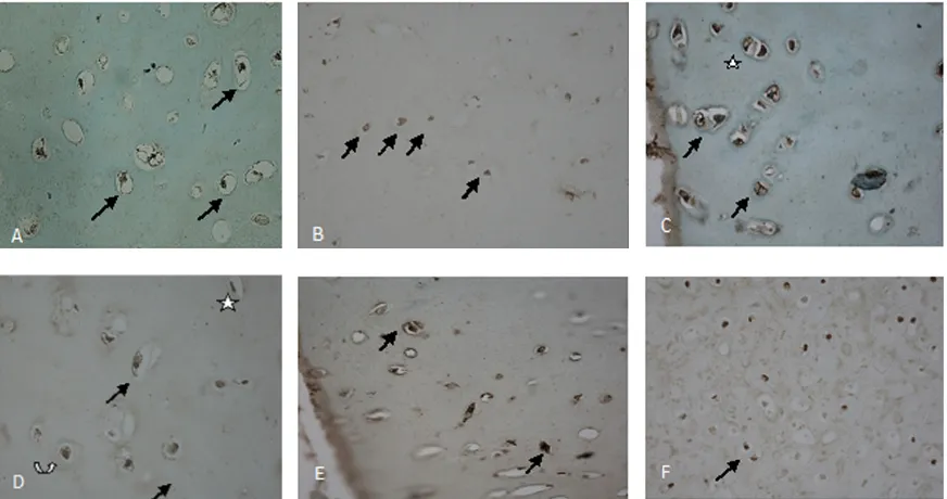
A) Chondrocyte of rib cartilage before grafting (phase1) = living cell, with green nucleus.
B) Autologous graft after 12 weeks grafting (phase 2) C) Autologous graft with FG, 12 weeks of grafting. Living chondrocyte with green nucleus ( ) apoptotic cell with brown nucleus ( ) D) Living hipertrophic chondrocyte. D) FG-DBM intervention shows chondrocyte apoptosis
clinical application of TGF
βeffect can be
used as the therapy.
14The use of TGF
βfamily
is demineralized bone matrix (DBM) which
is a homologous graft with osteoconductive
activity. DBM is made through bone
extraction by eliminating mineral component
and maintaining collagen and non-collagen
protein including TGF
β.
Fibrin glue or tissue adhesive contains
fibrinogen, in nature coagulation factor
XIII is substance which may strengthen
tissue contact and inhibit tissue leakage.
Fibrin glue (FG) may also play a role as the
conductive substance of growth factor into
the targeted delivery system. Cell culture
and histological evaluation shows that FG
does even possibly stimulate the healing
process. In clinical application, FG has been
used in maxillofacial reconstruction surgery
including those using flap and graft.
15Purpose of the research was to evaluate
and identify the success of autologous
cartilage graft with and without FG/DBM
which was assesed from chrondocyte
viability, hystomorphological change and
cartilage degradation process after 12 weeks
of grafting.
METHODS
This quasi experiment study was
approved by the Institutional Review
Board of Medical Faculty University of
Indonesia/Cipto Mangunkusumo Hospital
Jakarta, Indonesia. We evaluated the result
of cartilage graft using FG and/or DBM as
intervention group in 12 microtic ear which
underwent reconstructive surgery between
April 2008 and August 2009 at Plastics
Reconstructive Division, Department of Oto
laryngology,Ciptomangunkusumo Hospital.
Surgical procedure
Auricle reconstruction was performed
by harvesting 6th-7th rib cartilage graft,
then shaped and adjusted to the planned ear
shape. Helix is the highest part of aesthetic
unit while fossa triangularis, schapa and
concha are parts without cartilage. Carved
cartilage was then added with FG (Beriplast
®ZLB Behring GmbH Marburg, Alemania)
and implanted into a skinpocket under ear
region. The remaining cartilages which were
intervened with DBM, FG-DBM and control
group then inserted posterior to the ear
framework. All this piece of cartilage will be
used as buttress in second phase surgery.
After 12 weeks, ear projection was
performed starting with 1 cm curved incision
making a subcutaneous pocket. Fibrotic
capsule was maintained for covering the
ear framework. External ear canal was then
created and cartilage buttress placed on the
mastoid area. Retro auricular fascia flap was
undermined to cover ear buttress. Finally,
the posterior auricular region was veiled by
split thickness skin graft.
Cartilage tissue samples were examined
for biological characteristic of chondrocyte
apoptosis and viabilty, fibrosis, tissue
homogenity, collagen, proteoglycan and
TGFβ expression. Hystomorphology,
Safranin O staining, TUNEL and Elisa
were carried out in Department of Biology
Molecular Eijkman Institute Jakarta and
Department of Pathology Anatomy, Faculty
of Medicine Universitas Indonesia.
RESULTS
Evaluation of chondrocyte
The statistical analysis used pair t-test
for the asessment of apoptosis, living cells,
cell density between phase1-phase2 and
among the intervention groups (FG, DBM,
and FG-DBM and control group). Number
of apoptotic cells decreased in all groups
(p<0.001), accompanied by increased living
cells after grafting p<0.001). However, the
48.84 to 19.83 and number of living cells
increased from 51.16 to 79.23. before and
after grafting. There were no difference of
cell density and TGFβ in all groups (p>0.05).
In the DBM group, number of apoptotic
cell decreased from 48.84 to 27.03 (p=0.018).
There was no statistically different between
DBM and FG groups (p=0.592), however
significant different found in apoptosis
decrease of FG-DBM group. (p=0.008).
Table 1. TUNEL examination
Variabel N(12) Mean SD Min P25 Median P75 Max P* P 
Pᴲ P┴
Apoptotic
cells (%) PhaselControl FG
0,092 0,5920,008 0,123
Living
cells (%) PhaselControl FG P*Pair t test apoptosis, living cells among phase 1-phase 2, phase 1- FG, phase 1-DBM, and phase 1-FG-DBM
¶ Pair t test apoptosis , living cells among phase 2, FG, DBM, and FG-DBM ᴲ Pair t test apoptosis, living cell s among FG, DBM, and FG-DBM ┴ T test apoptosis in pair, living cells between DBM and FG-DBM
Wilcoxon test on cell density among phase1-phase2 and among phase2, FG,DBM,FGDBM, FG, DBM and FGDBM
Picture 1. A) Chondrocyte of rib cartilage before grafting (phase1) = living cell, with green nucleus.
resorption were statistically significant
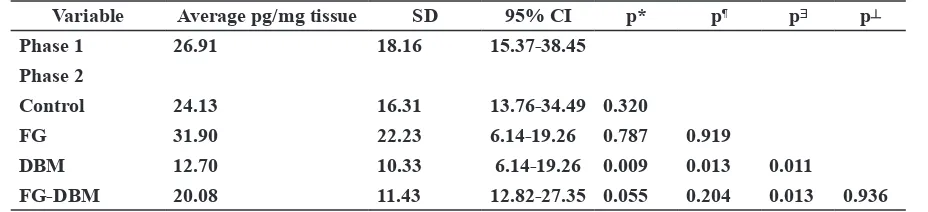
The decrease of TGF β expression in
significant difference between phase 1
and phase 2, 26.91pg/mg to 24,13 pg/
mg. FG intervention elevated TGF β level
from 26.91 to 31.90 pg/mg ( no significant
difference).
TGF β expression in DBM group
showed significant decreased from 26.91
to 12.70 pg/mg (p = 0.009; CI 6.14-19.26)
decrease TGF β expression in FG-DBM
intervention showed no significant decrease
compared with control group; but it was found
significant different from FG intervention
group (p=0,013; CI 12,82-27,35).
By Spearman correlation test, a
moderate correlation (r=0,70) was found
between apoptosis and TGF β expression in
control group. Weak correlations were found
TGF β expresion in allControl – FG, p<0.002 (Wilcoxon test) Control– DBM, p=0.034 (Wilcoxon test) Ccontrol-FG-DBM, p=0.317 (Wilcoxon test) FG-DBM, p=0.008 (Wilcoxon test)
FG-FG-DBM, p=0.002 (Wilcoxon test) DBM-FG-DBM, p=0.005 (Wilcoxon test)
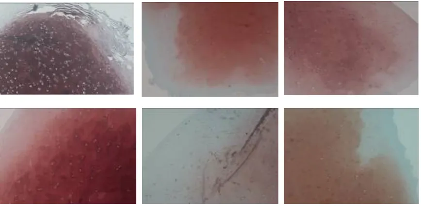
. A). Mankin score, total score as base line parameter. B.)Total score control group 7. Fibrosis 1, cell character 1, proteoglycan 2, homogenity 1, collagen 2,) C.) FG total score 2.( fibrosis 0, cell character 0, proteo glycan 1, homogenity 0, collagen 1,) D.) DBM Total score 5( Fibrosis 1, cell characyter 2, proteoglycan 1, homo genity 1, collagen 0,) . E). FG-DBM Total score 7. Fibrosis 1, cell characyter 1, proteoglycan 2, homogenity 1, collagen 2
Evaluation on cartilage degradation using
Safranin O and modified Mankin criteria
The paramaters used in the modified
Mankin criteria consist of cell character
(0-3), fibrosis (0-3), loss of safranin (0-3),
tissue homogenity 1) and collagen
(0-2). Maximal total score was 12. The results
were categorized into 0: Normal, 1-4: Mild
degradation, 5-8: moderate degradation,
9-12: severe degradation.
The evaluation of cell scores among the
treatment group shows that the FG group had
the best results with the lowest score (0.67)
while the DBM treated group had the score
of 1.25. The FG treated also shows the best
score for tissue structure 0.50 compared
to DBM 0.75. The loss of safranin O stain
indicated by the decrease of proteoglycan
expression. The use of FG provided the best
score (0.75) while DBM score was 1.17.
The evaluation score of tissue integrity, in
FG group was 0.58 and DBM group was
0,92 . The collagen score in the FG group
was 0.42. The FG administration prevented
the occurrence of tissue degradation with
the best score of 2.92, followed by the
DBM with score 4.75. In the control group,
the average cell character score was 1.33,
fibrosis 1.25, matrix homogenity 1.08 and
the total score was 7.25 which showed
equal with the FG-DBM score. Safranin
staining in the FG-DBM group showed that
cell character, fibrosis, proteoglycan, matrix
homogenity and collagen showed the worst
damage of extra cellular matrix, as those of
the control group.
There were statistically significant
differences on proteoglican, collagen and
total scores between the control and DBM
groups. However there was no significant
difference in total scores regarding cell
character, fibrosis and tissue homogenity
between FG and DBM groups.
Resorption analysis based on the tissue
degradation in the FG group was different
from the other groups. The least resorption
was in the FG group and it was statistically
significant different from DBM and
FG-DBM groups (<0.002). The FG-DBM, FG-FG-DBM
and control groups which showed moderate
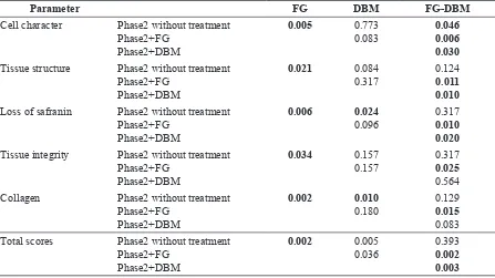
Table 2. Inter-groups evaluation in cell character, tissue structure, tissue integrity, proteoglycan and collagen
Parameter FG DBM FG-DBM
Cell character Phase2 without treatment Phase2+FG Tissue structure Phase2 without treatment
Phase2+FG Loss of safranin Phase2 without treatment
Phase2+FG Phase2+DBM
0.006 0.024
0.096 0.3170.010 0.020 Tissue integrity Phase2 without treatment
Phase2+FG Phase2+DBM
0.034 0.157
0.157 0.3170.025 0.564 Collagen Phase2 without treatment
Phase2+FG Total scores Phase2 without treatment
Phase2+FG Phase2+DBM
0.002 0.005
resorption were statistically significant
different from the FG group regarding tissue
degradation.
The decrease of TGF β expression in
various treatments showed no statistically
significant difference between phase 1
and phase 2, 26.91pg/mg to 24,13 pg/
mg. FG intervention elevated TGF β level
from 26.91 to 31.90 pg/mg ( no significant
difference).
TGF β expression in DBM group
showed significant decreased from 26.91
to 12.70 pg/mg (p = 0.009; CI 6.14-19.26)
,whereas there was no statistically different
decrease TGF β expression in FG-DBM
intervention showed no significant decrease
compared with control group; but it was found
significant different from FG intervention
group (p=0,013; CI 12,82-27,35).
By Spearman correlation test, a
moderate correlation (r=0,70) was found
between apoptosis and TGF β expression in
control group. Weak correlations were found
between apoptosis and
TGF β expresion in allTable 3. Cartilage degradation scores
Group
Cartilage degradation
Total Mild
1-4
Moderate 5-8
Severe 9-12
Control 0 (0%) 10 (83.3%) 2 (16.7%) 12 (100%)
FG 11 (91.7%) 1 (8.3%) 0 (0%) 12 (100%)
DBM 4 (33.3%) 8 (66.7%) 0 (0%) 12 (100%)
FG-DBM 0 (0%) 8 (66.7%) 4 (33.3%) 12 (100%)
Total 15 (31.3%) 27 (56.3%) 6 (12.5%) 48 (100%)
Control – FG, p<0.002 (Wilcoxon test) Control– DBM, p=0.034 (Wilcoxon test) Ccontrol-FG-DBM, p=0.317 (Wilcoxon test) FG-DBM, p=0.008 (Wilcoxon test)
FG-FG-DBM, p=0.002 (Wilcoxon test) DBM-FG-DBM, p=0.005 (Wilcoxon test)
Picture 2. A). Mankin score, total score as base line parameter. B.)Total score control group 7. Fibrosis 1, cell character 1, proteoglycan 2, homogenity 1, collagen 2,) C.) FG total score 2.( fibrosis 0, cell character 0, proteo
-glycan 1, homogenity 0, collagen 1,) D.) DBM Total score 5( Fibrosis 1, cell characyter 2, proteo-glycan 1, homo
chondrocytes and shows specific activity
(TGF β), cell viability and activity could be
In this study, TGF β1 expression has been
20,21
Collagen expression and excessive
fibrous tissue due to cartilage damage have
correlation with clinical finding after surgery.
22,23
TGF β expression. DBM can still be used
Dr. Endang Ch. Mangunkusumo,
SpTHT-KL(K), Department of Otorhinolaryngology
Universitas Indonesia/Dr. Cipto
Mangunkusumo Hospital Jakarta
1. Donald PJ. Cartilage grafting in facial irradiated graft. Laryngoscope. 1986;
786-2. Kridall RWH, Konior RJ. Irradiated Head Neck Surg. 1993; 119: 24-31.
3. Stucker FJ, Shaw GY. Biologic tissue implants. In: Papel ID, editor. Principles of York: Thieme; 2002. p.78-83.
typical microtia: Personal experience based on 352 microtic ear corrections. Scand J Plast Reconstr Hand Surg. 1998; 32: 35-47.
5. Wang TD. Auricular reconstruction. New York: Thieme; 2002. p.615-34
6. Britt JC, Park SS. Autogenous Head Neck Surg. 1998; 124: 671-7.
congenital microtia (grade III). Laryngoscope. 1996; 106(12): 1-26.
8. Schuller DE, Bardach J, Krause CJ. Irradiated restoration. Arch Otolaryngol. 1977; 103: 12-5.
9. Stucker FJ. Use of implantation in facial deformity. Laryngoscope. 1997; 87: 1523-7. 10. Goldring MB. Interleukin-1 Beta-Modulated
chondrocytes. J Clin Invest. 1994; 94: 2307. 11. Langer R, Vacanti JP. Tissue Eng. 1993;
260(5110): 920-6.
12. Naumann A, Aigner J, Staudenmaier R, Seeman M, Bruening R, Englmeier KH, et al. Clinical aspects and strategy for biomaterial
Otorhinolaryngol. 2003; 260: 568-75.
13. Lin Z, Willers C, Xu J, Zheng M-H. The Tissue Eng. 2006; 12(7): 1971-84.
14. Blom AB. BergWBvd. The Synovium and its role in osteoarthritis. In: Bronner F, Farach-Carson MC, editors. Bone and osteoarthritis. London: Springer; 2007. p.65-79.
DISCUSSION
FG plays a role as conductive substance
of growth factor to the targeted delivery
system. It served as scaffold which is an
application of tissue engineering, so that
cell viability and quality of extra cellular
matrix can be maintained.
16,17TUNEL on control group showed
increase of apoptosis into 48.48%+/-21.1
after 12 weeks. FG group showed decrease
of apoptosis into 19.83% +/-11.61. It might
be possibly caused by the existence of FG as
scaffold. FG will cause attachment between
graft and skin and other tissues. Such
as in physiological injury therapy, it will
produced fibroblast in proliferation phase.
During that time, various growth factors will
be produced.
FG (Beriplast
®) is available in local
market or, as an option we can use autologous
fibrin glue. The commercial preparation
consists of human plasma protein (fibrinogen,
active factor XIII, thrombine, apopthronine
and calcium chloride). In culture cell, FG
can be used to increase proliferation of
chondrocyte, and extra cell matrix such as
proteoglycan and collagen 4-8 times after
the 14
thday.
18In clinical experience, a reconstructive
surgeon can use FG mixed with a piece
of bone or cartilage, which filled into the
cartilage defect as chondrocyte conductive
material during the healing process.
16This study confirmed that FG is useful
as chondrocyte inductor and was able
to increase cell proliferation and matrix
regeneration in facilitating cartilage repair.
19In DBM there is a bone morphogenetic
protein (BMP), which is a derivate of TGF
β1 superfamily. It is a protein expressed by
intervention groups .
Resorption was evaluate among
intervention group to FG-DBM groups.
(<0.002). Mild resorption occured in FG
group followed by DBM group. FG-DBM
and control group showed moderate and
severe resorption. There were significant
differences in FG and DBM compare to
FG-DB control group.
TGF β level in various interventions
showed in table 5.8. TGF β level (tissue
pg/mg) was found in all tissue degradation
between phase 1 and phase 2 from 26.91
into 24, 13 tissue pg/mg (not significant).
FG group showed increased of TGF β level
from 26.91 into 31.90 tissue pg/mg (not
significant).
Table 4. TGF β level (tissue pg/mg) before and after 12 weeks grafting
Variable Average pg/mg tissue SD 95% CI p* p¶ pᴲ p┴ * paired t test TGF β level between phase 1 and phase2
chondrocytes and shows specific activity
to the target cells. This growth factor is
expressed during recovery process and
used in the management of cartilage or
bone defect. It has been proven in vitro
that by adding transforming growth factor
(TGF β), cell viability and activity could be
maintained, and matrix production increased.
In this study, TGF β1 expression has been
proven that it could prevent the occurence of
cartilage graft degradation.
20,21Collagen expression and excessive
fibrous tissue due to cartilage damage have
correlation with clinical finding after surgery.
Subcutaneous tissue thickening occurred and
covered detailed ear frame silhouette ear sub
unit. For this condition Siegert
22,23proposed
corrective subunit auricle surgery.
Fibrin Glue is highly recommended
to be used in microtia reconstruction in
order to increase chondrocyte viability with
minimal tissue degradation of autologous
cartilage graft. It correlates with increased
TGF β expression. DBM can still be used
alternative to maintain chondrocyte viability,
proteoglycans, and collagen.
Acknowledgements
Authors are grateful to:
Dr. Endang Ch. Mangunkusumo,
SpTHT-KL(K), Department of Otorhinolaryngology
Head and Neck Surgery, Faculty of
Medicine
Universitas Indonesia/Dr. Cipto
Mangunkusumo Hospital Jakarta
REFERENCE
1. Donald PJ. Cartilage grafting in facial reconstruction with special consideration of irradiated graft. Laryngoscope. 1986; 786-807.
2. Kridall RWH, Konior RJ. Irradiated cartilage graft in the nose. Arch Otolaryngol Head Neck Surg. 1993; 119: 24-31.
3. Stucker FJ, Shaw GY. Biologic tissue implants. In: Papel ID, editor. Principles of facial plastic and reconstructive surgery. New York: Thieme; 2002. p.78-83.
4. Firmin F. Ear reconstruction in cases of typical microtia: Personal experience based on 352 microtic ear corrections. Scand J Plast Reconstr Hand Surg. 1998; 32: 35-47.
5. Wang TD. Auricular reconstruction. Reconstructive Surgery of the Face and Neck. New York: Thieme; 2002. p.615-34
6. Britt JC, Park SS. Autogenous tissue-engineered cartilage. Arch Otolaryngol Head Neck Surg. 1998; 124: 671-7.
7. Aguilar EF. Auricular reconstruction of congenital microtia (grade III). Laryngoscope. 1996; 106(12): 1-26.
8. Schuller DE, Bardach J, Krause CJ. Irradiated homologous costal cartilage for facial contour restoration. Arch Otolaryngol. 1977; 103: 12-5.
9. Stucker FJ. Use of implantation in facial deformity. Laryngoscope. 1997; 87: 1523-7. 10. Goldring MB. Interleukin-1 Beta-Modulated
gene expression in immortalized human chondrocytes. J Clin Invest. 1994; 94: 2307. 11. Langer R, Vacanti JP. Tissue Eng. 1993;
260(5110): 920-6.
12. Naumann A, Aigner J, Staudenmaier R, Seeman M, Bruening R, Englmeier KH, et al. Clinical aspects and strategy for biomaterial engineering of an auricle based on three-dimensional stereolithography. Eur Arch Otorhinolaryngol. 2003; 260: 568-75.
13. Lin Z, Willers C, Xu J, Zheng M-H. The chondrocyte: biology and clinical application. Tissue Eng. 2006; 12(7): 1971-84.
15. Thorn, FoghW, MAndersen. Autologous fibrin glue with growth factors in reconstructive maxillofacial surgery. Int J Oral Maxillofac Surg. 2004; 33(2004): 95-100.
16. Arevalo-Silva CA, Cao Y, Vacanti M, Weng Y, Vacanti CA, Eavey RD. Influence of growth factors on tissue-engineered pediatric elastic cartilage. Arch Otolaryngol Head Neck Surg. 2000; 126:1234-8.
17. Bos PK, Osch GJVMv, Frenz DA, Verhaar JAN, Verwoerd-Verhoef HL. Growth factor expression in cartilage wound healing: temporal and spatial immunolocalization in a rabbit auricular cartilage wound model. Osteoarthritis Cartilage. 2001; 9:382-9. 18. Sah RLL, LM Schmidt TA Mankarious, Ska.
Effects of fibrin glue component on chondrocyte growth and matrix formation. 2007(49th annual meeting orthopedy society).
19. Hidaka C, Goodrich LR, Chen C-T, Warren RF, Crystal RG, Nixon AJ. Acceleration of Cartilage Repair by Genetically Modified Chondrocytes Over Expressing Bone Morphogenetic Protein-7. J Orthop Res. 2003; 21:573-83.
20. Fukui N, Sandell LJ. Anabolic mediators of cartilage healing. In: Bronner F, Farach-Carson MC, editors. Bone and osteoarthritis. London: Springer; 2007. p.97-108.
21. Bronner F, Chai DH,Steven AL,Grodzinky AJ. Biomechanical Aspect: Joint injury and osteoarthritis. In: Bronner F, Farach-Carson MC, editors. Bone and osteoarthritis.4th ed London: © Springer-Verlag 2007. p.165-80. 22. Siegert R, Magritz R. Reconstruction
of the auricle. GMS Current Topics in Otorchinolaryngology-Head and Neck Surgery. 2007; 6:1865-011.
23. Sulcus construction in microtia repair. A retrospective comparison of different technique. ShayI, Duvdevani, Ralph Magritz, Ralf Siegert. JAMA Facial Plast Surg. 2013; 15(1): 17-20