VOL. 3, NO. 1, pp. 58 – 62, January, 2013
Study of Necrosis in the Liver of Formaldehyde and Benzo(
α
)Pyrene
Exposured-Mice
Ahmad Soni, Sri Widyarti*, Aris Soewondo
Biology Department, Faculty of MathematicsandNatural Sciences, Brawijaya University, Malang, Indonesia
ABSTRACT
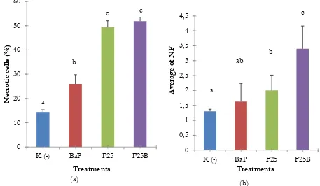
Formaldehyde and benzo(α)pyrene are compounds that harmful for health. Misapplication of this compound has an impact in the form of organ damage in the body. This study aims to determine t he impact of the treatment of the combined exposure of formaldehyde and benzo(α)pyrene to cell necrosis in the liver of mice (Mus musculus). Treatment of formaldehyde dose of 25 mg/kg BW to mice was given orally every day for 60 days. Treatment of benzo(α)pyrene via ip at a dose of 250 mg/kg BW were given after 30 days of incubation with four times injection with one day interval. Liver organ histological preparations were made through the HE staining and were observed by BX51 microcopy. The data obtained that is the percentage of cells necrosis and necrotic foci. This research used Completely Randomized Design (CRD) with 95% confidence interval. Liver organ preparations observations indicate that the percentage of necrosis in the untreated control, benzo(α)pyrene 250 mg/kg BW, formaldehyde 25 mg/kg BW, combination of formaldehyde 25 mg/kg BW with benzo(α)pyrene in a row that is equal to 14.43% ± 0.91; 26.05% ± 3.75; 49.38% ± 2.66; 51.86 ± 1.73. The mean of necrotic foci in liver organ formed in the untreatment control, benzo(α)pyrene 250 mg/kg BW, Formaldehyde 25 mg/kg BW, and the combination of formaldehyde 25 mg/kg BW with benzo(α)pyrene in a row, equal to 1.3 ± 0,07; 1.63 ± 0.61; 2 ± 0.51, and 3.4 ± 0.76. This suggests that the combined treatment had the highest level of toxicity compared with other treatments.
Keywords: benzo(α)pyrene, formaldehyde, liver, necrosis
INTRODUCTION
Formaldehyde are compound that is naturally produced by the body through the metabolic processes. However, the current misuse of formaldehyde had crossed the threshold, particularly in developing countries like Indonesia. Previous study, showed that treatment with formaldehyde may degrade the active cell to divide, inhibition of spermatogenesis process, and also the depletion of the basement membrane of the seminiferous tubules [1]. Exposure to formaldehyde also may increase the risk of changes in the gastric mucosa of mice (Mus musculus). Changes in the gastric mucosa of mice is shown by the widening of the parietal cells in gastric mucosa and the destruction of parietal cells and chief cells [2]. Benzo(α)pyrene
*
Corresponding author: Sri Widyarti
Biology Department, Faculty of Mathematics and Natural Sciences, Brawijaya University, Jl. Veteran, Malang, Indonesia 65145
Email: [email protected]
(BaP) are compound whose the structure has a five-ring polycyclic aromatic with hydrocarbons bonding. This substance is carcinogenic and can lead to cancer if these substances enter the cell. Benzo[α]pyrene can initiate the occurrence of mutations in the genes if exposed continuously [3]. The induction of benzo(α)pyrene in liver can induce changes in cell morphology, there is vakuolization, narrowing of the sinusoids and swollen nucleus. In the lungs there are change in the form of proliferation of bronchial epithelial layer [4]. Besides, the induction of benzo(α)pyrene in the liver can causes piknosis and karyolysis on the first day of incubation [5].
expression of these genes can initiate cell to undergoing necrosis that is concerned to have a greater likelihood for the occurrence of cancer compared to a tissue that has a normal PARP gene expression [6, 7].
Histological observations of the organs of mice that will prove an increase in the percentage of cells undergoing necrosis and necrotic foci in the liver organs of mice (Mus musculus) after exposure to formaldehyde and benzo(α)pyrene.
MATERIALS AND METHODS
All procedures were performed has been accepted by the Research Ethics Committee of the UB with the number 30-KE.
Animals Treatment
The animals that used in this study were two-months old male mice weighing between 20-30 grams. Animal treatment carried out for two months. The animals were divided into 4 groups i.e control (no treatment), formaldehyde, BaP, formaldehyde and BaP.Formaldehyde 25 mg/kg BW was given orally for 8 weeks everyday. Total dose of BaP 250 mg/kg BW was givenafter 30 days of incubation with four times the injection interval of one day.
Histological preparation
The first step taken is the organ liver of mice (Mus musculus) that have been exposed to formaldehyde, benzo(α)pyrene and the combination between formaldehyde and benzo(α)pyrene exposure were fixation first with 4% formaldehyde solution for 24 hours. Liver organ then soaked in 70% alcohol for 3 times for 10 minutes respectively while shaken. The specimen was then dipped in 70% alcohol for overnight. Followed by immersed in alcohol 80%, 3 times for 10 minutes respectively while shaken. The specimens then soaked with alcohol at a concentration of 90% and 96%. Followed by immersed in absolute alcohol and shaken for 10 minutes. Next, the specimens were soaked with xylol for ± 30 minutes. Infiltration process is carried out by comparison xylol : paraffin in a row equal to 3:1, 1:1, and 1:3 respectively for 30 min at 520 C. Continued with the provision of pure paraffin at a temperature of 56° C for 12 minutes. Next the specimens were blocking and sectioning with a chunk size of ± 5 microns. Tape the pieces placed on glass objects that have been given mayer albumin with a brush and put on the hot plate. Then, the specimens were stained by Haematoxylin-Eosin staining.
Observation
The first step is to perform observations and estimates of the percentage of necrosis in the field of view. Observations made using the Olympus BX51 binocular microscope with a magnification of 200x and 400x for liver organ specimens. In the liver organ, the observation is determined by taking five random fields of view, then in each field of view of the calculated number of total cells and subsequently in the average. The average results are then used as a reference the total number of cells in each field of view. Then performed counting the number of cells undergoing necrosis and the result is converted to a percentage.
Data Analysis
The data that was obtained will be sorted first by eliminating the outliers of data using the upper and lower limits based on data obtained from observations. Furthermore, this study used Completely Randomized Design (CRD) with 95% confidence interval. Analysis of significance by using ANOVA followed by Tukey-HSD test. The data used is the percentage of cells undergoing necrosis of the organ liver of mice exposed to benzo(α)pyrene and formaldehyde in toxic doses, and the average spotformed necrotic foci in liver organ of mice.
RESULTS AND DISCUSSION
Results
Necrosis and Necrotic Foci in Liver
Based on data analysis has been done, it is known that there is a difference between the control without treatment, and treatment of benzo(α)pyrene (BaP) with formaldehyde treatment dose of 25 mg/kg BW and the combined treatment of formaldehyde 25 mg/kg and benzo(α)pyrene in liver organ.
(a)
(b)
Figure 1. Data analysis of observations. (a) The percentage of cell necrosis in the liver organ. (b) Average of spots necrotic foci on liver organ. K (-): Control; BaP : Benzo(a)pyrene; F25: Formaldehyde 25 mg/kg BW; F25B: Combination Treatment.
Figure 2. Spot necrotic foci in the liver organ. (a) control without treatment, (b) treatment of BaP 250 mg/kg BW, (c)
treatment of formaldehyde 25 mg/kg BW, (d) the combined treatment of formaldehyde 25 mg/kg BW and BAP 250 mg/kg BW. Necrotic foci showed by the black arrows and whi te circle. (HE, Scale : 300 µm)
0 10 20 30 40 50 60
K (-) BaP F25 F25B
N
ec
roti
c
c
ells
(
%
)
Treatments a
c c
b
0 0,5 1 1,5 2 2,5 3 3,5 4 4,5
K (-) BaP F25 F25B
A
ve
rage
of
N
F
Treatments a
ab
b
Thus, the more extreme treatment can cause the more cells undergoing necrosis in liver of mice. Observations of liver organ preparations indicate there are formation of necrotic foci in each treatment, but with a different distribution (Figure 1). The mean necrotic foci are formed in the control treatment with no treatment, benzo(a)pyrene 250 mg/kg BW, Formaldehyde 25 mg/kg BW, and the combined formaldehyde 25 mg/kg BW with benzo(α)pyrene in a row, which is 1.3 ± 0.07 ; 1.63 ± 0.61; 2 ± 0.51, and 3.4 ± 0.76. Histological cross section of necrotic foci in liver organ preparations is shown in the Figure 2.
Discussion
Necrosis is a process of cell death that occurs as a result of exposure to toxic substances in high doses and occur suddenly. Necrosis also known as the end result of a catastrophic bioenergetic caused by ATP depletion due to the level that does not allow cells to repair themselves or apoptosis. The cause of ATP depletion itself due to the toxic substances that enter or the presence of physical damage. Necrosis can be characterized morphologically, that is the presence vakuolation the cytoplasm, plasma membrane rupture and induce inflammation in cells undergoing necrosis by removing debris of cells undergoing necrosis and release several proinflamatory molecules. Furthermore, a collection of debris cell and infiltration of lymphoid cells will form the necrotic foci [8]. Observations that have been done, show that the necrotic foci are formed mostly located around blood vessels, that is in the hepatocytes where located around the central vein. Necrotic foci formed by the cells undergoing lysis, so that the lysosomes of cells that have undergone cell lysis will be digestingaround its. This process will then result in cell debris, so can initiating infiltration of phagocytic cells [8].
Observations on liver organ showed that in each treatment reveal any cells undergoing necrosis, but in different percentages. This is due to necrosis is also a phenomenon that occurs in any organism if they get an infection, but in very small quantities [8], as well as in the control treatment. Some types of necrosis that can be found in the liver is the organ preparations piknosis, kariorrhexis, and karyolysis with different distributions in each treatment.
Cells undergoing necrosis and necrotic foci were also found in liver organ without treatment. This is due to necrosis is a phenomenon that
occurs in every living being, but the percentage is very small, so it doesn’t interfere with the metabolism in the body itself. It is highly related to the metabolism that occur in each organ. As is known, the liver is a detoxification organ where toxic substances that enter the body, so it is possible in these organs were also found to have necrosis of hepatocytes cells, although not treated with the treatment of toxic substances.
The previous research showed that there was increased expression of PARP-1 protein with increasing doses of benzo(α)pyrene and formaldehyde treatment given to the liver of mice. Based on the PARP protein fragment, there are indications that the event occurs cell necrosis in liver of mice [9]. DNA damage in the initial level will not lead to apoptosis or necrosis due to the presence of PARP-1 protein responsible for DNA repair process [10]. DNA damage levels are going to initiate cell apoptosis because the cells are not able to repair DNA damage that occurs, but cells in this case still has the ATP in sufficient quantities to perform apoptosis. However, DNA damage at higher levels will initiate the cells to undergo necrosis due to the over expression of PARP-1 resulting in ATP depletion. This resulted in ATP in the cell does not have sufficient numbers to perform apoptosis, so cells will lead to necrosis [7,11,12,13].
The cells undergoing DNA damage will take the process of DNA repair by activating protein PARP-1. Activation of this protein will lead to the occurrence of poly (ADP-ribosyl)ation there was a key process in DNA repair by using NAD+ as substrate. Then, NAD+ will be converted into nicotinamide and ADP-ribose. At low level of DNA damage, the cell will activate the protective homeostatic process that can repair the damage. If a strand of DNA damage has been repaired, the cell will remain alive and cellular NAD+ level will be increased again by converting nicotinamide. The conversion process requires two ATP molecules per one molecule of nicotinamide is converted to NAD+. If the repair process can not run properly, then the cell will make the process of apoptosis through caspase-dependent mechanism, for example is caspase 3 [14]. However, if the DNA damage that occurs is too severe, then the cell will not be able to repair such damage and this case can lead hyper activation of PARP-1 protein. This hyper activation process resulting in depletion of cellular NAD+ and ATP that direct cell death through the mechanism of necrosis [13].
treatments. This is evident from the distribution of necrotic foci formed in the liver organ of mice. In this study suggests that high exposure to formaldehyde can cause infiltration of lymphoid cells in liver organ around the spaces between cells, and also the occurrence of dilatation of the artery. According to previous research, showed that the benzo(α)pyrene may increase the accumulation of p53 protein. Increased accumulation of p53 protein is associated with an increased number of bonds between the BPDE-DNA, where this bond can lead to the dissolution of single chains of DNA. The breakdown of single chains of DNA can activate PARP-1 for DNA repair [15]. Furthermore, high doses of formaldehyde and benzo(α)pyrene treatment on this study can improve the over expression of the PARP protein itself, so as to direct the cells to undergo necrosis. This is what led to the combined treatment of formaldehyde 25 mg/kg BW with benzo(α)pyrene 250 mg/kg BW had a more severe effects than other treatments.
CONCLUSION
This research performed at each treatment reveal any organ like liver undergoing necrosis, but with different percentages. In general, the percentage of cells undergoing necrosis increased with increasing treatment extremities. The highest percentage of necrosis seen in the combined treatment of formalin 25 mg/kg BW with benzo(α)pyrene 250 mg/kg BW, while the highest rates of necrotic foci are formed in the combined treatment of formalin 25 mg/kg BW with a benzo(α)pyrene dose of 250 mg/kg BW.
REFERENCES
1. Golalipour MJ, Azarhoush R,Ghafari S,Gharravi
AM, Fazeli SA, Davarian A (2007) Formaldehyde
exposure induces histopathological and
morphometric changes in the rat testis. Folia Morphol. 66 (3) : 167-171.
2. Priambowo A (2010) Histopathology studies of
gastric mucosal mice (Mus musculus) after
exposure to subcronic formaldehyde. Skripsi Not
Published. Biology Department. Brawijaya
University. Malang.
3. Aygün SF, Kabadayi F(2005) Determination of
benzo(α)pyrene in charcoal grilled meat samples
by HPLC with fluorescence detection.
International Journal of Food Sciences and Nutrition. 56(8) : 581-585.
4. Yunus M(1996) Induction benzapiren influence
on liver histology and lung of white rat (Rattus
norvegicus). Skripsi not published. Biology Department. Brawijaya University. Malang. 5. Pinuji HM (1999) Liver histopathology and lien
of young Rattus norvegicus after induction
benzapiren. Skripsi not published. Biology Department. Brawijaya University. Malang.
6. Putri PF(2010) Gene expression of PARP-1
mRNA liver and lung of mice (Mus musculus) exposed to formalin and benzo(α)pyrene. Skripsi not published. Biology Department. Brawijaya University. Malang.
7. Filipovic DM, Meng X, Reeves WB (1999)
Inhibition of PARP prevents oxidant-induced necrosis but not apoptosis in LLC-PK1 Cells.Am J Physiol Renal Physiol. 277 : 438-476.
8. Edinger AL, Thompson CB(2004)death by
Design: apoptosis, necrosis and autophagy. Current Opinion in Cell Biology. 16: 663-669.
9. Maghfironi A(2010) Study of PARP-1 protein
expression in liver and lung of mice exposed to formalin and benzopiren. Skripsi not published. Department of Biology. Brawijaya University. Malang.
10. Wang ZQ, Stingl L, Morrison C, Jantsch M, Los M, Schulze-Osthoff K, Wagner EF(1997) PARP
is Important for genomic stability but
dispensable in apoptosis. Genes and
Development 11 : 2347-2358.
11. Bey EA, Bentle MS, Reinicke KE, Dong Y,
Chin-Yang R, Girard L, Minna JD, Bornmann WG, Gao J, Boothman DA(2007) An NQO1- and PARP-1-mediated cell death pathway induced in non-small-cell lung cancer cells by β-lapachone. National Academy of Sciences of USA. 28(104) : 1-6.
12. Erdelyi K, Bakondi E, Gergely P, Szabo C, Virag
L(2005) Poly-ADP-ribosylation in health and disease pathophysiologic role of oxidative
stress-induced poly (ADP-ribose) polymerase-1
activation: Focus on cell death and
transcriptional regulation. Cellular and Molecular Life Sciences. 62: 7-8.
13. Moubarak RS, Yuste VJ, Artus CD, Bouharrour
AD, Greer PA, Murcia JM,Susin SA (2007) Sequential activation of poly (ADP-Ribose) polymerase 1. calpains, and bax is essential in apoptosis-inducing factor-mediated programmed necrosis. Journal Molecular and Cellular Biology. 13(27): 4844-4862.
14. Chaitanya GV, Alexander JS, Babu PP(2010)
PARP-1 cleavage fragments: Signatures of cell-death proteases in neurodegeneration. Biomed Central. 8(31): 1-11.
15. Serpi R (2003) Mechanism of benzo(α)pyrene