Level of 25(Oh)D Serum, Expression of Interleukin 4 and Glucocorticoid Receptor of
Mononuclear Cell in Steroid Resistance Nephrotic Syndrome Children
Vonny Mariany Deckert1
, Krisni Subandiyah2*
, Loeki Enggar Fitri3
1
Magister Program in Biomedical Sciences, Faculty of Medicine, University of Brawijaya Malang
2Department of Pediatric, Saiful Anwar General Hospital, Malang 3Department of Parasitology, Faculty of Medicine, University of Brawijaya Malang
ABSTRACT
Nephrotic syndrome (NS) is an autoimmune disease and its steroid resistance status supposed correlate with 25(OH)D level and IL-4 expression. The aimed of this study was investigated 25(OH)D plasma level, IL-4 and GR expression of PBMC in steroid sensitive and resistant pediatric NS patients and the association of those parameters. 27 subjects were divided into three groups (control group, steroid resistant NS group, and steroid-sen-sitive NS group). Peripheral blood mononuclear cells (PBMCs) isolated using the Ficoll-Hypaque method. Plasma 25(OH)D level was measured using ELISA method. IL-4 and GR expression were measured using flow cytometry of PBMCs. This study showed that 25(OH)D level and GR expression were significantly different in the control group compared to steroid resistant NS group (p<0.05). Plasma 25(OH)D level, IL-4, and GR expression were not correlated each other in NS patients (p>0.05). Plasma 25(OH)D level, IL-4 and GR expression were not contrib-uted to steroid resistance in NS patients. However, GR expression has the highest contribution to steroid resis-tance of NS patient (Wald score 1.198). Plasma 25(OH)D level and GR expression were lower in steroid resistant NS group. GR expression has the highest contribution in steroid resistance of NS patients.
Keywords: 25(OH)D level, GR expression, IL-4 expression
Nephrotic syndrome (NS) defined as a glomerular disease that characterized by proteinuria, edema, and hypoalbuminemia. This disease could affect all ages, es-pecially 2-6 years old child with male: female ratio [1]. Nephrotic syndrome occurred 2-7 new case per 100.000 children [2]. Previous data had been reported that there was 101 case (34%) idiopathic NS from total 297 nephrology disease at Pediatric Department of Saiful Anwar Hospital [3].
Idiopathic NS is an autoimmune mediated disease that both steroid sensitive and steroid resistant pos-sessed T cell dysfunction, decreased of 25(OH)D level, and glucocorticoid receptor (GR) expression. Nephrotic syndrome caused by an imbalance of Th1 and Th2 cytokines that could trigger podocyte dysfunc-tion and massive proteinuria [4]. IL-4 and IL-13 could induce proteinuria in idiopathic NS patient [5].
A previous study showed that glucocorticoid recep-tor (GR) expression increase in the remission stage of NS and decreased GR expression in steroid resistant NS compared with steroid sensitive NS [6]. Further-more, GR expression negatively correlated with histopathological damage of glomerulus [7].
25(OH)D level in NS patients also decrease caused by renal function impairment. Takeda has been de-scribed that an active form of vitamin D3 could modu-late the immune response [8]. Vitamin D3 could re-verse GR downregulation induced by ligand through its interaction with GC signaling pathway [8]. The role of vitamin D3 in immune response had been studied in steroid resistant asthmatic patients. A study showed that administration active form of vitamin D3 could in-crease IL-4, IL-5, IL-13 expression by Th2 and IL-10 and TGF by Treg [9].
Previous study showed enhancement of various cy-tokines production such as IL-1 IL-2, IL-4 and TNF-α, and also decreased of IL-10 production in minimal changes NS patient’s peripheral blood mononuclear cells (PBMC) [10].
Its immunomodulatory effects of vitamin D could INTRODUCTION
*Corresponding author: Krisni Subandiyah
Department of Pediatric, Saiful Anwar General Hospital, Malang, Indonesia
suppress Th1: IFN-Ɣ; IL-2; Th17: IL-17 IL-1; IL-6; TNF-α; 12; 23 secretion, suppression of Treg: IL-10; TGF-β and also enhancement of Th2: IL-4; IL-5; IL-13 secretion [11]. In other hands IL-4 and IL-2 de-crease GRα without affect GRβ, which further con-cluded that steroid resistance induced by IL-4 and IL-2 and marked by decreased expression of GRα [12].
This study was conducted to investigate 25(OH)D plasma level, IL-4 and GR expression of PBMC in steroid sensitive and resistant pediatric NS patients and their association.
Study Design
This study was cross-sectionally designed to mea-sure vitamin D (25(OH)D) plasma level, GR and IL-4 expression of PBMC in steroid sensitive NS, steroid re-sistant NS, and control group. This study was con-ducted in nephrology policlinic and ward of Pediatric Department, Dr. Saiful Anwar Hospital Malang, and Biomedical Laboratory, Faculty of Medicine, Brawijaya University Malang. This study had been approved by Ethical Committee of Medical Faculty of Medicine, Brawijaya University.
Population and Subject
Research subject included in this study was nine subjects in each group. Inclusion criteria of topics are classified as steroid sensitive and steroid resistant NS aged 1-14 years old and allowed by his/her parents to join in this study (informed consent). Exclusion criteria of subjects is NS which classified as secondary NS (caused by extra-renal factor such as chemical/drugs, systemic disease such as SLE or Henoch Schonlein, in-fectious disease such as malaria, hepatitis), congenital/infantile NS, patient with autoimmune dis-ease (SLE, DM), patient with allergy (asthma, rhinitis, atopic dermatitis), patients with severe infection, liver dysfunction, and patient with vitamin D supplementa-tion.
PBMC Isolation
Blood sample in Vacutainer EDTA/heparin mixed properly, and then mixed with PBS (1:1). Blood sam-ple-PBS take using micropipette and then overlaid to falcon tube which already contains Ficoll-Hipaque d=1.077 g/mL through its wall (volume sample: Ficoll-Hipaque 1:1). This mixture then centrifuged at room temperature 1000 rpm for 30 min. After centrifuged, this mixture will form 4 layer (plasma, PBMC, Ficoll-Hipaque, and granulocyte or erythrocyte). PBMC ring
has taken slowly using micropipette and then placed in new 15 mL centrifuge bottle. PBMC solution and then washed with PBS 10 mL and centrifuged at room temperature, 1200 rpm for 10 min. Pellet washed again with PBS and then centrifuged at room temperature, 1200 rpm for 10 min (this process is repeated two times). PBMC formed as a pellet in the bottom of the bottle.
Measurement of GR Expression in CD3
PBMC’s pellet was added with PE-conjugated anti-human CD357 (GITR) antibody (catalog number 311604) and then incubated for 15 min in the dark room. After that, the pellet was centrifuged at 2500 rpm for 3 min. Then, the pellet was added to one mL fixation buffer solution in each tube, centrifuged at 2500 rpm, 4°C for 3 min. The supernatant was re-moved. Cell pellet that has been fixated then resus-pended by added 1x permeabilization washed buffer and centrifuged at 2500 rpm, 4°C for 3 min. After this process, the cell pellet was resuspended in added 0,5 ml cell staining buffer and then analyzed using flow cy-tometry. Expression of GR analyzed using software BD Cell Quest Pro.
Measurement of IL-4 Expression
PBMC’s pellet was added with PerCP/Cy5.5 anti-human IL-4 (catalog number MP 4-25D2) and then in-cubated in the dark room for 15 min. After that, the pellet was added to 1 mL cell staining buffer and then centrifuged for 3 min at Mystral-1000 rt and hatch cen-trifuge at 2500 rpm, room temperature continuously. Supernatant was removed, then added with 1 ml fixa-tion buffer, incubated for 15 min in dark room, room temperature, and centrifuged for 3 min in Mystral-1000 rt and hettch centrifuge at 2500 rpm at room tempera-ture continuously.
The supernatant was removed, the pellet was added with permeable wash buffer and then incubated for 15 min in a dark room at room temperature. After this process, centrifuged again for 3 min in Mystral-1000 rt and hatch centrifuge at 2500 rpm at room temperature continuously. The supernatant was removed, and the pellet was added with cell staining buffer 500 µl, and then placed in a sample tube for flow cytometry ma-chine, BD Facs Calibur. IL-4 expression analyzed using software BD Cell Quest pro.
Measurement of Plasma 25(OH)D Level
solution was added to all tube, and then vortex for 10 seconds. Each of calibrator dilution added 200 µl, con-trol or sample in each appropriate well according to an antibody-coated plate/microplate. The plate was cov-ered with plastic and then incubated in 18-250C for
two hours. All well then washed three times using wash solution. 200 µl conjugate were added to each well using multichannel pipette. Plate covered again with plastic, incubated in 18-250
C for 30 min (this process repeat 2 times). 200 µl TMB substrate was added into each well, and then covered with plastic, in-cubated in 18-250
C for 30 minutes. 100 µl stop solution was added to each well. Absorbance was measured in each well at 450 nm using microplate reader in 30 min after added stop solution.
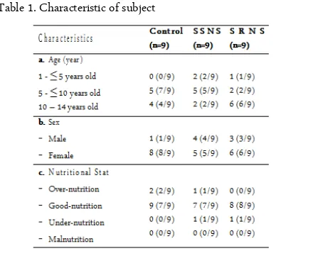
Characteristics of Subjects
This study involved 27 subjects divided into three groups: control group, steroid resistant NS group, and steroid-sensitive NS group. The subject characteristic such as sex, age, nutrition status described in Table 1.
Level of Plasma 25(OH)D
Figure 1 showed 25(OH)D level was 23.86 ng/mL for the control group, 15.50 ng/mL for steroid sensitive NS group, and 10.88 ng/mL for steroid-resistant NS group. One way ANOVA showed significant differ-ences between groups (p<0.05). Post Hoc Tukey test showed significant differences between the control group and steroid resistant group (p = 0.003).
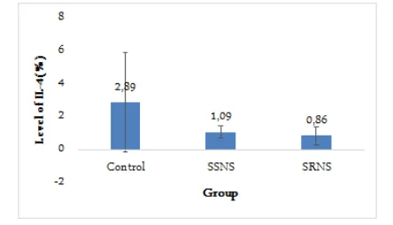
IL-4 Expression
Figure 2 showed IL-4 expression was 2.89% for the control group, 0.86% for steroid-resistant NS group, and 1.09% for steroid-sensitive NS group. Non-para-metric test Kruskal-Wallis showed no significant differ-ences among groups (p = 0.095).
GR Expression
Figure 3 showed GR expression was 3.86% for the control group, 1.74% for steroid-resistant NS group, and 2.66% for steroid-sensitive NS group. One way ANOVA showed significant differences between groups (p<0.05). Post Hoc Tukey test showed signifi-cant differences between the control group and steroid-resistant group (p = 0.008).
Correlation of Plasma 25(OH)D Level, IL-4 and GR Expression
Pearson correlation test showed no significant
cor-relation between 25(OH)D level and IL-4 expression (p>0.05, r = 0.382), 25(OH)D level and GR expression (p>0.05, r = 0.417), IL-4 expression and GR expression (p>0.05, r = 0.271). Logistic regression test showed 25(OH)D level, IL-4 expression, and GR expression has no significant contribution for steroid resistance. However, from 3 parameters, GR expression has highest Wald score (1.198) which mean GR expression is the most significant predictor for steroid resistance in NS patients.
Characteristics of Subjects
Data showed a range of patient age was 5-10 years old for steroid sensitive NS, 10-14 years old for steroid-resistant NS. This result consistent with the previous study reported that 75% NS aged less than 10 (ten) years old [2].
This study showed in NS groups dominated by the female. Epidemiological data showed in children aged younger than 8 (eight) years old at the onset of disease, male: female ratio varied from 2:1 until 3:2. However, in older children, male: female ratio almost same. Other data indicated that 66% patients with MCNS (minimal changes nephrotic syndrome) or FSGS (focal segmental glomerulosclerosis) was male, but 65% pa-tients with MPGN was female [16]. Berg and Wening [10] speculated that a higher prevalence of NS in the male might be correlated with abnormal T cell clone that frequently occurred in thymus gland [10].
Nutritional status showed in all three groups has good nutrition status. NS patients possessed higher risk factor for developmental impairment caused by NS itself or its complication [13].
Plasma 25(OH)D Level, IL-4 and GR Expression Data showed that plasma 25(OH)D level in the control group significantly higher than NS groups. RESULTS AND DISCUSSION
Furthermore, a post hoc test showed that plasma 25(OH)D level significantly different between a control group and steroid-resistant group, but no significant differences between steroid-resistant and steroid-sensi-tive groups. This results indicated that vitamin D has a role in the development of NS. However, because of limitation of study design (observational), we cannot conclude whether vitamin D caused NS or NS caused vitamin D dysfunction. This result also indicated that vitamin D might be a role for steroid resistance in NS patient.
Cheng et al. [14] reported that dexamethasone combined with vitamin D3 protect podocyte from ex-perimental injury. Colin et al. [15] showed that 1,25(OH)2D3 could inhibit tumor necrosis factor (TNF), interleukin-17 (IL-17) and interferons (IFN) production, and also triggered IL-4, IL -5, and IL-10 expression in PBMC or CD4T-cell.
Calcitriol administration in T cell culture could in-hibit proinflammatory cytokine production in Th1, Th9, and Th22 and enhance production of anti-inflam-matory cytokines in Th2. Clinically, 1,25(OH)2D3 has the beneficial effect of reducing proteinuria and altered renin-angiotensin-aldosterone (RAAS) system. Histopathologically, 1,25(OH)2D3 could preserve the structural and functional integrity of podocytes and also suppress renin expression directly in transcrip-tional stage [16].
This study also showed that IL-4 expression lower in the control group compared with NS group and sig-nificantly different with steroid resistant NS group. This data indicated that NS group showed activation of Th2-cells. Kanai described that in NS occurred imbal-ance of Th1 and Th2 ratio whereas Th2 more domi-nant and cause increased expression of IL-4 and IL-13
[17]. In relapse NS, activated T cell induce IL-4 and IL-13 secretion, which in turn triggered IgE isotype switching [17].
Higher IgE group showed increased of IL-4 and IL-5 expression significantly [25]. [25] further explains that increased of IL-4 and IL-5 production in idiopathic NS could indicate several cytokines involved in IgE production. Allergic treatment with single agent could not induce NS remission, suggested that increased of IgE did not cause NS, but reflected allergic response toward their humoral immunity perturbation [10,25,26].
Pan et al. and [18] consistently agree with Hafez [4] about imbalance of Th1/Th2 and domination of Th2 response in NS [4,18,27]. In MCNS, there was an increased of IL-4, IL-8 and GRO-Ɣ (growth regulated protein) production. IL-8 caused altered permeability of glomerular basement membrane (GBM) through a production of HSPGs (Heparan Sulfate Proteoglycans) which in turn could induce proteinuria in a rat model. The IL-4 and IL-13 receptor also found in podocytes and enhanced expression of both receptors found in MCNS. Overexpression of IL-13 in the animal model caused podocyte damage through transduction signal and activation of the transcriptional pathway. However, the role of IL-4 in podocyte damage still can not be understood [4].
In this study, lower expression of IL-4 in NS groups might be caused by administration of glucocor-ticoid and cytostatic drugs. The subject included in this study has been a routine visit to pediatric nephrology policlinic and hospitalized for cytostatic drugs therapy. Because of this, IL-4 expression mean still did not re-flect steroid resistant NS, which newly diagnosed. Cy-tostatic drugs that act in T lymphocyte and usually used in NS is cyclophosphamide, chlorambucil or cyclosporine. Cyclophosphamide and chlorambucil are alkylating agents and cross-linked DNA during all cell Figure 1. Mean of Plasma 25(OH)D level (ng/mL) in control,
steroid resistant NS, and steroid-sensitive NS group. * Post Hoc Tukey showed significant differences with the control group.
cycle phase. Cyclosporine is calcineurin inhibitor that blocked the production of several cytokines such as IL-2, IL-3, IL-4 and interfered IL-2 receptor expression (CD25) which in turn prevent T cell activation [20].
Other possible cause-related with the role of Th1/Th2 ratio in the pathogenesis of NS. Glucocorti-coid treatment could change Th1/Th2 ratio through suppression of proinflammatory cytokines and induced of anti-inflammatory cytokines as described by Barnes (2010) [19]. Barnes stated that GC treatment could trans-repressed inflammatory cytokines (2, 3, IL-4, IL-5, IL-6, IL-23, IL-15, TNF-α, GM-CSF, SCF, TSLP); chemokines (CCL1, CCL5, CCL11, CXCL8); inflammatory enzymes: iNOS (inducible nitric oxide synthase), COX-2 (inducible cyclo-oxygenase), cPLA2 (inducible phospholipase A2); inflammatory peptides (endothelin-1); mediator receptor: NK1 (neurokinin), bradykinin (B2)-receptor; adhesion molecules (iCAM-1, VCAM-1) and also trans-activated lipocortin-1; Β 2-Adrenoceptors; Secretory leukocyte inhibitory protein; IκB-α (inhibitor NF-κB); MKP1 (inhibitor MAP ki-nase); Glucocorticoid inducible leucine zipper (GILZ); anti-inflammatory cytokines or inhibitor: IL-10, IL-12, IL-1 receptor antagonist [19].
This study also showed lower GR expression in steroid-resistant NS group compared with control and steroid-sensitive NS group. Among three variables, GR has the highest contribution to steroid resistance in NS patients. This data are consistent with the previous study conducted by [21] which reported that GR den-sity in peripheral T cell was important factor deter-mined GC sensitivity. GR expression could be used as the predictor for steroid responsivity rate in adult MCNS [28]. Wasilewska et al. [6] stated that lower expression of GR caused a worse response to GC
treatment. Carlotti et al. [22] showed that GR might be mediated the occurrence of glucocorticoid resistance in idiopathic NS.
Other study showed that autoregulation of GR was not related with GC response in idiopathic NS patients [23]. GC sensitivity depends on functionality and ex-pression of its receptor. GR expressed almost in all cells, include convoluted distal tubule cells, collecting duct cells, and all part of glomerular cells. GC cellular response directly correlated with the hormone-binding capacity [24].
This study was designed as observational (cross-sec-tional) that has a limitation in investigating the causal effects. Because of that, we suggested to do further re-search which developed experimentally both in vivo or in vitro to investigate the role of IL-4, vitamin D, and GR in NS (especially in newly diagnosed steroid resis-tant NS patients and focused on Th1/Th2 ratio). Fur-ther investigation in GR isomer (GRα and GRβ) and their role in steroid resistance of NS should be per-formed. Development knowledge in the pathophysiol-ogy of steroid resistance especially in NS could be fol-lowed by the development of drugs targeting in mole-cules that act as the predictor in steroid resistance.
We conclude that plasma 25(OH)D level, IL-4 and GR expression was lower in steroid resistant NS group. GR expression has the highest contribution to steroid resistance of NS patients.
We would like to thank the Departement of Child Health, Faculty of Medicine, University of Brawijaya/dr. Saiful Anwar General Hospital, Malang, Indonesia for providing the grant to accomplish this research. We also thank Wahyuda Ngatiril Lady S.Si, from Biomedical of Medical Faculty, Brawijaya Univer-sity for his excellent assistance in flow cytometry.
1. Davin J-C, Rutjes NW (2011) Nephrotic Syndrome in Children: From Bench to Treatment. Int J Nephrol. 372304.
2. Roth KS, Amaker BH, Chan JCM (2002) Nephrotic Syn-drome: Pathogenesis and Management. Pediatr Rev 23: 237-47.
3. Krisni (2007) Pola penyakit ginjal pada anak di RS Saiful Anwar Malang tahun 2002-2006. Disampaikan pada Pertemuan Ilmiah Tahunan Ilmu Kesehatan Anak Ikatan Dokter Anak Indonesia (PIT IKA-IDAI), Yogyakarta. ACKNOWLEDGMENT
REFERENCES CONCLUSIONS Figure 3. Mean of PBMC’s GR expression (%) in control,
4. Hafez MA, Shimada M, Lee PY, Johnson RJ, Garin EH (2009) Idiopathic Nephrotic Syndrome and Atopy: Is There a Common Link?, American Journal of Kidney Dis-eases. 54(5): 945–53.
5. Araya C, Diaz L, Wasserfall C, Atkinson M, Mu W, John-son R, Garin E (2009) T regulatory cell function in idio-pathic minimal lesion nephrotic syndrome. Pediatr Nephrol. 24(9): 1691–8.
6. Wasilewska A, Zoch-Zwierz W, Tomaszweska B, Wiercin-ski R, Stasiak-Barmuta A (2003) Expression of Glucocorti-coid Receptors in Mononuclear Cells in Nephrotic Syn-drome. Pediatr Nephrol. 18(8): 778-82.
7. He XJ, Yi ZW, Dang XQ, Zhang HQ, He QN, Mo SH, Bai HT, Geng WM, Yang HB (2005) Effect of glucocorti-coid on glucocortiglucocorti-coid-resistant children with primary nephrotic syndrome. Zhonghua Er Ke Za Zhi. 43(2): 109-12.
8. Takeda M, Yamashita T, Sasaki N, Nakajima K, Kita T, Shinohara M, Ishida T Hirata K (2010) Oral Administra-tion of an Active Form of Vitamin D3 (Calcitriol) De-creases Atherosclerosis in Mice by Inducing Regulatory T Cells and Immature Dendritic Cells With Tolerogenic Functions. Arterioscler Thromb Vasc Biol. 30: 2495-503. 9. Mathieu C (2011) Vitamin D and the immune system:
Getting it right. IBMS BoneKEy. 8: 178–186.
10. Berg JG, Weening JJ (2004) Role of the immune system in pathogenesis of idiopathic nephrotic syndrome. Clinical Science. 107: 125-36.
11. Song Y, Hong J, Liu D, Lin Q, Lai G (2013) 1,25dihy -droxyvitamin D3 inhibits nuclear factor kappa B activa-tion by stabilizing inhibitor IκBα via mRNA stability and reduced phosphorylation in passively sensitized human airway smooth muscle cells. Scand J Immunol. 77: 109-16. 12. Vazquez-Tello A, Halwani R, Hamid Q, Al-Muhsen S
(2013) Glucocorticoid receptor-beta up-regulation and steroid resistance induction by IL-17 and IL-23 cytokine stimulation in peripheral mononuclear cells. J Clin Im-munol. 33(2): 466-78.
13. Eddy AA, Symons JM (2003) Nephrotic syndrome in Childhood. Lancet. 362: 629-39.
14. Cheng X, Zhao X, Khurana S, Bruggeman LA, Kao HY (2013) Microarray Analyses of Glucocorticoid and Vita-min D3 Target genes in Differentiating Cultured Human Podocytes. PloS ONE. 8: 1-11.
15. Colin EM, Asmawidjaja PS, Van Hamburg JP, Mus AMC, Van Driel M, Hazes JMW, Van leeuwen JPTM, Lubberts E (2010) 1,25-Dihydroxyvitamin D3 Modulates Th17 po-larization and interleukin-22 expression by Memory T Cells From patients with Early Rheumatoid arthritis.
Arthritis & Rheumatism. 62: 132-42.
16. Lang CL, Wang MH, Chiang CK, Lu KC (2014) Vitamin D and the Immune System from the Nephrologist’s View-point. ISRN Endocrinology. 2014: 1-1.
17. Kanai T, Shiraishi H, Yamagata T, Ito T, Odaka J, Saito T, et al. (2010) Th2 cells predominate in idiopathic steroid-sensitive nephrotic syndrome. Clin Exp Nephrol. 14: 578-83.
18. Bhimma R (2014) Steroid sensitive nephrotic syndrome in children. J. Nephrol Therapeutic. S11: 003.
19. Barnes PJ (2010) Glucocorticosteroids: current and future directions. BJP. 163: 29-43.
20. Brogan PA, Dillon MJ (2000) The use of immunosuppre-sive and cytotoxic drugs in non malignant disease. Arch Dis Child. 83: 259-264
21. Coutinho AE, Chapman KE (2011) The anti-inflamma-tory and immunosuppressive effects of glucocorticoids, re-cent developments and mechanistic insights. Molecuar and Cellular Endocrinology. 335: 2-13.
22. Carlotti P, franco PB, Ellas LL, fancicani I, Costa C, Foss N, Moreira AC, Castro M (2004) Glucocorticoid recep-tors, in vitro sensitivity, and cytokine secretion idiopathic nephrotic syndrome. Kidney Int. 65: 403-8.
23. Chen P, Jiang T, Ouyang J, Cui Y (2011) Glucocorticoid receptor autoupregulation and its relation with glucocorti-coid sensitivity in idiopathic nephrotic syndrome. Int Urol Nephro. 43: 167-74.
24. Gross KL, Lu NZ, Cidlowski JA (2009) Molecular mecha-nisms regulating glucocorticoid sensitivity and resistance. Mol Cell Endocrinol. 300: 7-16.
25. Youn YS, Lim HH, Lee JH (2012) The Clinical Character-istics of Steroid Responsive Nephrotic Syndrome and Children according to the Serum Immunoglobulin E Lev-els and Cytokines. Yonsei Med J. 53(4): 715-722.
26. Fuke Y, Endo M, Ohsawa I, Satomura A, Hidaka M, Fu-jita T, Ohi H (2002) Implication of Elevated Serum IgE Levels in Minimal Change Nephrotic Syndrome. Nephron. 91: 769-770.
27. Pan QJ, Wu J, Tao J, Chen Y, Li L, Deng Z, Liu W, Liu H (2014) Role of Basophils in the Pathogenesis of Minimal Change Nephrotic Syndrome: A Literature Review. Exp Ther Med. 8(4): 1027-1031.