Humibacter ginsengiterrae
sp. nov., and
Humibacter ginsengisoli
sp. nov., isolated
from soil of a ginseng field
Eul-Kon Kim,
13
Van-An Hoang,
13
Yeon-Ju Kim,
1Ngoc-Lan Nguyen,
1Johan Sukweenadhi,
2Jong-Pyo Kang
1and Deok-Chun Yang
1,2Correspondence Deok-Chun Yang [email protected] Yeon-Ju Kim [email protected]
1Department of Oriental Medicinal Material and Processing, College of Life Science, Kyung Hee University, Seocheon-dong, Giheung-gu, Yongin-si, Gyeonggi-do 446-701, Republic of Korea 2
Graduate school of Biotechnology, Ginseng Genetic Resource Bank, College of life science, Kyung Hee University, Yongin, 446-701, Republic of Korea
Two novel Gram-staining-positive bacteria, designated DCY60Tand DCY90T, were isolated from soil of a ginseng field in the Republic of Korea. 16S rRNA gene sequence comparisons showed the two novel strains were closely related to members of the genusHumibacterwith greatest similarity toHumibacter antriKCTC 33009T(98.8 and 98.4 % for DCY60Tand DCY90T, respectively). The predominant menaquinones present were MK-11 and MK-12. The major fatty acids were anteiso-C17 : 0and summed feature 8 containing C18 : 1v7cand/or C18 : 1v6c. The DNA G+C contents of strains DCY60Tand DCY90Twere 62.8 and
66.8 mol%, respectively. The peptidoglycan of both strains contained the amino acids ornithine, 2,4-diaminobutyric acid, alanine, glutamic acid and glycine. The cell-wall sugars of strain DCY60Tcomprised glucose, galactose, rhamnose and xylose, while strain DCY90Tcontained glucose, galactose, rhamnose and ribose. The major polar lipids of both strains were
phosphatidylglycerol, an unidentified glycolipid, and an unknown phospholipid. On the basis of the phenotypic analysis strains DCY60Tand DCY90Trepresent novel species of the genus Humibacter,for which namesHumibacter ginsengiterraesp. nov. (type strain DCY60T5KCTC 33520T
5JCM 30079T) and
Humibacter ginsengisolisp. nov. (type strain DCY90T 5KCTC 33521T
5JCM 30080T) are proposed.
Ginseng (Panax ginseng C.A. Meyer) is mainly cultivated in China, Korea and Canada. It has been regarded as one of the most important remedies in oriental medicine for more than 1000 years (Yuet al., 2003). The ginseng plant grows best under conditions that simulate its natural habi-tat. It requires 70–90 % artificial or natural shade. Ginseng thrives in a climate with 4–10 cm of annual precipitation and a temperature from 16–188C (Yang, 1974). It requires several weeks of cold temperature for adequate dormancy. Ginseng generally prefers a deep (20–30 cm), well-drained,
loamy soil with a high organic content and a pH near 5.5 (Hong, 1978). Very sandy soils tend to produce plants with long, slender roots of inferior quality. Ginseng requires 3 to 5 years to produce a marketable crop. Many novel bacterial species have been found in ginseng soil, belonging to four phylaProteobacteria, Bacteroidetes, Fir-micutes, and Actinobacteria. In this study, we have charac-terized two ginseng soil isolates, strains DCY60T and DCY90Twith phenotypic, chemotaxonomic and phyloge-netic analyses and found the affiliation of these isolates to the genus Humibacter. Strains DCY60T and DCY90T are classified as representing two distinct novel species based on the evidence presented below.
The genusHumibacterwas first established by Vaz-Moreira
et al.(2008) with Humibacter albus as the type species. At the time of writing, the genus Humibacter includes two recognized species (http://www.bacterio.net/ humibacter.html), the type species and Humibacter antri
(Lee, 2013), isolated from a cave clay soil sample. Species of the genus Humibacter are Gram-stain-positive, motile or non-motile, with short rod-like shape. Members of the 3These authors contributed equally to this manuscript.
Abbreviations:ML, maximum-likelihood; MP, maximum-parsimony; NJ, neighbour-joining.
The GenBank/EMBL/DDBJ accession numbers for the 16S rRNA gene sequences ofHumibacter ginsengiterraesp. nov. DCY60Tand
Humi-bacter ginsengisolisp. nov. DCY90T are JQ010859 and KF915800,
respectively.
genus have ornithine and 2,4-diaminbutyric acid as diag-nostic diamino acids in their cell-wall peptidoglycan with
N-acetylated murein, while the major menaquinones are MK-11 and MK-12. The major fatty acids are cyclohexyl-C17 : 0, anteiso-C17 : 0, iso-C16 : 0 and anteiso-C15 : 0. The polar lipids comprise phosphatidylglycerol, glycolipid and phospholipid.
Strains DCY60T and DCY90Twere isolated from ginseng soil samples (sample Y1 obtained from Yeoncheon County and sample G1 from Gochang County, Republic of Korea) by plating of serial dilutions up to 1024 on R2A agar (Difco). Single colonies were selected and trans-ferred onto new plates for purification. Routine cultivation was performed on R2A agar at 308C and the isolates were stored at2808C in R2A broth (Difco) supplemented with 25 % (v/v) glycerol. For further testing, we used tryp-ticase soy agar (TSA; Difco) for optimal growth of both strains.
Colony morphology was observed after cultivation on TSA for 4 days at 308C. Cell size, shape, morphology and flagel-lation of strains DCY60T and DCY90T were observed by phase-contrast microscopy (61000 magnification, Nikon
Optiphot-2) and by transmission electron microscopy after growth on TSA for 1 day at 308C. Motility was deter-mined by the hanging-drop technique (Bernardet et al., 2002). Gram staining was performed using a bioMe´rieux Gram stain kit according to the manufacturer’s instruc-tions. Catalase activity was analysed by the measurement of bubble production after application of 3 % (v/v) hydrogen peroxide solution. Oxidase activity was determined by using 1 % (w/v) N,N,N,N,-tetramethyl-1,4-phenylenediamine reagent (bioMe´rieux) according to the manufacturer’s instructions. Growth of the strains was assessed on differ-ent media: nutridiffer-ent agar (NA; Difco), TSA, R2A, MacCon-key (Difco) and Luria–Bertani (LB; Difco) at 308C. The temperature range for growth was tested by checking growth in trypticase soy broth (TSB) and on TSA at 4, 10, 15, 25, 30, 37 and 408C. Tolerance for salinity was eval-uated in TSB supplemented with [0–10.0 % (w/v) NaCl, at 1.0 % intervals] at 308C. The pH range for growth was examined from pH 3.0–10.0 using 1.0 pH unit intervals in TSB adjusted with 10 mM phosphate/citrate buffer (for pH 4.0–5.0), MES buffer (pH 6.0), HEPES buffer (pH 7.0–8.0), Tris buffer (pH 9.0) and NaHCO3/Na2CO3 (pH 10.0). For pH values from 3.0, TSB was adjusted by the addition of 1 M HCl after sterilization. H2S production was evaluated on triple-sugar iron agar. Indole production was analysed using Kova´cs’ reagent in 1 % tryptone broth. Nitrate reduction was tested in nitrate broth containing 0.2 % KNO3 (Skerman, 1967). Urease activity was evalu-ated in Christensen’s medium (Christensen, 1946). DNase activity, hydrolysis of casein, gelatin and aesculin were also checked by the standard described methods (Prescott & Harley, 2001). Carbon utilization and enzyme pro-duction were examined using the API 20NE, API 50CH and API ZYM strips (bioMe´rieux) according to the manu-facturer’s instructions. The results of API 50CH and API
20NE strips were recorded after 24 h and those of API ZYM strips were recorded after incubation for 6 h. Anti-biotic susceptibilities were checked according to the Kirby–Bauer method (Bauer et al., 1966). The following antibiotics were tested: tetracycline (30 mg), neomycin
(30mg), erythromycin (15 mg), oleandomycin (5mg),
cef-tazidime (30 mg), rifampicin (5mg), novobiocin (34mg),
carbenicillin (100mg), penicillin (10 mg), cephazolin
(30mg) (Oxoid). Inhibition zones were interpreted
accord-ing to the manufacturer’s manual.
Strain DCY60Tformed ivory, circular and smooth colonies that were approximately 0.3–1.0 mm in diameter after 4 days cultivation on TSA at 308C, while strain DCY90T formed white–cream, circular, entire and smooth colonies that were approximately 0.5–1.3 mm in diameter after 4 days cultivation on TSA at 308C. Both strains were oxidase-negative, catalase-positive, Gram-reaction-positive, non-motile, non-flagellated, rod-shaped, and approximately 0.561.3mm for strain DCY60T and approximately
0.460.8mm for strain DCY90T (Fig. S1, available in the
online Supplementary Material). Both strains grew at 15–378C (optimum 308C). Strain DCY60T exhibited growth with NaCl concentrations of 0–5.0 %; no growth was observed with 6 % NaCl. Strain DCY90T exhibited growth with NaCl concentrations of 0.0–4.0 %; no growth was observed with 5 % NaCl. The pH range for growth of the strains was pH 4.0–8.0 (optimum pH 5.0–6.0). Both strains grew on NA, TSA, R2A and LB but not on MacConkey agar at 30 8C. Both strains could not hydrolyse Tweens 20 and 80, casein, starch, cellulose and gelatin. Strain DCY90Thydrolysed DNA, but strain DCY60Tdid not. The results of other physiological and biochemical analyses are summarized in Table 1 and the species descriptions.
determined by the method of Mesbah et al. (1989). The obtained nucleoside mixture was separated by HPLC [NS-6000A, Futecs; reversed-phase column YMC-Triart C18 (4.66250 mm65mm). The mobile phase used was 20
mM NH4H2PO4/acetonitrile (20 : 1; v/v)]. Genomic DNA fromEscherichia colistrain B (D4889; Sigma-Aldrich) was used as a standard. DNA–DNA hybridization was performed fluorometriccally, according to the method of Ezaki et al.
(1989), using photobiotin-labelled DNA probes and micro-dilution wells with optimal hybridization temperatures of strain DCY60Tand DCY90T, which were 50.1 and 51.78C, respectively. The hybridization was conducted in five repli-cations for each sample. The highest and lowest values obtained for each sample were excluded, and the mean of the remaining three values are quoted as DNA–DNA related-ness values.
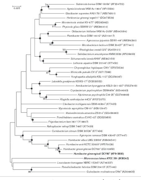
Phylogenetic analysis, based on 16S rRNA gene sequences, showed that strains DCY60Tand DCY90Tbelonged to the genus Humibacter (Fig. 1, Fig. S2). Strains DCY60T and DCY90T were most closely related to Humibacter antri
KCTC 33009T, sharing 98.9 % and 98.3 % 16S rRNA gene sequence similarity, respectively. Strains DCY60T and DCY90Tformed a reliable and monophyletic cluster with
H. antriKCTC 33009Tin the ML and MP algorithms with high bootstrap values (Fig. 1). The similarity value between strains DCY60Tand DCY90Twas 98.5 %. The DNA G+C contents of strains DCY60T and DCY90T were 62.8 and 66.8 mol%, respectively. The DNA–DNA relatedness values between strain DCY60Tand strain DCY90T,H. albusLMG 23996TandH. antri KCTC 33009Twere 47.6¡0.9, 26.2¡ 0.6 and 15.6¡0.9 %, respectively. Meanwhile, strain DCY90T showed DNA–DNA relatedness values of 47.5¡ 1.0, 32.1¡0.7 and 20.0¡1.1 % with strain DCY60T,
H. albusLMG 23996TandH. antriKCTC 33009T, respectively.
For isoprenoid quinone analysis, cell biomass was grown in R2A broth medium (Difco) at 308C for 48 h and then dried. Menaquinones were extracted from freeze-dried cells (50 mg) with chloroform/methanol (2 : 1, v/v), purified using Sep-Pak Vac 6cc silica cartridge (Waters) and subsequently analysed by HPLC as described by Collins (1985). Cellular fatty acids were saponified, methylated and
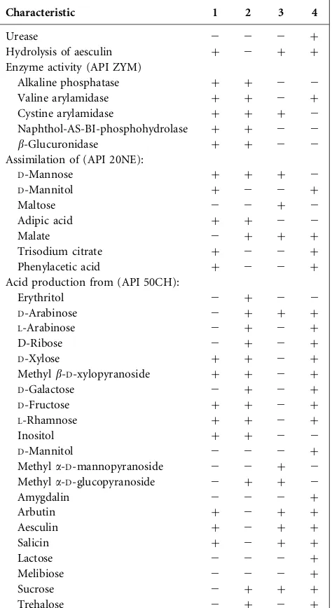
Table 1. Differential characteristics of strains DCY60T and DCY90T and type strains of other related species of the genusHumibacter
Strains: 1,Humibacter ginsengiterraesp. nov. DCY60T; 2,Humibacter gin-sengisolisp. nov. DCY90T; 3,H. antriKCTC 33009T; 4,H. albusLMG 23996T. All strains are positive for Gram-staining, nitrate reduction to nitrites, and activity of esterase (C4), lipase (C8), leucine arylamidase, acid phosphatase,a-galactosidase,b-galactosidase,a-glucosidase,b -glu-cosidase,N-acetyl-b-glucosaminidase,a-mannosidase anda-fucosidase. All strains are negative for lipase (C14), trypsin,a-chymotrypsin, gelati-nase, arginine dihydrolase, indole production and glucose fermentation. All strains assimilatedD-glucose andN-acetylglucosamine, but not pot-assium gluconate, capric acid orL-arabinose (API ZYM and API 20NE). All strains produced acid fromD-glucose,D-mannose,N -acetylglucosa-mine and cellobiose, but not from glycerol,L-xylose,D-adonitol,L -sor-bose, dulcitol,D-sorbitol, inulin, melezitose, starch, glycogen,D-tagatose, L-arabitol, potassium 2-ketogluconate, potassium 5-ketogluconate (API 50CH).+, Positive;2, negative.
Characteristic 1 2 3 4
Urease 2 2 2 +
Hydrolysis of aesculin + 2 + +
Enzyme activity (API ZYM)
Alkaline phosphatase + + 2 2
Valine arylamidase + + 2 +
Cystine arylamidase + + + 2
Naphthol-AS-BI-phosphohydrolase + + 2 2
b-Glucuronidase + + 2 2
Assimilation of (API 20NE):
D-Mannose + + + 2
D-Mannitol + 2 2 +
Maltose 2 2 + 2
Adipic acid + + 2 2
Malate 2 + + +
Trisodium citrate + 2 2 +
Phenylacetic acid + 2 2 +
Acid production from (API 50CH):
Erythritol 2 + 2 2
D-Arabinose 2 + + +
L-Arabinose 2 + 2 +
D-Ribose 2 + 2 +
D-Xylose + + 2 +
Methylb-D-xylopyranoside + + 2 +
D-Galactose 2 + 2 +
D-Fructose + + 2 +
L-Rhamnose + + 2 +
Inositol + + 2 2
D-Mannitol 2 2 2 +
Methyla-D-mannopyranoside 2 2 + 2 Methyla-D-glucopyranoside 2 + + 2
Amygdalin 2 2 2 +
Arbutin + 2 + +
Aesculin + 2 + +
Salicin + 2 + +
Lactose 2 2 2 +
Melibiose 2 2 2 +
Sucrose 2 + + +
Trehalose 2 + 2 +
Table 1.cont.
Characteristic 1 2 3 4
Raffinose 2 + 2 2
Xylitol 2 2 + +
Gentiobiose 2 + 2 +
Turanose 2 + 2 2
D-Lyxose 2 + 2 2
D-Fucose 2 2 + +
L-Fucose + + 2 2
extracted according to the protocol of the Sherlock Microbial Identification System (Sasser, 1990). The fatty acid methyl esters were analysed by GC (Hewlett Packard 6890) with
Sherlock MIDI software (version 6.0) and a TSBA database (version 6.0). The polar lipids of strains DCY60T, DCY90T andH. albusLMG 23996Twere extracted and analysed by Subtercola boreus DSM 13056T (AF224722)
Agreia bicolorata VKM Ac-1804T (AF15936)
Glaciibacter superstes AHU1791T (AB378301)
Herbiconiux ginsengi wged11T (DQ473536)
Microterricola viridarii KV-677T (AB282862)
Phycicola gilvus SSWW-21T (AM286414)
Okibacterium fritillariae VKM Ac-2059T (AB042094)
Plantibacter flavus DSM 14012T (AJ310417)
Agrococcus jejuerisis SSW1-48T (AM396260)
Microbacterium lacticum DSM 20427T (X77441)
Rhodoglobus vestalii LV3T (AJ459101)
Salinibacterium amurskyense KMM 3928 (AF539699)
Schumannella luteola KHIAT (AB362159)
Leifsonia aquatica DSM 20146T (X77450)
Chryseoglobus frigidaquae CW1T (EF373534)
Microcella putealis CV-2T (AJ717388)
Yonghaparkia alkaliphila KSL-113T (DQ256087)
Labedella gwakjiensis KSW2-17T (DQ533552)
Amnibacterium kyonggiense KSL51201-037T (FJ527819)
Cryobacterium psychrophilum DSM4854T (AJ544063)
Alpinimonas psychrophila Cr8-25T (GU784868)
Klugiella xanthotipulae 44C3T (AY372075)
Clavibacter michiganensis DSM 46364T (X77435)
Mycetocola saprophilus CM-01T (AB012647)
Marisediminicola antarctica ZS314T (GQ496083)
Frondihabitans australicus E1HC-02T (DQ525859)
Frigoribacterium faeni 801T (Y18807)
Rathayibacter rathayi DSM 7485T (X77439)
Curtobacterium citreum DSM 20528T (X77436)
Agromyces ramosus DSM 43045T (X77447)
Humibacter albus LMG 23996T (AM494541)
Humibacter antri KCTC 33009T (HF570028)
Humibacter ginsengiterrae DCY60T (JQ010859)
Humibacter ginsengisoliDCY90T (KF915800)
Micrococcus luteusATCC 381 (M38242)
Leucobacter komagatae NBRC 15245T (AJ746337)
Pseudoclavibacter helvolus DSM 20419T (X77440)
Gulosibacter molinativorax ON4T (AJ306835) 78
97
92
99
87
85
77
96
99
78
88 0.005
two-dimensional thin layer chromatography. For the pre-sence of all lipids, TLC plates were sprayed with 5 % molyb-dophosphoric acid followed by charring at 1208C for 15 min. Amino lipids were detected by spraying with 0.2 % ninhydrin and charring at 1108C for 10 min. Glycolipids were detected by spraying with 5 %a-naphthol and charring at 1108C for 10 min. Phospholipids were detected by spray-ing with molybdenum blue reagent (Minnikinet al., 1984). Cell-wall sugars of strains DCY60Tand DCY90Twere deter-mined by TLC according to Beckeret al.(1965). For this, 50 mg freeze-dried cells were hydrolysed in 1 ml H2SO4 (1 M) at 958C for 2 h. The pH of the samples was then adjusted to pH 5.0–5.5 with saturated Ba(OH)2. Cen-trifugation (1500 r.p.m., 5 min) precipitated the resultant BaSO4. The liquid phase was evaporated with a rotary evap-orator and the residue was dissolved in 4 ml H2O, and then centrifuged to remove particles. Cell-wall sugar extraction was determined by spotting samples onto TLC cellulose (Merck, 20620 cm). The solvent system used was ethylacetate/
pyridine/water (50 : 30 : 20, by vol.). Sugars were detected by spraying with anilinphthalate [0.8 g phthalic acid (Sigma Aldrich, P/N 402915), 2 ml distilled water, 24 ml 1-butanol, 24 ml diethylether and 0.5 ml aniline (Sigma Aldrich, P/N 242284)]. Peptidoglycan of strains DCY60T and DCY90T were determined by TLC according to McPherson & Popham (2003). Freeze-dried cells (200 mg) were dissolved in 50 mM phosphate buffer (pH 7.2), and then sonicated three times (10 min) in ice water. Cell suspensions were centrifuged at 4000g for 20 min to remove unlysed cells. The supernatants were col-lected in new tubes and centrifuged again at 40 000gfor 25 min. The supernatants were discarded and pellets which contain the peptidoglycan were collected. The resi-dues were suspended with 4 % SDS solution (6 ml) in a glass tube and heated at 1008C in an oven until it became colourless. The samples were then centrifuged again at 40 000gfor 25 min at room temperature. The col-lected pellets were washed three times with distilled water and centrifuged at 40 000gfor 25 min at room tempera-ture. The pellet was dried at 508C and then 5 mg material was hydrolysed in 6 M HCl at 1008C for 16 h. The hydro-lysed peptidoglycans were centrifuged at 13 000gfor 5 min and the samples were then filtered through a 0.2mm-filter.
HCl was removed using a vacuum evaporator, and dried samples were finally dissolved in 0.3 ml water. The chemi-cal content of the hydrolysed peptidoglycan was deter-mined by spotting the sample on TLC cellulose Merck KGaA plates (20620 cm). The solvent system used
was methanol/pyridine/HCl/water (100 : 12.5 : 6 : 32.5, by vol.).
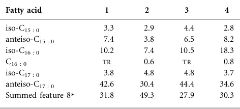
The predominant isoprenoid quinones of strain DCY60T were MK-11 (79.1 %) and MK-12 (20.9 %), while for strain DCY90T were MK-11 (74.2 %), MK-12 (17.1 %) and MK-10 (8.7 %). The major polar lipids of the novel isolates consisted of diphosphatidylglycerol, phosphatidyl-glycerol and an unidentified glycolipid. Both DCY60T and DCY90T showed polar lipid profiles similar to that
ofH. albusLMG 23996T. An unidentified aminoglycolipid (AGL) was found in strain DCY90Tthat was not found in strain DCY60TorH. albusLMG 23996T(Fig. S3). The major cellular fatty acids (.10 %) of strain DCY60Twere anteiso-C17 : 0, summed feature 8 (containing C18 : 1v7c and/or C18 : 1v6c) and iso-C16 : 0, while those of strain DCY90T were summed feature 8 (containing C18 : 1v7c and/or C18 : 1v6c) and anteiso-C17 : 0. The fatty acid profiles of the two strains showed similarity with the type strains of species of the genus Humibacter (Table 2). The peptidoglycan of strains DCY60T and DCY90T contained the amino acids ornithine, 2.4-diaminobutyric acid, alanine, glutamic acid and glycine. This profile was similar to that of described species of the genus Humibacter. The cell-wall sugars of strain DCY60Tcontained glucose, galactose, rhamnose and xylose, while strain DCY90T contained glucose, galactose, rhamnose and ribose.
The results of the phylogenetic and chemotaxonomic ana-lyses suggested the two isolated strains belong to the genus
Humibacter. The DNA–DNA relatedness among strains DCY60T, DCY90Tand related species of the genus Humi-bacterwere lower than 50 %; these values are well-below the 70 % threshold proposed for species delineation (Wayne
et al., 1987). The results of physiological and biochemical tests enabled strains DCY60T and DCY90T to be distin-guished from other members of genusHumibacter. There-fore, it proposed that strains DCY60T and DCY90T be classified as representatives of two novel species of the genusHumibacter, for which the namesHumibacter ginsen-giterrae sp. nov. and Humibacter ginsengisoli sp. nov., respectively, are proposed.
Table 2. Cellular fatty acid profiles of strains DCY60T and DCY90T and type strains of other related species of the genusHumibacter
Strains: 1,Humibacter ginsengiterraesp. nov. DCY60T; 2,Humibacter ginsengisolisp. nov. DCY90T; 3,H. antriKCTC 33009T; 4,H. albus LMG 23996T. All type strains were collected after 24 h growth on TSA medium (Difco) at 308C. All data are from this study. Fatty acids,0.5 % are not listed.TR, Traces (,0.5 %).
Fatty acid 1 2 3 4
iso-C15 : 0 3.3 2.9 4.4 2.8
anteiso-C15 : 0 7.4 3.8 6.5 8.2
iso-C16 : 0 10.2 7.4 10.5 18.3
C16 : 0 TR 0.6 TR 0.8
iso-C17 : 0 3.8 4.8 4.8 3.7
anteiso-C17 : 0 42.6 30.4 44.4 34.6 Summed feature 8* 31.8 49.3 27.9 30.3
Description ofHumibacter ginsengiterraesp. nov.
Humibacter ginsengiterrae(gin.sen.gi.ter9rae. N.L. n. ginsen-gumginseng; L. n.terra -aesoil; N.L. gen. n.ginsengiterrae
from soil of a ginseng field).
Colonies are ivory, circular, smooth and approximately 0.3–1.0 mm in diameter after 4 days cultivation on TSA agar medium at 30uC. Cells are oxidase-negative, catalase-positive, Gram-reaction-positive, non-motile, non-flagellated, rod-shaped and approximately 0.5
|
1.3 mm.Grows at 15–37uC (optimum 30uC), and with NaCl concen-tration of 0–5.0 %; no growth is observed with 6 % NaCl or higher. The pH range for growth is pH 4.0–8.0 (optimum pH 5.0–6.0).Growth occurs on NA, TSA, R2A and LB agars, but not on MacConkey agar at 30uC. Does not hydrolyse Tweens 20 and 80, casein, starch, cellulose, gelatin or DNA. According to the API ZYM test, positive for esterase (C4), lipase (C8), leucine arylamidase, valine aryla-midase, cystine arylaaryla-midase, acid phosphatase, naphthol-AS-BI-phosphohydrolase, a-galactosidase, b-galactosidase,
a-glucosidase, b-glucosidase, N-acetyl-b-glucosaminidase,
a-mannosidase,a-fucosidase, alkaline phosphatase and b -glucuronidase activities, but negative for lipase (C14), tryp-sin and a-chymotrypsin activities. According to the API 20NE test, positive for nitrate reduction to nitrites, aesculin hydrolysis, b-galactosidase and assimilation of D-glucose, D-mannose,D-mannitol,N-acetylglucosamine, adipic acid, trisodium citrate and phenylacetic acid; negative for glucose fermentation, gelatinase, arginine dihydrolase, urease, indole production, and assimilation ofL-arabinose, maltose, pota-ssium gluconate, capric acid and malate. According to the API 50 CH test, positive for acid production from D -xylose, methyl b-D-xylopyranoside, D-glucose, D-fructose, D-mannose, L-rhamnose, inositol, N-acetylglucosamine, arbutin, aesculin, salicin, cellobiose, maltose and L-fucose, but negative for acid production from glycerol, erythritol, D-arabinose, L-arabinose, D-ribose, L-xylose, D-adonitol, D-galactose, L-sorbose, dulcitol, D-mannitol, D-sorbitol, methyla-D-mannopyranoside, methyla-D-glucopyranoside, amygdalin, lactose, melibiose, sucrose, trehalose, inulin, mele-zitose, raffinose, starch, glycogen, xylitol, gentiobiose, tura-nose, D-lyxose, D-tagatose, D-fucose, L-arabitol, D-arabitol, potassium gluconate, potassium 2-ketogluconate and potass-ium 5-ketogluconate. The predominant menaquinones are MK-11 and MK-12. The cell-wall peptidoglycan contains the amino acids ornithine, 2.4-diaminobutyric acid, alanine, glutamic acid and glycine. The cell-wall sugars contain glucose, galactose, rhamnose and xylose. The major polar lipids are diphosphatidylglycerol, phos-phatidylglycerol and an unidentified glycolipid. The major cellular fatty acids (w10 %) are anteiso-C17 : 0, summed feature 8 (containing C18 : 1v7cand/or C18 : 1v6c) and iso-C16 : 0.
The type strain is DCY60T (5KCTC 33520T5JCM 30079T), isolated from soil of a ginseng field in Yeoncheon county (38u049000N 126u579000E), Republic of Korea. The DNA G+C content of the type strain is 62.8 mol%.
Description ofHumibacter ginsengisolisp. nov.
Humibacter ginsengisoli (gin.sen.gi.so9li. N.L. n.ginsengum
ginseng; L. n. solum soil; N.L. gen. n. ginsengisoli of soil of a ginseng field).
Colonies are white–cream, circular, smooth and approxi-mately 0.5–1.3 mm in diameter after 4 days cultivation at 30uC. Cells are oxidase-negative, catalase-positive, Gram-reaction-positive, non-motile, non-flagellated, rod-shaped and approximately 0.4
|
0.8mm. Grows at 15–37uC (optimum 30uC), at pH 4.0–8.0 (optimum pH 5.0–6.0) and with NaCl concentrations of 0–4.0 %. Grows on NA, TSA, R2A and LB agars, but not on Mac-Conkey agar at 30uC. DNA is hydrolysed, but Tweens 20 and 80, casein, starch, cellulose and gelatin are not. Accord-ing to the API ZYM test, positive for esterase (C4), lipase (C8), leucine arylamidase, valine arylamidase, cystine aryla-midase, acid phosphatase, naphthol-AS-BI-phosphohydrolase,
a-galactosidase,b-galactosidase,a-glucosidase,b-glucosidase,
N-acetyl-b-glucosaminidase,a-mannosidase, a-fucosidase, alkaline phosphatase and b-glucuronidase activities, but negative for lipase (C14), trypsin and a-chymotrypsin activities. According to the API 20NE test, positive for b -galactosidase, nitrate reduction to nitrites, and assimilation ofD-glucose, malate, adipic acid,N-acetylglucosamine and D-mannose; negative for gelatinase, indole production, urease, aesculin hydrolysis, arginine dihydrolase, glucose fermentation, and assimilation of trisodium citrate, pheny-lacetic acid, maltose, potassium gluconate, capric acid, L-arabinose and D-mannitol. According to the API 50CH test, positive for acid production from erythritol, D-arabinose, L-arabinose, D-ribose, D-xylose, methyl
b-D-xylopyranoside, D-galactose, D-glucose, D-fructose, D -mannose,L-rhamnose, inositol, methyla-D-glucopyranoside,
N-acetylglucosamine, cellobiose, maltose, sucrose, trehalose, raffinose, gentiobiose, turanose, D-lyxose and D-fucose; negative for acid production from glycerol, L-xylose, D -adonitol,L-sorbose, dulcitol,D-mannitol,D-sorbitol, methyl
a-D-mannopyranoside, amygdalin, arbutin, salicin, lactose, melibiose, inulin, melezitose, starch, glycogen, aesculin, xyli-tol, D-tagatose, L-fucose, L-arabitol, D-arabitol, potassium gluconate, potassium 2-ketogluconate and potassium 5-ketogluconate. The predominant menaquinones are MK-10, MK-11 and MK-12. The cell-wall peptidoglycan contains the amino acids ornithine, 2.4-diaminobutyric acid, alanine, glu-tamic acid and glycine; the cell-wall sugars contain glucose, galactose, rhamnose and ribose. The major polar lipids consist of diphosphatidylglycerol, phosphatidylglycerol and an uni-dentified glycolipid. The major cellular fatty acids are summed feature 8 (containing C18 : 1v7cand/or C18 : 1v6c) and anteiso-C17 : 0.
Acknowledgements
This research was supported by the Korea Institute of Planning & Evaluation for Technology in Food, Agriculture, Forestry & Fisheries (KIPET no.: 313038-03-1-SB010).
References
Anzai, Y., Kim, H., Park, J. Y., Wakabayashi, H. & Oyaizu, H. (2000).
Phylogenetic affiliation of the pseudomonads based on 16S rRNA sequence.Int J Syst Evol Microbiol50, 1563–1589.
Bauer, A. W., Kirby, W. M. M., Sherris, J. C. & Turck, M. (1966).
Antibiotic susceptibility testing by a standardized single disk method. Am J Clin Pathol45, 493–496.
Becker, B., Lechevalier, M. P. & Lechevalier, H. A. (1965).Chemical composition of cell-wall preparations from strains of various form-genera of aerobic actinomycetes.Appl Microbiol13, 236–243.
Bernardet, J. F., Nakagawa, Y., Holmes, B. & Subcommittee on the taxonomy of Flavobacterium and Cytophaga-like bacteria of the International Committee on Systematics of Prokaryotes (2002).
Proposed minimal standards for describing new taxa of the family Flavobacteriaceaeand emended description of the family.Int J Syst Evol Microbiol52, 1049–1070.
Christensen, W. B. (1946). Urea decomposition as a means of differentiatingProteusand paracolon cultures from each other and fromSalmonellaandShigellatypes.J Bacteriol52, 461–466.
Collins, M. D. (1985).Isoprenoid quinone analysis in classification and identification. In Chemical Methods in Bacterial Systematics, pp. 267–287. Edited by M. Goodfellow & D. E. Minnikin. London: Academic Press.
Ezaki, T., Hashimoto, Y. & Yabuuchi, E. (1989). Fluorometric deoxyribonucleic acid-deoxyribonucleic acid hybridization in micro-dilution wells as an alternative to membrane filter hybridization in which radioisotopes are used to determine genetic relatedness among bacterial strains.Int J Syst Bacteriol39, 224–229.
Felsenstein, J. (1985).Confidence limits on phylogenies: an approach using the bootstrap.Evolution39, 783–791.
Fitch, W. M. (1971). Toward defining the course of evolution: minimum change for a specific tree topology.Syst Zool20, 406–416.
Hong, S. K. (1978).Ginseng cultivation. InKorean ginseng, 2nd edn, pp. 245–278. Edited by H. W. Bae. Korea ginseng Research Institute.
Kim, O. S., Cho, Y. J., Lee, K., Yoon, S. H., Kim, M., Na, H., Park, S. C., Jeon, Y. S., Lee, J. H. & other authors (2012).Introducing EzTaxon-e: a prokaryotic 16S rRNA gene sequence database with phylotypes that represent uncultured species.Int J Syst Evol Microbiol62, 716–721.
Kimura, M. (1983). The Neutral Theory of Molecular Evolution. Cambridge: Cambridge University Press.
Lane, D. J. (1991). 16S/23S rRNA sequencing. In Nucleic Acid Techniques in Bacterial Systematics, pp. 115–176. Edited by E. Stackebrandt & M. Goodfellow. Chichester: Wiley.
Lee, S. D. (2013). Humibacter antri sp. nov., an actinobacterium isolated from a natural cave, and emended description of the genus Humibacter.Int J Syst Evol Microbiol63, 4315–4319.
McPherson, D. C. & Popham, D. L. (2003).Peptidoglycan synthesis in the absence of class A penicillin-binding proteins inBacillus subtilis. J Bacteriol185, 1423–1431.
Mesbah, M., Premachandran, U. & Whitman, W. B. (1989).Precise measurement of the G+C content of deoxyribonucleic acid by high-performance liquid chromatography.Int J Syst Bacteriol39, 159–167.
Minnikin, D. E., O’Donnell, A. G., Goodfellow, M., Alderson, G., Athalye, M., Schaal, A. & Parlett, J. H. (1984). An integrated procedure for the extraction of bacterial isoprenoid quinones and polar lipids.J Microbiol Methods2, 233–241.
Prescott, L. M. & Harley, J. P. (2001). Laboratory exercises in Microbiology, 5th edn. New York: McGraw-Hill.
Saitou, N. & Nei, M. (1987).The neighbor-joining method: a new method for reconstructing phylogenetic trees.Mol Biol Evol4, 406–425.
Sasser, M. (1990).Identification of bacteria by gas chromatography of cellular fatty acids. MIDI Technical Note 101. Newark, DE: MIDI Inc.
Skerman, V. B. D. (1967).A Guide to the Identification of the Genera of Bacteria, 2nd edn. Baltimore: Williams & Wilkins.
Tamura, K., Peterson, D., Peterson, N., Stecher, G., Nei, M. & Kumar, S. (2011).MEGA5: molecular evolutionary genetics analysis using maxi-mum likelihood, evolutionary distance, and maximaxi-mum parsimony methods.Mol Biol Evol28, 2731–2739.
Thompson, J. D., Gibson, T. J., Plewniak, F., Jeanmougin, F. & Higgins, D. G. (1997). The CLUSTAL_X windows interface: flexible strategies for multiple sequence alignment aided by quality analysis tools.Nucleic Acids Res25, 4876–4882.
Vaz-Moreira, I., Nobre, M. F., Ferreira, A. C., Schumann, P., Nunes, O. C. & Manaia, C. M. (2008).Humibacter albusgen. nov., sp. nov., isolated from sewage sludge compost. Int J Syst Evol Microbiol58, 1014–1018.
Wayne, L. G., Brenner, D. J., Colwell, R. R., Grimont, P. A. D., Kandler, O., Krichevsky, M. I., Moore, L. H., Moore, W. E. C., Murray, R. G. E. & other authors (1987). International Committee on Systematic Bacteriology. Report of the ad hoc committee on reconciliation of approaches to bacterial systematics.Int J Syst Bacteriol37, 463–464.
Yang, Y. Y. (1974).The effects of different shading or mulching on yield of root and quality inPanax ginseng.Proceeding of International Ginseng Symposium. The Central Res Inst office of Monool1, 137–146.
Yu, W. J., Lee, B. J., Nam, S. Y., Yang, D. C. & Yun, Y. W. (2003).