Corresponding author: [email protected]
The influence of long-term diabetes
mellitus (DM) on pain response in mice:
in
vivo
models of painful diabetic neuropathy
(PDN)
Fajrin FA1,2*, Susilowati R3,Nurrochmad A4, Nugroho AE4
1Postgraduate Programme, Faculty of Pharmacy, Universitas Gadjah Mada, Yogyakarta
2Departement of Clinical and Community Pharmacy, Faculty of Pharmacy, Jember
University, Jember, 3Departement of Histology and Cell Biology, Faculty of Medicine,
Universitas Gadjah Mada, Yogyakarta, 4Department of Pharmacology and Clinical
Pharmacy, Faculty of Pharmacy, Universitas Gadjah Mada, Yogyakarta, Indonesia
DOI: http://dx.doi.org/10.19106/JMedSci004903201701
ABSTRACT
Painful diabetic neuropathy (PDN) is a complication of long-term diabetes mellitus (DM) characterized by hyperalgesia and allodynia. In streptozotocin (STZ)-induced diabetic mice, higher dose of STZ and lengthened hiperglycemic condition results in better model of PDN. However, higher dose of STZ tend to induce mortality. The aim of the study was to evaluate the doses of STZ that caused PDN with less mortality rate and the timing of pain behavior development in mice model of PDN. Balb/c mice were divided into non-diabetic and STZ-induced diabetic group. The doses of STZ were started from 180 mg/kg BW i.p. Serum glucose levels were measured 7 days after induction. Mice with glucose levels ≥ 200 mg/dL were considered as diabetic. Pain behaviour was determined by four method i.e. hot plate, tail lick test, von Frey illament and Randall Selitto, measured on week-0 (baseline), 1, 2, 3, 4 and 5. Data were presented as mean ± SEM. The mean differences between weeks were evaluated by One-Way ANOVA and the mean differences between two groups by independent t-test. STZ doses 180, 150 and 120 mg/kg BW caused 100% death and STZ 90 mg/kg BW failed to induce diabetic condition. STZ 110 mg/kg BW resulted in 0% mortality while it induced diabetes in 100% mice. Latency time toward thermal stimulus decreased to 5.8 ± 0.4 sec at 1st week after the mice become diabetes
(p<0.05) and it was continued decrease until 4th week. The same result was also showed
in tail lick test and Randal Selitto. The pain sensitivity determined by von Frey ilament decreased to 1.37 ± 0.12 g at 1st week (p<0.05) and continued decrease until 5th week.
In conclusion, optimum dose of STZ to induce PDN was 110 mg/kg BW. Pain behaviour of diabetic group was observed at 1st week after diabetes and continued until 4th week.
ABSTRAK
yang menyebabkan NND dengan tingkat kematian rendah dan waktu timbulnya rasa nyeri pada model mencit NND. Mencit Balb/c dibagi menjadi kelompok tikus tidak DM dan tikus DM akibat induksi STZ. Dosis STZ dimulai dengan 180 mg/kg yang diberikan secara intraperitonial. Kadar gula darah diukur 7 hari setelah induksi. Mencit dengan kadar gula darah ≥ 200 mg/dL ditetapkan sebagai mencit DM. Perilaku nyeri ditetapkan menggunakan 4 metode yaitu metode hot plate, tail lick, von Frey illament dan Randall
Selitto yang diukur pada minggu ke 0 (data awal), 1, 2, 3, 4 dan 5. Data disajikan sebagai rerata ± SEM. Perbedaan rerata antara perilaku nyeri setiap minggu dievaluasi dengan ANOVA satu jalan dan perbedaan antara 2 kelompok dievaluasi dengan uji t. Dosis STZ 180, 150 dan 120 mg/kg BB menyebabkan 100% kematian dan dosis STZ 90 mg/kg BW gagal menginduksi diabetes. Dosis STZ 110 mg/kg BW menghasil 0% kematian dan menginduksi diabetes 100% mencit. Waktu latensi terhadap stimulus panas menurun 5,8 ± 0,4 detik pada minggu pertama setelah mencit diabetes (p<0,05) dan berlanjut menurun hingga minggu ke 4. Hasil yang sama juga ditunjukkan pada uji dengan tail ick
dan Randal Selitto. Sensitivitas nyeri yang diukur dengan von Frey ilament menurun menjadi 1,37 ± 0,12 g pada minggu pertama (p<0,05) dan berlanjut menurun hingga minggu ke 5. Dosis optimum STZ untuk menginduksi NND adalah 110 mg/kg BW. Perilaku nyeri kelompok diabetes teramati pada minggu pertama setelah diabetes dan berlanjut hingga minggu ke 4.
Keywords: PDN, hot plate, tail lick test, von Frey illament, Randall Selitto
INTRODUCTION
Diabetes mellitus (DM) is the leading cause of degenerative diseases with the highest mortality rate.1 Diabetes mellitus is caused by disturbance in insulin secretion, insulin action, or both that characterized by hyperglycemia.2 Since DM is a chronic degenerative disease, diabetic patients suffer from its complication such as painful diabetic neuropathy (PDN). Approximately, PDN occurs in 25-50% patient with DM.3 Painful diabetic neuropathy is the most common complication of DM and actually results from ectopic pain signal
system in our body3 due to peripheral nerve
damage. Patient with PDN usually complains chronic pain with hyperalgesia and allodynia
symptoms.4 The pain in PDN results in
impairment of their daily activities, then reduce the quality of life.3,4
Various pathogenic pathways are involv-ed in development of PDN. Prolonginvolv-ed hyperglycemia that causes accumulation of reactive oxygen species (ROS), is one of
the main pathway in PDN pathogenesis.5
Hyperglycemia, thus affects to cellular surface proteins, resulting in progressive pathologic changes and diabetic complications.6 Strepto-zotocin (STZ) is one of the chemical substances that widely used to induce diabetes in animal model.7 Previous studies showed that different doses of STZ differentially induced PDN. Some studies reported development of PDN at 2-4 weeks after DM,7 but others showed that it needed more than 4 weeks to develop PDN
symptoms.8-10 On the other hand, prolonged
DM at more than 6 weeks caused hypoalgesia,
related to loss of nerves ibers.7
with less mortality rate and the timing of pain behavior development in mice model of PDN.
MATERIALS AND METHODS
Materials
All the reagents and chemical used for this research were analytical grade. Streptozotocin was purchased from Biolab (USA), Biochemical diagnostic such as GOD-POD was purchased from DyaSis (Germany) and citrate buffer pH 4.5 was from Sigma (Singapore). The pain behavior was determined using hot plate (UgoBasile, Italy),
tail lick test (Columbus Instrument, USA), von Frey ilament (Aesthesio, USA) and
Randall Selitto (UgoBasile, Italy).
Preparation of STZ
Streptozotocin 1.1% was dissolved in freshly prepared ice cold citrate buffer (pH 4.5) before used.
Preparation of experimental animals Sixteen of male Balb/c strain mice (25-35 g, 8-10 w) were obtained from Faculty of Pharmacy, Universitas Gadjah Mada, Yogyakarta, Indonesia. Mice were placed in clean cages and maintained at temperature
25±1 °C with 12 hours’ light/dark cycle.
Mice had free access on food and water
ad libitum. Ethical clearance was obtained from the Medical and Health Research Ethic
Committee, Faculty of Medicine, Universitas
Gadjah Mada-Dr. Sardjito General Hospital, Yogyakarta number Ref. KE/FK/559/
EC/2016.
Experimental protocol
Mice were fasted overnight for 12 hours, before the experiment. Each group of mice were given a single-high-dose of STZ (180,
150, 120, 110 or 90 mg/kg BW) i.p. The fasting-blood glucose levels were checked 7 days after STZ induction using GOD-POD kit. Mice with fasting-blood glucose
levels ≥200 mg/dL were considered diabetes.
Measurement of blood glucose levels were conducted once every week until week 5.
Pain behavioral assays
Pain behavior assays were measured before STZ induction and at week 1, 2, 3, 4 and 5 after diabetes. All experiments were taken in three replications.
Hot plate test
Each mouse was placed individually on a
hot plate that adjusted at 50±0.5°C. The latency
time toward thermal stimulus was evaluated when the mice showed pain responses such as
licking, jumping, tapping, rearing, lattering and frizzing (whatever which came irst).
The cut off time was 30 sec in order to avoid damage of the nerves.
Tail lick test
The tail of each mouse was exposed to nichrome-radiant heat. The intensity of the radiant heat was adjusted to give a basal latency of 6-8 sec in both diabetic and
non-diabetic groups. Tail lick latency was time interval that taken by mice to lick its tail after
the exposure of radiant heat. The maximum cut-off time was 15 sec to prevent tissue damage.
von Frey ilament Test
The sensitivity of mechanical stimulus
was measured by von Frey ilament. Mice
were placed in an individual glass-caged that completed by wire-mesh-bottom. A von Frey
ilament was applied 10 times for 5 sec to the
positive response (such as jumping, scratching or scraping) was recorded. The increasing of sensitivity was interpreted as more than 5 positive responses from 10 times test.
Randall Selitto test
Randall Selitto test applied pressure to the dorsal surface of the hind paws via conical probe with 250 g cut-off. The pressure was increased until the mouse withdrew the paw, squeaked or struggled.
Statistical analysis
Data was statistically analyzed using SPSS version 20. All values were described as mean ± SEM. The mean differences of pain behaviour measurement between weeks were compared using One-Way ANOVA and between two groups (diabetic and non-diabetic) using independent t-test. The
signiicance level were set at 95%.
RESULTS
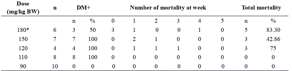
The result of the diabetic induction and subsequent survival of the mice after STZ administration of different dose of STZ is presented in TABLE 1. At day 7 after STZ administration, doses 180, 150, 120 and 110 mg/kg BW resulted in 100% diabetic induction. Lower STZ dose at 90 mg/kg BW failed to induce diabetic condition in any mice. Although STZ dose 180, 150 and 120 mg/kg BW were successful in inducing diabetes, the mice survival was a problem. Most of the mice were died before week 3 and all of the mice from 180 and 120 mg/kg BW groups were died at week 7. The mean blood glucose level of the died mice at week 1 were
378.77±74.45 mg/dL, higher than the mean
of blood glucose level of the surviving mice
(281.64±66.84 mg/dL). However, it was not
signiicantly different (p >0.05).
TABLE 1. The percentage of diabetic mice and mortality in various doses of STZ induction until 5 weeks
Dose
(mg/kg BW) n DM+ Number of mortality at week Total mortality
n % 0 1 2 3 4 5 n %
180* 6 3 50 3 1 0 0 1 0 5 83.30
150 7 7 100 0 2 1 0 0 0 3 42.86
120 4 4 100 0 1 1 1 0 0 3 75
110 8 8 100 0 0 0 0 0 0 0 0
90 10 0 0 0 0 0 0 0 0 0 0
* 3 mice died before the irst examination of blood glucose level
STZ dose 110 mg/kg BW induced diabetes at 100% rate, but the dose did not cause any mortality. All of the mice in this group survived until week 7. Since, the STZ dose 110 mg/kg BW gave the best result, this group was used in subsequent analysis. In this group, the blood glucose level reached the
# indicated signiicant differences compared to non-diabetic group
FIGURE 1. Blood glucose levels between diabetic and non-diabetic groups at weeks-0 (baseline), 1, 4 and 5.
FIGURE 2. The body weight of diabetic and non-di-abetic groups at weeks-0 (baseline), 1, 2, 3, 4 and 5
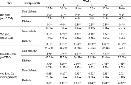
TABLE 2. Pain behaviour assessment in non-diabetic and diabetic groups after STZ induction dose 110 mg/kg BW.
Test Groups (n=8)
Week-0 1 2 3 4 5
Data analysis between groups using independent t test (p<0.05). Different letter indicated signiicant differences of pain be -haviour between diabetic and non-diabetic groups of each day. # indicated no signiicant differences compared to baseline
Pain behaviour in mice was evaluated using four different methods. Hyperalgesia
was determined by hot plate, tail lick test and
Randall Selitto. Allodynia was determined
by von Frey ilament. The pain behaviour of
diabetic mice and control mice are described in TABLE 2. Diabetic mice shortened latency time toward thermal stimulus using hot plate
from 10.3 sec at baseline to 5.8 sec in the irst
week after STZ induction and lasted up fourth week with consecutive latency time 4.9, 3.9,
and 3.3 sec. This result was signiicantly
different with non-diabetic group, which maintained the latency time arround 10 sec
(p>0.05). At week-5, latency time toward
thermal stimulus become 4.6 sec, longer than previous week but still lower than non-diabetic group and not returned yet to baseline.
The pain behavior evaluation using other
tests such as tail lick and Randall Selitto
gave similar results. Tail latency time using
tail lick test shortened from 7.95 s to 5.04 at
week 1 after STZ induction. This condition lasted up 4th week. Decreasing of withdrawal load threshold using Randall Selitto test was observed from 97.29 g at baseline to 53.75 g at week 1 after STZ induction and had been reducing until week 4. At the end study (week-5), the pain responses were less severe than the previous week.
In contrast to the other tests, measurement
of pain behaviour using von Frey ilament
showed slighlty different outcome. The tactile sensitivity towards mechanical stimulus was
continuously decreased until 5th week from
9.33±0.62 g to 1.37 ± 0.12, 0.67 ± 0.05, 0.36 ± 0.04, 0.10 ± 0.01 and 0.10 ± 0.02 g.
DISCUSSION
The dose of STZ 110 mg/kg BW was found to be the most effective in inducing diabetes and prevent mortality of the mice. Our STZ
dose result was lower than previous studies that used the high dose of STZ such as 150, 180 and 200 mg/kg BW.11-13 Administration of higher dose of STZ induced the higher level of blood glucose level more than 450 mg/dL. In our study, mouse with blood glucose levels up to 450 mg/dL was more vulnerable to die. When we used STZ dose 110 mg/dL, the blood glucose levels were found in range
231.89-413.96 mg/dL (mean 281.64±66.84 mg/dL).
This result might give explaination about why this dose successful to maintain survival life of mice during the experiments. The used of STZ-induced diabetic animals seen the
similar result with human.14 STZ works by
selectively accumulated in pancreatic beta
cells via the low-afinity GLUT-2 glucose
transporter in the plasma membrane. The transfer of the methyl group from STZ to the DNA molecule causes damage resulting in the fragmentation of the DNA. This mechanism will diminish cellular NAD+, and furthermore impact to ATP stores. The depletion of cellular energy stores subsequently result in beta cell necrosis or apoptosis.15 The inal result of STZ
induction was increasing of blood glucose levels. There are various test to measure pain behaviour. Based on Sandkuhler,16 STZ-induced PDN shows mechanical allodynia, mechanical hyperalgesia, heat hyperalgesia and cold hyperalgesia.
At week-0 (baseline), there were no
signiicant differences between non-diabetic
and diabetic groups. This result indicated that mice were in the same condition, at the beginning of the study. Heat hyperalgesia occurs in early stages of PDN. STZ-induced dibetic mice shows heat hyperalgesia within one week after induction and sustains for up to two weeks.16 In this study, the latency time toward thermal stimulus using hot plate
and tail lick test shows the signiicant result
induction. The time to response after heat stimulus declined, means that mice started in hyperalgesia state. The previous studies also gave the same statement as ours, that heat hyperalgesia happens in early week of diabetic state.17,18 A study conducted by Tian
et al.19 found that the pain behaviour using
hot plate signiicant changing in six days after
considered as diabetes. It is concluded that in mice model, hyperalgesia condition may occur within a week after diabetic state.
Previous studies reported that the mechanical hyperalgesia and tactile allodynia were occurred within 1-8 weeks after STZ induction.20-23 In this study, both of mechanical hyperalgesia test using Randall Selitto and
tactile allodynia using von Frey ilament were
developed one week after STZ induction, as
same as the other prior research.19 Another
study conducted by Yadav et al.,24 showed
different result, that mechanical hyperalgesia was happened in 6th week after diabetic state and this condition continued until 10-11 weeks. It means that mechanical-induced pain
was dificult to distinguish the progressivity
of either hyperalgesia or hypoalgesia. Otherwise, thermal stimulus effected more complicated responses included hypoalgesia and hyperalgesia. Referred to our result, in week-5, latency time using both hot plate
and tail lick were signiicantly longer than
week-4. It can be explained that heat stimulus might be more sensitive to detect hypoalgesia and hyperalgesia. Meanwhile, von Frey
ilament was used to conirm tactile allodynia.
Our result indicated that allodynia state,
detected using von Frey ilament have been
occurred since one week after STZ induction,
conirming other studies.21-23 Other report showed that mechanical allodynia could be happened more than one week in 5-6 weeks after diabetic.24
CONCLUSION
Optimum dose of STZ to induce PDN is 110 mg/kg BW. Pain behaviour of diabetic group is reached at 1st week after diabetic and continued until 4th week.
ACKNOWLEDGEMENT
The authors wished to thank Dr. D. Pakaya for excellent technical assistance.
REFERENCES
1. World Health Organization (WHO).
Non-communicable diseases country proiles.
Geneva: WHO Publishing, 2011.
2. International Diabetes Federation. Atlas Diabetes, 6th Edition. Brussels: International
Diabetes Federation, 2013.
3. Alleman CJ, Westerhout KY, Hensen M, Chambers C, Stoker M, Long S, et al.
Humanistic and economic burden of painful diabetic peripheral neuropathy in europe: a
review of the literature. Diabetes Res Clin
Pract 2015; 109(2):215-25. http://dx.doi. org/10.1016/j.diabres.2015.04.031
4. Haanpaa M, Hietaharju A. Halting the march of painful diabetic neuropathy. IASP: Pain
Clinical Update 2015; 23(2):1-8.
5. Singh R, Kishore L, Kaur N. Diabetic peripheral neuropathy: current perspective and future directions. Pharmacol Res 2014; 80:21-35. http://dx.doi.org/10.1016/ j.phrs.2013.12.005
6. Mohini A, Phansea SB, Padhyeb MJ, Patila
AR, Bafnaa AR, Takawalec VV. Thespesia populnea extract attenuates thermal hyperalgesia in diabetic mouse model
of neuropathic pain. IJPS Autumn 2010;
6(4):269-76.
7. Shaikh AS, Somani RS. Animal models and biomarkers of neuropathy in diabetic rodents.
8. Zangiabadi N, Mohtashami H, Shabani
M, Jafari M. Neuroprotective effect of
cerebrolysin on diabetic neuropathy: a study
on male rats. J Diabetes Metab 2014;
5(4):1-7.http://dx.doi.org/10.4172/2155-6156. 1000355
9. Raposo D, Morgado C, Pereira-Terra P,
Tavares I. Nociceptive spinal cord neurons of laminae I-III exhibit oxidative stress damage during diabetic neuropathy which is prevented by early antioxidant treatment
with epigallocatechin-gallate (EGCG). Brain
Res Bull 2015; 110:68-75. http://dx.doi. org/10.1016/j.brainresbull.2014.12.004
10. Reda HM, Zaitone SA, Moustafa YM. Effect of levetiracetam versus gabapentin on peripheral neuropathy and sciatic degeneration in streptozotocin-diabetic mice:
inluence on spinal microglia and astrocytes. Eur J Pharmacol 2016; 771:162-72. http://
dx.doi.org/10.1016/j.ejphar.2015.12.035 11. Tanaka K, Nakanishi Y, Sekino S, Ikegami
M, Ikeda H, Kamei J. Fentanyl produces
an anti-hyperalgesic effect through the suppression of sodium channels in mice with
painful diabetic neuropathy. Eur J Pharmacol
2014; 733:68-74. http://dx.doi.org/10.1016/j. ejphar.2014.03.042
12. Lennertz RC, Medler KA, Bain JL, Wright DE, Stucky CL. Impaired sensory nerve
function and axon morphology in mice with
diabetic neuropathy. J Neurophysiol 2011;
106(2):905-14. http://dx.doi.org/10.1152/ jn.01123.2010
13. Micov A, Tomic M, Pecikoza U, Ugresic N, Stepanovic-Petrovic R. Levetiracetam synergises with common analgesics in producing antinociception in a mouse model of painful diabetic neuropathy. Pharmacol Res 2015; 97:131-42. http://dx.doi.org/10.1016/j. phrs.2015.04.014
14. Sytze Van Dam P, Cotter MA, Bravenboer B, Cameron NE. Pathogenesis of diabetic
neuropathy: focus on neurovascular
mechanism. Eur J Pharmacol 2013;
719(1-3):180-6. http://dx.doi.org/10.1016/j.ejphar. 2013.07.017
15. Janes K, Neumann WL, Salvemini D.
Anti-superoxide and anti-peroxynitrite strategies in pain suppression. Biochim Biophys Acta 2012; 1822(5):815-21. http://dx.doi. org/10.1016/j.bbadis.2011.12.008
16. Sandkuhler J. Models and mechanism of
hyperalgesia and allodynia. Physiol Rev 2009; 89(3):707-58. http://dx.doi.org/10.1152/ physrev.00025.2008
17. Parkar N, Addepalli V. Effect of nobiletin on diabetic neuropathy in experimental rats.
Austin J Pharmacol Ther 2014; 2(5):1-5.
18. Hajializadeh Z, Nasri S, Kaeidi A, Sheibani V, Rasoulian B, Esmaeili-Mahani S. Inhibitory effect of Thymus caramanicus
Jalas on hyperglycemia-induced apoptosis
in in vitro and in vivo models of diabetic
neuropathic pain. J Ethnopharmacol 2014;
153(3):596-603. http://dx.doi.org/10.1016/j. jep.2014.02.049
19. Tian R, Yang W, Xue Q, Gao L, Huo J, Ren D,
et al. Rutin ameliorates diabetic neuropathy by lowering plasma glucose and decreasing oxidative stress via Nrf2 signaling pathway in
rats. Eur J Pharmacol 2016; 771:84-92. http://
dx.doi.org/10.1016/j.ejphar.2015.12.021
20. Dyck PJ, Dyck PJ, Larson TS, O’Brien PC, Velosa JA. Patterns of quantitative sensation
testing hypoesthesia and hyperalgesia are predictive on diabetic polyneuropathy: a study of three cohorts. Nerve growth factor
study group. Diabetes Care 2000;
23(4):510-7. http://dx.doi.org/10.2337/diacare.23.4.510
21. Calcutt NA, Jorge MC, Yaksh TL, Chaplan
22. Courteix C, Eschalier A, Lavarenne J. Streptozocin-induced diabetic rats:
behavioural evidence for a model of chronic pain. Pain 1993; 53(1):81-8. http://dx.doi. org/10.1016/0304-3959(93)90059-X
23. Fox A, Eastwood C, Gentry C, Manning D, Urban L. Critical evaluation of the
streptozotocin model of painful diabetic neuropathy in the rat. Pain 1999;
81(3):307-16. http://dx.doi.org/10.1016/S0304-3959 (99)00024-X
24. Yadav SK, Nagori BP, Desai PK. Pharmaco-logical characterization of different fractions of Calotropis procera (Aslepiadaceae) in streptozotocin induced experimental model of
diabetic neuropathy. J Ethnopharmacol 2014;