Curcumin decreases renal triglyceride accumulation through AMPK
–
SREBP signaling
pathway in streptozotocin-induced type 1 diabetic rats
☆
Vivian Soetikno
a, b, Flori R. Sari
a, Vijayakumar Sukumaran
a, Arun Prasath Lakshmanan
a, Meilei Harima
a,
Kenji Suzuki
c, Hiroshi Kawachi
d, Kenichi Watanabe
a,⁎
a
Department of Clinical Pharmacology, Faculty of Pharmaceutical Sciences, Niigata University of Pharmacy and Applied Life Sciences, Niigata City 956-8603, Japan
b
Department of Pharmacology, Faculty of Medicine, University of Indonesia, Jakarta 10430, Indonesia
c
Department of Gastroenterology and Hepatology, Niigata University Graduate School of Medical and Dental Sciences, Niigata 951-8510, Japan
dDepartment of Cell Biology, Institute of Nephrology, Niigata University Graduate School of Medical and Dental Sciences, Niigata 951-8510, Japan
Received 8 August 2011; received in revised form 20 April 2012; accepted 26 April 2012
Abstract
Diabetic kidney disease has been associated with the presence of lipid deposits. We assumed that curcumin, a polyphenol, would attenuate the tissue dyslipidemic condition through activation of 5' adenosine monophosphate (AMP)-activated protein kinase (AMPK) phosphorylation and suppression of sterol regulatory element-binding protein (SREBP)-1c in the kidney and would prevent renal progression in experimental type 1 diabetic rats. Diabetes was induced with streptozotocin (STZ) (55 mg/kg) by intraperitoneal injection in male Sprague–Dawley rats. Three weeks after STZ injection, rats were divided into three groups, namely, control, diabetic and diabetic treated with curcumin (100 mg/kg/day) by gavage for 8 weeks. We found that curcumin decreased plasma triglyceride and the amount of renal triglyceride significantly. Furthermore, treatment of diabetic rats with curcumin increased the phosphorylation of AMPK and prevented the increased renal expression of SREBP-1c and, as a result, decreased the expression of acetyl CoA carboxylase and fatty acid synthase as well as adipose differentiation-related protein, a marker of cytoplasmic droplets. We also demonstrate that curcumin significantly suppressed the increased expression of transforming growth factorβ, vascular endothelial growth factor and extracellular matrix proteins such as type IV collagen and fibronectin. In addition, curcumin treatment increased nephrin expression to near-normal levels in diabetic rats. These results demonstrated that curcumin protects against the development of diabetic nephropathy through the AMPK–SREBP pathway and the reduction of renal triglyceride accumulation which could be a possible mechanism by which curcumin preserves renal function in diabetes.
© 2012 Elsevier Inc. All rights reserved.
Keywords:Curcumin; Diabetic nephropathy; AMPK; SREBP; Dyslipidemia; Streptozotocin-induced diabetes
1. Introduction
Diabetic nephropathy (DN) is characterized by early vascular
dysfunction and increasing matrix accumulation in the kidney,
eventually leading to proteinuria, glomerulosclerosis and interstitial
fi
brosis
[1]. Since the description by Kimmelstiel and Wilson
[2]
of
classical nodular glomerulosclerosis and the presence of lipid deposits
in diabetic kidney, several investigators have shown the presence of
lipid deposits in the kidney of diabetic humans and experimental
animals, and they have proposed that these deposits may play an
important role in the pathogenesis of diabetic kidney disease in
addition to the notorious role played by hyperglycemia, hypertension,
growth factors, in
fl
ammatory cytokines, oxidative stress and
ad-vanced glycation end products
[3,4]. That the deposition of lipids per
se may mediate diabetic renal disease is supported by up-regulated
renal expression of the transcriptional factor sterol regulatory
element-binding protein (SREBP)-1c, which causes increased fatty
acid synthesis and accumulation of triglycerides, lipid bodies and
cholesterol in the kidneys
[5,6].
The SREBPs have been described as master regulators of both fatty
acid and cholesterol metabolism
[7
–
9]. Studies have shown that
increased renal expression of a transcriptional factor, SREBP-1,
resulted in increased synthesis and accumulation of triglyceride and
was correlated with renal sclerosis and proteinuria
[6,10]. The role of
SREBP-1 in DN was demonstrated in a study using SREBP-1c knockout
mice. The absence of SREBP-1c completely abolished the increase in
urinary albumin excretion induced by diabetes in the streptozotocin
(STZ)-treated mice, revealing the crucial role of SREBP-1c in DN
[11].
Another important modulator of lipid metabolism is 5
′
adenosine
monophosphate (AMP)-activated protein kinase (AMPK). AMPK is a
heterotrimeric enzyme complex consisting of a catalytic subunit (
α
)
and two regulatory subunits (
β
and
γ
)
[12]. Previous studies
demonstrated that increased phosphorylation and activation of
AMPK lead to suppression of SREBP-1c protein expression and, as a
Journal of Nutritional Biochemistry xx (2012) xxx
–
xxx
☆ This research was supported by a Yujin Memorial Grant, Ministry of
Education, Culture, Sports and Technology of Japan, and by a grant from the Promotion and Mutual Aid Corporation for Private Schools, Japan.
⁎ Corresponding author.
E-mail address:[email protected](K. Watanabe).
result, down-regulation of target genes of SREBP-1c which
preferen-tially controls the expression of genes involved in triglyceride
synthesis and accumulation, such as acetyl-CoA carboxylase (ACC)
and fatty acid synthase (FAS)
[13,14]. It has been reported that AMPK
is regulated by cytokines in the control of whole-body energy balance,
by antidiabetic drugs and by natural plant products such as berberine,
resveratrol and green tea catechin epigallocatechin-3-gallate
[12].
Previous investigations have shown that curcumin, a yellow
coloring component of the rhizome of
Curcuma longa
L, has complex
pharmacological effects, such as antiin
fl
ammatory, antioxidant,
antic-arcinogenic and hypolipidemic activities
[15,16]. Study showed that
some genes, such as low-density-lipoprotein receptor, SREBP and
CD36, involved in cholesterol homeostasis were in
fl
uenced by
curcumin
[17
–
20]. In an
in vitro
study, curcumin activated AMPK
and suppressed hepatic gluconeogenic gene expression
[21].
Further-more, curcumin has been shown to protect against nephropathy,
retinopathy and islet damage in STZ-induced diabetic rats
[22,23].
However, the molecular mechanisms that mediate the
triglyceride-lowering activity of curcumin are poorly understood. In this study, we
investigated the effect of curcumin on activating AMPK, decreasing
tissue triglyceride as well as SREBP-1c expression in the kidney and on
the attenuation of renal dysfunction in STZ-induced type 1 diabetes.
2. Methods and materials
2.1. Animal model
Male Sprague–Dawley rats (250–280 g; obtained from Charles River Japan Inc.) were housed in colony cages, maintained on a 12-h light/12-h dark cycle. Rats in the STZ group were injected intraperitoneally with 55 mg/kg body weight of 20 mM sodium citrate solution (pH 4.5), and rats in the control group were injected with 20 mM sodium citrate solution. Tail vein blood glucose levels were measured every week, and rats with blood glucose of greater than 300 mg/dl were considered diabetic. After 3 weeks of induction of diabetes, rats were randomized to receive either vehicle or curcumin (100 mg/kg body weight; Sigma-Aldrich, Tokyo, Japan) by oral gavage and followed for 8 weeks (n= 5 per group). Curcumin was suspended in 0.5% carboxymethyl cellulose. Before sacrifice, individual rats were placed in metabolic cages to collect 24-h urine samples for the measurement of urine protein and creatinine concentrations, which were determined by the Bradford and Jaffe methods, respectively[24,25]. All animal studies were performed in accordance with protocols approved by the Institutional Animal Care and Use Committee of Niigata University.
2.2. Estimation of biochemical parameters
Blood samples were drawn via heart puncture in each rat at the time of death into EDTA Vacutainer tubes. EDTA blood was centrifuged at 3000g, 4°C, for 15 min for separation of plasma. The plasma was used for the estimation of plasma lipids and creatinine. Creatinine clearance was calculated in individual rats as follows: creatinine clearance=urine creatinine×urine volume/plasma creatinine×time[26].
2.3. Lipid extraction and triglyceride analysis
Briefly, total lipids were extracted from rat kidney by the method of Bligh and Dyer [27]. Triglyceride in the kidneys was analyzed using a triglyceride determination kit.
2.4. Preparation of kidney homogenates, membrane fraction and nuclear extracts
Kidney cortex was homogenized in ice-cold buffer A (20 mmol/L Tris-HCl, pH 7.5; 2 mmol/L EDTA; 10 mmol/L EGTA; and 0.25 mol/L sucrose containing complete protease and phosphatase inhibitors). To prepare membrane fractions, tissue homogenates were centrifuged at 1000g for 10 min at 4°C, and the supernatant was then ultracentrifuged at 100,000gfor 1 h at 4°C. This supernatant was retained as the cytosolic fraction, and the pellet was resuspended in buffer B (buffer A with 1% Triton X-100) and ultracentrifuged at 100,000gfor 1 h at 4°C. This supernatant was retained as the membranous fraction[28]. To prepare nuclear fraction, kidney cortex was homogenized in ice-cold HEPES buffer (10 mM HEPES, 0.2% Triton X-100, 50 mM NaCl, 0.5 mM sucrose, 0.1 mM EDTA, and protease and phosphatase inhibitors) and homogenized with an ice-chilled Dounce homogenizer at 4°C. This was spun at 10,000 rpm for 10 min. The pellet was suspended in ice-cold buffer (10 mM HEPES, 500 mM NaCl, 10% glycerol, 0.1 mM EDTA, 0.1 mM EGTA, 0.1% IGEPAL, and protease and phosphatase inhibitors), vortexed at 4°C for 15 min and centrifuged for 10 min at 14,000 rpm. The resulting supernatant was aliquoted and stored as the nuclear extract at−80°C. The absence of cross-reactivity withβ-actin in Western blots confirmed the
purity of nuclear extracts. A small aliquot of kidney homogenate, membrane fraction and nuclear extract was kept at 4°C for protein estimation[29].
2.5. Western blot analyses
Protein samples were subjected to sodium dodecyl sulfate polyacrylamide gel electrophoresis and then transferred to nitrocellulose membranes. Membranes were blocked in 5% powdered milk in Tris-buffered saline with Tween (0.2% Tween 20 in 1 × Tris-buffered saline) (TBS-T) and incubated using the following antibodies: Antibodies against SREBP-1c, AMPKα, p-AMPKα(Thr 172), adipose differentiation-related protein (ADRP) and vascular endothelial growth factor (VEGF) were purchased from Santa Cruz Biotechnology, Inc. (Santa Cruz, CA, USA). Antibody against transforming growth factor (TGF)-βwas obtained from Promega. Antibody against nephrin was obtained from ProSci. All antibodies were used at a dilution of 1:1000. After washing for three times with TBS-T, incubation with appropriate horseradish-peroxidase-conjugated secondary antibodies was performed for 1 h at room temperature. Next, samples were washed three times with 1 × Tris-buffered saline and then developed using a chemiluminescence detection system (Amersham Biosciences, Buckinghamshire, UK). The signals were quantified with densitometric analysis using Scion Image program (Epson GT-X700, Tokyo, Japan). Antibodies to Lamin A (Santa Cruz) andβ -actin (Cell Signaling) served as housekeeping proteins for nuclear and cytosolic target proteins, respectively.
2.6. Quantitative real-time polymerase chain reaction (PCR)
Kidney tissues were preserved by immersion in RNAlater (Ambion Inc., Austin, TX, USA) immediately after sampling. Total RNA was extracted after homogenization using Ultra TurraxT8 (IKA Labortechinik, Staufen, Germany) in TRIzol reagent (Invitrogen Corp., Carlsbad, CA, USA) according to the standard protocol. Then, cDNA was synthesized with Invitrogen SuperScript preamplification system. To investigate the expression of ACC and FAS mRNA, real-time PCR (QIAcube real-time analyzer, Qiagen, Germantown, MD, USA) was performed with TaqMan probe (TaqMan Gene expression assays; Applied Biosystems, Foster City, CA, USA) according to the following protocol: 95°C for 3 min, 95°C for 15 s, 60°C for 30 s, 72°C for 30 s, 40 cycles. Primer sequences were as follows: ACC (forward), CCCAGCAGAATAAAGCTACTTTGG, (reverse), TCCTTTTGTGCAACTAGGAACGT; FAS, (forward), CCTGGATAGCATTCCGAACCT, (re-verse), AGCACATCTCGAAGGCTACACA; GAPDH, (forward), GCTCATTTCCTGGTATGA-CAACG, (reverse), AGGGGTCTACATGGCAACTG. Relative standard curves representing several 10-fold dilutions (1:10:100:1000:10,000:100,000) of cDNA from kidney tissue samples were used for linear regression analysis of other samples. Results were normalized to GAPDH mRNA as an internal control and are thus shown as relative mRNA levels.
2.7. Histology and oil red O staining
Paraffin sections were stained with periodic acid–Schiff, Azan–Mallory, and hematoxylin and eosin (H&E). The stained kidney sections were imaged with an Olympus microscope, and 100 glomeruli showing global and/or segmental sclerosis and tubules showing dilatation and epithelial hyperplasia were scored semiquantita-tively in a blinded manner. Frozen sections were used for oil red O staining to determine renal accumulation of neutral fats[30]. Briefly, kidney transverse frozen sections were cut at 8μm and placed on glass slides. To determine lipid deposition, sections werefixed in 10% formalin for 10 min, rinsed in distilled water and stained with oil red O solution (Muto Pure Chemicals, Tokyo, Japan) which was preheated to 60°C, for 5 min. Sections were rinsed with 60% isopropanol for 2 min and distilled water and counterstained with Mayer's hematoxylin for 1 min. These were subsequently used to assess the area of lipid deposition.
2.8. Immunofluorescence staining for fibronectin, type IV collagen and ADRP
temperature for 60 min. Sections were then washed in phosphate-buffered saline and incubated with FITC-conjugated rabbit anti-goat secondary antibody (1:50; Sigma-Aldrich, Tokyo, Japan). After extensive washing with phosphate-buffered saline, slides were mounted in 3:1 Vectashield:DAPI and examined with an Olympus CIA-102 fluorescence microscope equipped with an Olympus camera. The integrated fluorescence intensity was quantified and measured with the HPIAS-1000 Image-Analysis System. The relativefluorescence intensity was calculated by dividing the total integrated optical density by the total number of cells in each field. Mean fluorescence intensity measurements were obtained from six separate experiments in each group[31–33].
2.9. Statistical analysis
Data are shown as mean±S.E.M. and were analyzed using one-way analysis of variance followed by Tukey's methods for post hoc analysis and two-tailedttest when appropriate. A value ofPb.05 was considered statistically significant. For statistical analysis, GraphPad Prism 5 software (San Diego, CA, USA) was used.
3. Results
3.1. Biochemical parameters in experimental animals
As shown in
Table 1, the mean plasma glucose levels were
signi
fi
cantly higher in vehicle-treated diabetic rats than in control
nondiabetic rats (
P
b
.01). Chronic treatment with curcumin in diabetic
rats altered plasma glucose levels signi
fi
cantly compared with those
in vehicle-treated diabetic rats. To evaluate the effect of curcumin on
preventing hyper
fi
ltration induced by STZ, we measured 24-h urinary
protein excretion. Although vehicle-treated diabetic rats showed a
marked elevation of 24-h urinary protein excretion, curcumin
treatment signi
fi
cantly improved this alteration. Moreover, plasma
triglyceride was signi
fi
cantly increased in the vehicle-treated diabetic
rats compared with that in the control nondiabetic rats. The
administration of curcumin markedly decreased plasma triglyceride
levels. In addition, vehicle-treated diabetic rats also exhibited
increased plasma creatinine and plasma urea, and decreased
creatinine clearance, which were all ameliorated by curcumin
treatment. At 11 weeks of STZ-induced type 1 diabetes, kidney
weight corrected for body weight was signi
fi
cantly increased by 45%
in vehicle-treated diabetic rats (Fig. 1A;
P
b
.01), which was prevented
by curcumin treatment.
3.2. Curcumin inhibits lipid accumulation in kidney
There were signi
fi
cant increases in renal triglyceride content in
vehicle-treated diabetic rats, which were reversed by curcumin
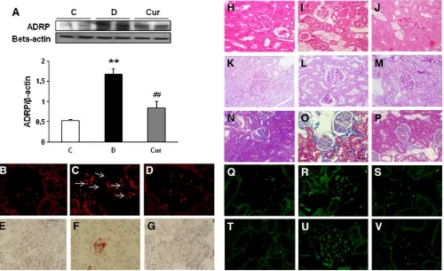
treatment (Fig. 1B). ADRP is a marker of lipid droplets. Western blot
analysis clearly showed a 2.2-fold increase of ADRP protein
expression in vehicle-treated diabetic rats (Fig. 2A), which was
attenuated by curcumin treatment. In agreement with Western blot
analysis, immuno
fl
uorescence staining of ADRP revealed typical lipid
droplets as ring-shaped in the tubules of vehicle-treated diabetic rats
(Fig. 2C). In contrast, there were almost no lipid droplets in the
curcumin-treated diabetic rats (Fig. 2D). Likewise, there is very strong
staining of oil red O in the glomerular and tubulointerstitial cells of
vehicle-treated diabetic rats, which indicates accumulation of neutral
lipids deposits (Fig. 2F) and almost no staining in the
curcumin-treated diabetic rats (Fig. 2G). Lipid accumulation was also shown by
the unstained area in renal tissue under H&E staining in the
vehicle-treated diabetic rats (Fig. 2I) which was reduced by curcumin
treatment (Fig. 2J). These data strongly indicate that there were
excessive amounts of lipid deposits in the kidneys of vehicle-treated
diabetic rats. Curcumin treatments improved this lipid accumulation
in the kidneys of diabetic rats.
3.3. Renal histologic change in experimental animals
Periodic acid
–
Schiff staining reveals ~56% sclerosis of glomeruli in
vehicle-treated diabetic rats (Fig. 2L), whereas the curcumin-treated
diabetic rats developed only 28.3% (
P
b
.05) (Fig. 2M) when compared
with control nondiabetic rats (Fig. 2K). In addition to pronounced
glomerular sclerosis, severe tubulointerstitial changes were observed
in the vehicle-treated diabetic rats. Histological evaluations revealed
that the vehicle-treated diabetic rats had 58.3% dilated tubules
(Fig. 2L). Curcumin treatment signi
fi
cantly reduced this to 34%
(
P
b
.05) compared with that in vehicle-treated diabetic rats (Fig. 2M).
Interstitial collagen deposition was studied in Azan
–
Mallory-stained
renal sections as an index of interstitial
fi
brosis. Vehicle-treated
diabetic rats showed higher interstitial
fi
brosis (Fig. 2O) than control
nondiabetic rats (Fig. 2N), which was improved in the
curcumin-treated diabetic rats (Fig. 2P). Immuno
fl
uorescence microscopy with
anti-
fi
bronectin and anti-type IV collagen antibodies revealed
increased intensity of immuno
fl
uorescence in the glomeruli and
tubulointerstitial cells in vehicle-treated diabetic rats (Fig. 2R,
2U),
indicating accumulation of extracellular matrix (ECM) proteins and
glomerulosclerosis and tubulointerstitial
fi
brosis, whereas curcumin
Table 1Biochemical parameters of the experimental animals at the end of intervention study (n= 5)
C D Cur
24-h urine protein (g/day) 12.5±0.7 42±5.2⁎ 15.2±3.7⁎⁎⁎ Plasma glucose (mg/dl) 118.6±6 744.4±9.3⁎ 597.1±9.9⁎,⁎⁎⁎ Plasma urea (mg/dl) 21.9±0.6 45±1.9⁎ 33.2±2.5⁎,⁎⁎⁎ Plasma creatinine (mg/dl) 0.3±0.01 2.1±0.1⁎ 0.8±0.2⁎⁎,⁎⁎⁎ Creatinine clearance (ml/min) 3.9±0.5 0.8±0.1⁎ 2.7±0.5⁎,⁎⁎⁎ Plasma triglyceride (mg/dl) 104.8±2.1 320.3±2.1⁎ 114±3.7⁎⁎⁎
Data are means±S.E.M. ⁎ Pb.01.
⁎⁎ Pb.05 vs. C. ⁎⁎⁎ Pb.01 vs. D.
Fig. 1. (A) Effect of curcumin treatment on the kidney weight to body weight ratio. (B) The vehicle-treated diabetic rats showed statistically significant increases in renal triglyceride content, which were reduced by curcumin treatment. Results are expressed as mean±S.E.M.n=5 per group. C, age-matched normal rats; D, diabetic-treated rats administered with vehicle; Cur, diabetic rats diabetic-treated with curcumin 100 mg/kg/day.⁎⁎Pb.01 vs. C;##
suppressed the increased intensity of
fi
bronectin and type IV collagen
expression in the diabetic rats (Fig. 2S, V).
3.4. Effect of curcumin on renal expression of AMPK
To understand the mechanism by which curcumin attenuated
lipid accumulation in the kidneys of diabetic rats, we measured the
effect of curcumin on AMPK activation on renal tissues. Since
phosphorylation of the catalytic subunit AMPK-
α
at Thr172 position
is essential for AMPK activation, AMPK activation was monitored by
using a speci
fi
c antibody that recognizes the phosphorylated AMPK-
α
at Thr172. As shown in
Fig. 3A, AMPK phosphorylation was reduced in
the vehicle-treated diabetic rats by nearly 60% (
P
b
.01). Curcumin
prevented the reduction in AMPK phosphorylation in diabetic rats,
suggesting that it may protect the AMPK activity.
3.5. Effect of curcumin on renal expression of nuclear receptors and
genes that regulate triglyceride and cholesterol metabolism
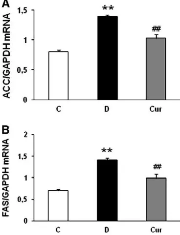
SREBP-1c is a transcription factor that activates the transcription
of genes involved in fatty acid and triglyceride synthesis, such as ACC
and FAS
[34]. To assess the SREBP-1c precursor and the nuclear form,
cell lysates were fractionated into a separate membrane fraction
containing SREBP precursors and a nuclear fraction containing active
nSREBP
[35].
Fig. 3
(B and C) shows that both precursor (2.6-fold) and
mature forms (2.7-fold) of SREBP-1c were increased in the
vehicle-treated diabetic rats. It is noteworthy that curcumin administration
signi
fi
cantly reduced both precursor and mature forms of SREBP-1c
and was well correlated with mRNA encoding of the SREBP-1c target
genes, ACC and FAS (Fig. 4A and B).
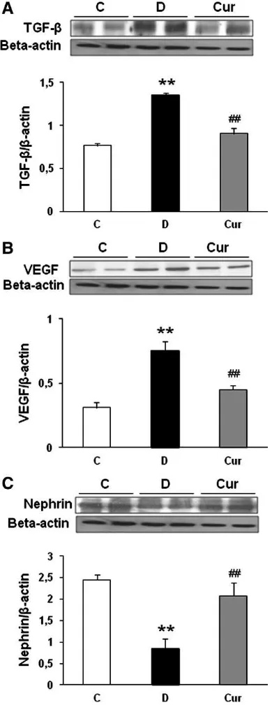
3.6. Curcumin decreases proteinuria and renal expression of profibrotic
growth factors
Because mesangial expansion and accumulation of ECM proteins
are mediated by increased expression of growth factors such as TGF-
β
[36]
and VEGF
[37], we measured the expression of TGF-
β
1 and VEGF
in the kidney cortex of experimental animals. There were a 1.8-fold
increase in TGF-
β
1 (Fig. 5A) and a 2.4-fold increase in VEGF (Fig. 5B)
expression, and curcumin treatment ameliorated these increases by
1.2- and 1.4-fold, respectively. Next, we analyzed the expression
pattern of the slit diaphragm protein nephrin.
Fig. 5C shows that the
down-regulation of nephrin occurred in the vehicle-treated diabetic
rats compared with that in the control nondiabetic rats. This decrease
in renal nephrin protein expression was markedly increased by
curcumin treatment (
P
b
.05).
4. Discussion
The most important
fi
ndings of this study were as follows: (1)
Curcumin can activate AMPK by increasing AMPK phosphorylation in
renal tissue in STZ-induced type 1 diabetes. (2) This increase of AMPK
Fig. 2. (A) Western blot and densitometric quantification of ADRP. (B–D) Immunofluorescence staining of kidney for ADRP (glomeruli and tubules) (×400). (E–G) Representative photomicrograph of the oil-red-O-stained renal tissues. Lipid droplets appear as red spots. There is increased oil red O staining in the glomerular and tubular cells of diabetic rats, and treatment with curcumin 100 mg/kg/day reverses these changes (×400). (H–J) Hematoxylin and eosin staining of the cross-sectional tissue slices of kidney depicted lipid accumulation, which was indicated by the unstained area in renal tissues (×200). (K–M) Periodic acid–Schiff staining of kidney depicted glomerular sclerosis and tubular dilatation (×200). (N–P) Azan–Mallory staining forfibrosis of the cross-sectional tissue slices of kidney. Fibrosis is indicated by the blue area (×200). (Q–S) Immunofluorescence microscopy of frozen sections stained withfibronectin antibody (×400). (T–V) Immunofluorescence microscopy of frozen sections stained with type IV collagen antibody (× 400). Kidney tissues were harvested from age-matched normal rats as a control (B, E, H, K, N, Q, T), diabetic-treated rats administered with vehicle (C, F, I, L, O, R, U) and diabetic rats treated with curcumin 100 mg/kg/day (D, G, J, M, P, S, V).n=5 rats in each experimental group.⁎⁎Pb.01 vs. C;##activation, consecutively, sharply suppresses renal SREBP-1c and the
target genes such as ACC and FAS. (3) As a result, curcumin attenuates
proteinuria, glomerulosclerosis, and tubulointerstitial dilatation and
fi
brosis. In the present study, we also found that, in STZ-induced type
1 DN rats, there were excessive amounts of lipid deposits in the
kidney sections, as shown by oil red O and H&E staining, the presence
of lipid bodies, as shown by ADRP immuno
fl
uorescence microscopy
and ADRP protein levels, using Western blot analysis; and signi
fi
-cantly higher renal content of triglyceride compared with those in
control nondiabetic rats; all of these abnormalities were ameliorated
by curcumin treatment.
Accumulating evidence indicates that curcumin
(diferuloyl-methane), a polyphenol natural product of the plant
C. longa
, exerts
effects on a wide range of molecular targets controlling lipid
accumulation and has antidiabetic and anticancer activities
[15,16].
Our previous study in STZ-induced type 1 DN showed that curcumin
exhibited renoprotective effects to lessen proteinuria and diabetic
pathologic changes through inhibition of macrophage in
fi
ltration,
which suggests that curcumin has an anti
fi
brotic and anti-in
fl
amma-tory effect
[38]. Diabetic rats with curcumin diet showed that
curcumin has a hypocholesterolemic action via stimulation of hepatic
cholesterol-7a-hydroxylase and HMG-CoA reductase
[16]. Curcumin
also inhibits ox-LDL-induced cholesterol accumulation in cultured
vascular smooth muscle cells through inhibition of nuclear
translo-cation of SREBP-1
[18]. It has also been shown that curcumin
improved muscular insulin resistance through the up-regulation of
phosphorylated AMPK
[39]. Furthermore, curcumin increases the
phosphorylation of AMPK and its downstream target as well as
suppresses gluconeogenic gene expression in hepatoma cells
[21,40].
Although several lines of evidences strongly suggest that curcumin
could regulate glucose and lipid homeostasis, the potential
mecha-nism of curcumin in regulating triglyceride accumulation in type 1 DN
has not been studied. In the present study, we show for the
fi
rst time
that curcumin protects renal triglyceride accumulation in
STZ-induced type 1 diabetes through activation of AMPK and suppresses
lipogenic gene expression.
AMPK is a heterotrimeric protein consisting of a combination of
α
1/2-,
β
1/2- and
γ
1/2/3-subunits
[41], with the
α
1- rather than
α
2-subunit expressed in the kidney
[42]. The enzymatic activity of AMPK
is dependent on phosphorylation of Thr172 of the
α
-subunit
[43]. On
Fig. 3. (A) Representative Western blots and group data depicting protein abundance of the phosphorylated AMPK in the renal tissues. (B) Western blots analysis of the expression of SREBP-1c nuclear extracts and membrane (precursor) (C) isolated from kidney tissues. STZ-induced type 1 diabetes causes increased SREBP-1c expression in both nuclear and membrane extracts, and these changes were reversed upon treatment with curcumin. Data are means±S.E.M.n= 5 per group. C, age-matched normal rats; D, diabetic-treated rats administered with vehicle; Cur, diabetic rats treated with curcumin 100 mg/kg/day.⁎⁎Pb.01 vs. C;##
Pb.01 vs. D.
Fig. 4. (A and B) Effects of curcumin on renal mRNA levels of ACC and FAS in rats with DN were determined by quantitative reverse transcriptase PCR. The expression level of each sample is expressed relative to the expression level of GAPDH gene. Data are means±S.E.M.n= 5 per group. C, age-matched normal rats; D, diabetic-treated rats administered with vehicle; Cur, diabetic rats treated with curcumin 100 mg/kg/day. ⁎⁎Pb.01 vs. C;##
activation, AMPK inhibits energy-consuming reactions, such as
synthesis of fatty acids and sterols, and activates
adenosine-triphosphate-generating processes, such as fatty acid oxidation
[44].
A very recent study also demonstrated that activation of AMPK
inhibits hepatic fatty acid synthesis by suppressing SREBP-1c, a
master regulator of lipogenic gene expression, in rat hepatoma cells
[45]. Lee et al. have shown that, in STZ-induced insulin-de
fi
cient
diabetes, a model of type 1diabetes, AMPK phosphorylation was
reduced at the time when the kidneys were hypertrophic
[46]. In line
with previous reports, we have also observed that both total and
phosphorylated AMPK expressions were reduced in vehicle-treated
diabetic rats (Fig. 3A) and that these decreases under diabetic
conditions were ameliorated by curcumin treatment, which in turn
attenuated the expression of SREBP-1c in the renal tissue of type 1
diabetic rats and, as a consequence, caused decreases in the mRNA
abundance of two SREBP-1-activated enzymes that mediate fatty acid
and triglyceride synthesis, ACC and FAS.
Abnormal lipid deposits have been observed in tubules of human
diabetic kidney
[47]
and have been proposed to participate in the
pathogenesis of DN and other proteinuric renal diseases
[3]. Lipid
droplets were also detected in the cortical tubules of the STZ-induced
diabetic rats
[5]. Increased deposition of lipid in the kidney
contributes to cellular damage and to the progression of diabetic
kidney disease. ADRP, also called adipophilin in humans, is a ~ 50-kDa
fatty-acid-binding protein that is transcriptionally activated when
preadipocytes differentiate into mature adipocytes
[48]. ADRP
belongs to the perilipin family of lipid storage proteins
[49]
and,
similar to other perilipin members, localizes to the cytoplasmic
surface of lipid storage droplets in distinct cell types. Induction of the
ADRP gene is apparently an important aspect of the response to
diabetes and formation of lipid storage droplets in the diabetic kidney.
We found that the ADRP protein (Fig. 2A) and ADRP immuno
fl
uo-rescence (Fig. 2C) were signi
fi
cantly induced in vehicle-treated
diabetic rat kidneys and corresponded with the presence of lipid
droplets in oil red O (Fig. 2F) and H&E staining (Fig. 2I) and increase in
renal triglyceride content (Fig. 1B), and that these increases under
diabetic conditions were ameliorated by curcumin treatment.
In vivo
and
in vitro
studies have shown that low-density
lipoprotein or very low density lipoprotein induces up-regulation of
growth factors, including TGF-
β
and platelet-derived growth factor,
ECM
[50], lipid peroxidation and glycoxidation
[51], and change in
expression of podocyte-speci
fi
c proteins such as nephrin
[52],
indicating a direct role for lipids in activating the mediators of
glomerulosclerosis and proteinuria. Ishigaki et al. indicated that
activation of renal SREBP-1c was consistently associated with
pathohistological
fi
brosis and TGF-
β
activation both
in vivo
and
in vitro
, accompanying induction of
fi
brosis markers such as type IV
collagen and
fi
bronectin
[11]. It has also been reported that renal
hypertrophy in STZ-induced type 1 diabetic rats was associated with
reduction in AMPK phosphorylation
[53]. Our study indicates that
accumulation of renal triglyceride in STZ-treated diabetic rats caused
increased expression of TGF-
β
and VEGF as well as ECM proteins such
as type IV collagen and
fi
bronectin and decreased expression of
nephrin, resulting in glomerulosclerosis and proteinuria, respectively.
Curcumin treatment markedly decreased TGF-
β
and VEGF and
increased nephrin expression in renal tissue of diabetic rats.
Curcumin also reduced the augmented expressions of type IV
collagen and
fi
bronectin. This effect might be mediated by curcumin's
function as an AMPK activator, which further down-regulated the
SREBP-1c expression.
We have shown that curcumin inhibits renal triglyceride
accu-mulation in STZ-induced type 1 DN by activation of AMPK and
suppresses SREBP-1c expression, as well as inhibits the expression of
lipogenic genes, which may explain some of the lipid-lowering effects
of curcumin. Since AMPK is considered one of the preventive or
therapeutic targets for metabolic diseases such as diabetes, obesity
and cancer, the effective modulation of AMPK by curcumin could be a
promising effective approach for treating these diseases.
References
[1] Catania JM, Chen G, Parrish AR. Role of matrix metalloproteinases in renal pathophysiologies. Am J Physiol Renal Physiol 2007;292:F905–11.
[2] Kimmelstiel P, Wilson C. Intercapillary lesions in the glomeruli of the kidney. Am J Pathol 1936;12:83–98.
[3] Keane WF. The role of lipids in renal disease: future challenges. Kidney Int Suppl 2000;75:S27–31.
Fig. 5. Representative Western blots and group data depicted protein abundance of TGF-β(A), VEGF (B) and nephrin (C) in the renal tissues of diabetic rats, curcumin-treated diabetic rats and control nondiabetic rats.n= 5 in each group. Data are means±S.E.M.⁎⁎Pb.01 vs. C;##
[4] Oda H, Keane WF. Lipids in progression of renal disease. Kidney Int Suppl 1997;62: S36–8.
[5] Sun L, Halaihel N, Zhang W, et al. Role of sterol regulatory element-binding protein 1 in regulation of renal lipid metabolism and glomerulosclerosis in diabetes mellitus. J Biol Chem 2002;277:18919–27.
[6] Wang Z, Jiang T, Li J, et al. Regulation of renal lipid metabolism, lipid accumulation, and glomerulosclerosis in FVBdb/db mice with type 2 diabetes. Diabetes 2005;54: 2328–35.
[7] Brown MS, Goldstein JL. The SREBP pathway: regulation of cholesterol metabolism by proteolysis of a membrane-bound transcription factor. Cell 1997; 89:331–40.
[8] Horton JD, Shimomura I. Sterol regulatory element-binding proteins: activators of cholesterol and fatty acid biosynthesis. Curr Opin Lipidol 1999;10:143–50. [9] Rawson RB. Control of lipid metabolism by regulated intramembrane proteolysis
of sterol regulatory element binding proteins (SREBPs). Biochem Soc Symp 2003;70:221–31.
[10] Proctor G, Jiang T, Iwahashi M, et al. Regulation of renal fatty acid and cholesterol metabolism, inflammation, and fibrosis in Akita and OVE26 mice with type 1 diabetes. Diabetes 2006;55:2502–9.
[11] Ishigaki N, Yamamoto T, Shimizu Y, et al. Involvement of glomerular SREBP-1c in diabetic nephropathy. Biochem Biophys Res Commun 2007;364:502–8. [12] Hardie DG. AMP-activated protein kinase as a drug target. Annu Rev Pharmacol
Toxicol 2007;47:185–210.
[13] Zhou G, Myers R, Li Y, et al. Role of AMP-activated protein kinase in mechanism of metformin action. J Clin Invest 2001;108:1167–74.
[14] Yang J, Craddock L, Hong S, et al. AMP-activated protein kinase suppresses LXR-dependent sterol regulatory element-binding protein-1c transcription in rat hepatoma McA-RH7777 cells. J Cell Biochem 2009;106:414–26.
[15] Goel A, Kunnumakkara AB, Aggarwal BB. Curcumin as "Curecumin": from kitchen to clinic. Biochem Pharmacol 2008;75:787–809.
[16] Babu PS, Srinivasan K. Hypolipidemic action of curcumin, the active principle of turmeric (Curcuma longa) in streptozotocin induced diabetic rats. Mol Cell Biochem 1997;166:169–75.
[17] Peschel D, Koerting R, Nass N. Curcumin induces changes in expression of genes involved in cholesterol homeostasis. J Nutr Biochem 2007;18:113–9.
[18] Yuan HY, Kuang SY, Zheng X, et al. Curcumin inhibits cellular cholesterol accumulation by regulating SREBP-1/caveolin-1signaling pathway in vascular smooth muscle cells. Acta Pharmacol Sin 2008;29:555–63.
[19] Fan C, Wo X, Dou X, et al. Regulation of LDL receptor expression by the effect of curcumin on sterol regulatory element pathway. Pharmacol Rep 2006;58:577–81. [20] Dou X, Fan C, Wo L, et al. Curcumin up-regulates LDL receptor expression via the sterol regulatory element pathway in HepG2 cells. Planta Med 2008;74:1374–9. [21] Kim T, Davis J, Zhang AJ, et al. Curcumin activates AMPK and suppresses gluconeogenic gene expression in hepatoma cells. Biochem Biophys Res Commun 2009;388:377–82.
[22] Kowluru RA, Kanwar M. Effects of curcumin on retinal oxidative stress and inflammation in diabetes. Nutr Metab (Lond) 2007;4:8.
[23] Meghana K, Sanjeev G, Ramesh B. Curcumin prevents streptozotocin-induced islet damage by scavenging free radicals: a prophylactic and protective role. Eur J Pharmacol 2007;577:183–91.
[24] Bradford MM. A rapid and sensitive method for the quantitation of microgram quantities of protein utilizing the principle of protein-dye binding. Anal Biochem 1976;72:248–54.
[25] Husdan H, Rapoport A. Estimation of creatinine by the Jaffe reaction. A comparison of three methods. Clin Chem 1968;14:222–38.
[26] Bagheri F, Gol A, Dabiri S, et al. Preventive effect of garlic juice on renal reperfusion injury. Iran J Kidney Dis 2011;5:194–200.
[27] Bligh EG, Dyer WJ. A rapid method of total lipid extraction and purification. Can J Biochem Physiol 1959;37:911–7.
[28] Thallas-Bonke V, Thorpe SR, Coughlan MT, et al. Inhibition of NADPH oxidase prevents advanced glycation end product-mediated damage in diabetic nephrop-athy through a protein kinase C-alpha-dependent pathway. Diabetes 2008;57: 460–9.
[29] Ghosh SS, Massey HD, Krieg R, et al. Curcumin ameliorates renal failure in 5/6 nephrectomized rats: role of inflammation. Am J Physiol Renal Physiol 2009;296: F1146–57.
[30] Jiang T, Wang Z, Proctor G, et al. Diet-induced obesity in C57BL/6J mice causes increased renal lipid accumulation and glomerulosclerosis via a sterol regulatory element-binding protein-1c-dependent pathway. J Biol Chem 2005;280: 32317–25.
[31] Gao J, Serrero G. Adipose differentiation related protein (ADRP) expressed in transfected COS-7 cells selectively stimulates long chain fatty acid uptake. J Biol Chem 1999;274:16825–30.
[32] Xia Z, Kuo KH, Nagareddy PR, et al. N-acetylcysteine attenuates PKCbeta2 overexpression and myocardial hypertrophy in streptozotocin-induced diabetic rats. Cardiovasc Res 2007;73:770–82.
[33] McManaman JL, Palmer CA, Wright RM, et al. Functional regulation of xanthine oxidoreductase expression and localization in the mouse mammary gland: evidence of a role in lipid secretion. J Physiol 2002;545:567–79.
[34] Shimano H. Sterol regulatory element-binding proteins (SREBPs): transcriptional regulators of lipid synthetic genes. Prog Lipid Res 2001;40:439–52.
[35] DeBose-Boyd RA, Ou J, Goldstein JL, et al. Expression of sterol regulatory element-binding protein 1c (SREBP-1c) mRNA in rat hepatoma cells requires endogenous LXR ligands. Proc Natl Acad Sci U S A 2001;98:1477–82.
[36] Basile DP. The transforming growth factor beta system in kidney disease and repair: recent progress and future directions. Curr Opin Nephrol Hypertens 1999;8:21–30.
[37] de Vriese AS, Tilton RG, Elger M, et al. Antibodies against vascular endothelial growth factor improve early renal dysfunction in experimental diabetes. J Am Soc Nephrol 2001;12:993–1000.
[38] Soetikno V, Sari FR, Veeraveedu PT, et al. Curcumin ameliorates macrophage infiltration by inhibiting NF-κB activation and proinflammatory cytokines in streptozotocin induced-diabetic nephropathy. Nutr Metab (Lond) 2011;8:35. [39] Na LX, Zhang YL, Li Y, et al. Curcumin improves insulin resistance in skeletal
muscle of rats. Nutr Metab Cardiovasc Dis 2011;21:526–33.
[40] Kanitkar M, Gokhale K, Galande S, et al. Novel role of curcumin in the prevention of cytokine-induced islet death in vitro and diabetogenesis in vivo. Br J Pharmacol 2008;155:702–13.
[41] Carling D. The AMP-activated protein kinase cascade—a unifying system for energy control. Trends Biochem Sci 2004;29:18–24.
[42] Fraser S, Mount P, Hill R, et al. Regulation of the energy sensor AMP-activated protein kinase in the kidney by dietary salt intake and osmolality. Am J Physiol Renal Physiol 2005;288:F578–86.
[43] Hawley SA, Davison M, Woods A, et al. Characterization of the AMP-activated protein kinase kinase from rat liver and identification of threonine 172 as the major site at which it phosphorylates AMP-activated protein kinase. J Biol Chem 1996;271:27879–87.
[44] Merrill GF, Kurth EJ, Hardie DG, et al. AICA riboside increases AMP-activated protein kinase, fatty acid oxidation, and glucose uptake in rat muscle. Am J Physiol 1997;273:E1107–12.
[45] Yap F, Craddock L, Yang J. Mechanism of AMPK suppression of LXR-dependent Srebp-1c transcription. Int J Biol Sci 2011;7:645–50.
[46] Lee MJ, Feliers D, Mariappan MM, et al. A role for AMP-activated protein kinase in diabetes-induced renal hypertrophy. Am J Physiol Renal Physiol 2007;292: F617–27.
[47] Ong AC, Moorhead JF. Tubular lipidosis: epiphenomenon or pathogenetic lesion in human renal disease? Kidney Int 1994;45:753–62.
[48] Jiang HP, Serrero G. Isolation and characterization of a full-length cDNA coding for an adipose differentiation-related protein. Proc Natl Acad Sci U S A 1992;89: 7856–60.
[49] Londos C, Brasaemle DL, Schultz CJ, et al. Perilipins, ADRP, and other proteins that associate with intracellular neutral lipid droplets in animal cells. Semin Cell Dev Biol 1999;10:51–8.
[50] Nishida Y, Oda H, Yorioka N. Effect of lipoproteins on mesangial cell proliferation. Kidney Int Suppl 1999;71:S51–3.
[51] Lee HS, Kim BC, Kim YS, et al. Involvement of oxidation in LDL-induced collagen gene regulation in mesangial cells. Kidney Int 1996;50:1582–90.
[52] Garman JH, Mulroney S, Manigrasso M, et al. Omega-3 fatty acid rich diet prevents diabetic renal disease. Am J Physiol Renal Physiol 2009;296:F306–16. [53] Lee MJ, Feliers D, Sataranatarajan K, et al. Resveratrol ameliorates high