THE PERFORMANCE OF CHITOSAN-FERRIHIDRYTE MEMBRANE FOR PHOSPHATE
UPTAKE
Barlah Rumhayati*, Chasan Bisri, and Wahyu O. Fajarina
Department of Chemistry, Brawijaya University, Jl. Veteran, Malang 65145
Received March 27, 2009; Accepted October 21, 2009
ABSTRACT
The uptake of orthophosphate onto the chitosan-ferrihydrite membrane has been studied. The membrane was proposed as a new binding layer of DGT technique. Membrane was prepared from a mixture of chitosan solution with ferrihydrite (FeOOH) paste and was crosslinked with glutaraldehyde. As a result, the uptake of orthophosphate was slow. The maximum adsorption capacity was reached at pH 5.0 and 8.0. It was mainly due to electrostatic attraction of phosphate ions to the protonated free amino groups of chitosan and to active sites of ferrihydrite.
Desorption could be occurred optimally using 0.3 M of sulphuric acid. The absorption of SO4
ion to the protonated amino groups and the formation of ionic crosslinking could exchange and desorb phosphate ions. Ferrihydrite was eluted also by the acid. However, the eluted ferrihydrite readsorbed phosphate ions, resulted in minimizing the analysed free phosphate.
Keywords: adsorption, phosphate, chitosan-ferrihydrite/glutaraldehide membrane, DGT technique
INTRODUCTION
Phosphorus is an essential nutrient for all life forms. It has an important role in fundamental biochemical reactions [1] involving genetic material (DNA, RNA) and energy transfer (ATP), and in the structural support of organisms provided by membranes (phospholipids) and bone (the bio-mineral hydroxyapatite). In addition, this compound is responsible in the productivity of freshwater and terrestrial ecosystems. In the hydrosphere, the predominant source of phosphorus input into freshwater systems is from geological deposits and surface soils. Groundwater influxes or atmospheric deposits contribute only minor amount of phosphate. In urban areas, industrial and domestic sewage effluents are the major sources of phosphate loading into many rivers and lakes [2]. Human activities such as agriculture, the mining of phosphate rock and deforestation have enhanced phosphorus transport from terrestrial to aquatic systems.
As an essential nutrient for photosynthesis, phosphorus is an important cause of eutrophication in numerous water systems. Even though the external sources of phosphate in water system may have been controlled, phosphate exchange between the overlying water and sediments can continue to support the growth of algae and eutrophication [3-4]. Therefore, studies on phosphorus exchange between sediment and overlying waters are of much significance for eutrophication control. The exchanged phosphorus species should be also characterized to gain better understanding of the biogeochemical behaviour of phosphorus in
environmental. The concentration of phosphorus species often found at low level due to phosphorus uptake by phytoplankton in freshwater. Even though the phosphorus analysis which is based on the formation of phosphomolybdenum blue complex [5] is a sensitive method, the available techniques including batch method [5] and flowing system (FIA) [6], could not detect the low level of phosphorus. On the other hand, the filterable reactive phosphate (FRP) which can be directly detected by the phosphomolybdenum blue method is prone to change during sample storage [7] because of the dynamic exchange of this compound with biota and surfaces. Hence, a simple but more sensitive procedure that could be used for phosphorus routine monitoring is desirable.
Since last decade, a sensitive and fully quantitative technique for measuring phosphorus species in situ in aquatic systems has been developed [8], called the Diffusive Gradient in Thin Films (DGT) technique. A significant benefit arising from the in-situ application of the DGT technique comes from the binding gel which continuously accumulates solute, which then enables measurement of low concentrations of solute. In addition, the technique may provide an in situ measurement of the rate constant for transfer from solid phase of sediment to solution [9]. The time averaged concentration of available reactive phosphate could be also gained by in situ deploying of the DGT probes in the field during a known time instead of assessing of available reactive phosphate comparatively by grab sampling [8]. In addition, analysis errors induced by storage of samples prior to
* Corresponding author. Tel/Fax : +62-341-575838 Email address : [email protected]
Barlah Rumhayati et al.
analysis due to rapid change of phosphorus species could be minimized by in situ DGT technique.
A DGT technique is based on the principle of diffusive gradients in thin films. A probe of DGT consists of three layers, i.e. a filter which directly contact with sample solution, a diffusive layer based on acrylamide as a second layer and a binding layer which consists of binding agent imbedded in acrylamide polymer [10]. The successfulness of DGT measurement is determined by the performance of the binding gel which is determined by the binding agent. At present, ferrihydrite (FeOOH) [8] and La(OH)3 [11] imbedded in acrylamide gel have been
used as a binding agent of the DGT technique for phosphate (orthophosphate and organic phosphates) monitoring. Even though, those gels showed good performance for phosphate uptake, acrylamide as base material of binding gel is environmental unfriendly material. The DGT with La(OH)3 binding gel could be
used for phosphate measurement at pH 4-9. This gel would not be suitable for use at the pH of intended use, if it fell outside the pH range 4-9, e.g. acid mine drainage, or in waters with high pH because the gel will change dimensions during deployment [10]. On the other hand, the ferrihydrite binding gel showed good agreement when used for phosphate uptake at pH 2-10 [8].
Chitosan biopolymer that is consisted of glucosamine monomer units [12] may be used for an environmental friendly base material of binding layer. Chitosan can be modify to form a membrane by diluting chitosan flake into acetic acid followed by casting [13]. In this condition, the amino groups are easily protonated which may cause electrostatic attraction of anionic compounds [14], such as phosphate ions. The chemical modification of chitosan membrane can be performed heterogeneously by immersing the membrane into crosslinker solution, such as glutaraldehyde. The modification may prevent the dissolving of the polymer when anion sorption is performed in acidic solutions, prevent swelling, and enhance the pore size distribution [13,15]. However, the adsorption capacity of anions on to the glutaraldehyde crosslinked chitosan membrane will be decreased due to only few free protonated amino groups are still available for anion attraction after crosslinking. Hence, a specific adsorbent such as ferrihydrite should be imbedded into chitosan polymer matrix to enhance the adsorption capacity of membrane.
This research was aimed to investigate a new binding layer of DGT technique. Chitosan was chosen as a base material for producing binding layer with ferrihydrite as binding agent. The performance of chitosan-ferrihydrite on the phosphate uptake was investigated in this research at various time and pH solution.
EXPERIMENTAL SECTION
Material
Chitosan gel solution was prepared by diluting chitosan powder (teknis, ~80% deacetylation) in acetic acid (~100%). Glutaraldehyde (Merck) was used as crosslinker. Ferrihydrite was produced through precipitation process between NaOH (Merck) and Fe(NO3)3. Synthetic sample of phosphate was
prepared from KH2PO4 (BDH). Reactive phosphate was
detected as phosphomolybdenum blue complex which was produced by mixing the colouring reagents of ammonium molybdate tetrahydrate in acidic solution (H2SO4, 95-97%)) and reducing agent of ascorbic acid.
Instruments
Volumetric glasses were used for preparing reagents, chitosan gel solution and ferrihydrite (FeOOH) precipitate. Chitosan membrane was drying mounted in a Memmert oven. Phosphate was uptake onto the chitosan-ferrihydrite membrane by shaking with an Edmund Buhler SM 25 shaker. The pH of phosphate solution was measured by Schott Gerate CG 820 pH meter. For assessing the crosslinking process, Infrared spectrophotometer (Shimadzu) was used to determine the functional groups of modified chitosan membrane compared to the pristine one. Phosphate concentrations in bulk solution at initial and final uptake, and phosphate eluted concentration from the membrane were detected by Shimadzu UV-Vis Spectrophotometer.
Procedure
Preparation of chitosan-ferrihydrite membrane
The production of chitosan membrane solution followed the previous procedure [13]. Chitosan membrane solution 2.5% (w/v) (solution A) was prepared by dissolving 2.5 g chitosan powder into 100 mL of acetic acid 3% (v/v). On the other hand, ferrihydrite (FeOOH) paste was formed through precipitation reaction between 10 mL of Fe(NO3)3 0.2 M
Barlah Rumhayati et al.
neutralize the amino groups. After 24 h, the membranes were exhaustively washed with distilled water until all alkali was removed. The membranes were stored in water and cut into disks. At least 3.0 g of wet membrane disks were crosslinked heterogeneously in 50 mL of aqueous glutaraldehyde solution 0.75% (w/w) for 2 h at room temperature without agitation. Unreacted glutaraldehyde residue was removed by rinsing the membranes with deionized water. The IR spectra of modified membranes were evaluated to predict the amount of free protonated amino groups which is responsible for phosphate sorption.
Phosphate uptake experiment
Batch kinetic experiments were carried out by soaking wet membrane disks in 10 mL of orthophosphate solution (5 mg P/L) at pH 5 under stirring at 200 rpm. The membranes were withdrawn at 30, 60 seconds, and 1 to 24 h at fixed time intervals. The amount of phosphate adsorbed at fixed time was calculated as differences between the initial mass and the final mass of bulk solution.
The adsorption capacity was determined from batch equilibrium experiments. These experiments were conducted by soaking membrane disks in 10 mL of orthophosphate solution (ranging from 2 to 25 mg P/L) at pH 2.0, 5.0, and 8.0 for 24 h at room temperature under stirring. The adsorption capacity of chitosan-ferrihydrite membrane was calculated based on the difference of phosphate concentration in bulk solution before and after uptake. The equilibrium isotherms were adjusted by Langmuir model, given by Eq. 1 and is arranged to Eq. 2.
Q is the amount of phosphate adsorbed by the membrane (mg/g), Qmax is the maximum amount
adsorbed within a monolayer (mg/g), Ce is the final
phosphate concentration (mg P/L) and K (L/mg) is the Langmuir dissociation constant, which is related to the adsorption energy.
Desorption experiments
Desorption of phosphate from the chitosan-ferrihydrite membrane was performed using sulfuric acid as eluent solution. These experiments were performed by immersing the membranes, which has adsorbed phosphate (5 mg P/L at pH 5 for 24 h), in 10 mL of H2SO4 at various concentration, i.e. 0.1, 0.25 and 0.3 M,
for 2 h with stirring at 200 rpm. The bulk phosphate concentration in solution was measured
spectrophotometrically. The extent of desorption was calculated from Eq. 3.
desorbed amount of phosphate
Desorption(%) = x 100
adsorbed amount of phosphate (3)
Determination of phosphate concentration
Concentrations of phosphate, either in the bulk solution of adsorption and eluted solution, were determined spectrophotometrically based on the formation of the phosphomolybdenum blue complex which can be measured at 816.5 nm. Phosphomolybdenum complex was performed through reaction between orthophosphate and molybdate reagent. This reagent was prepared by dissolving 20 g of ammonium molybdate tetrahidrat into 100 mL distilled water. 20 mL of sulfuric acid was added into solution and then was diluted into 500 mL. The phosphomolybdate complex was reduced into the blue complex by the reducing reagent, i.e. ascorbic acid which was prepared from 1.76 g of ascorbic acid into 100 mL distilled water. Before measurement, the mixed reagent was prepared by mixing 50 mL sulphuric acid 2.5 M, 15 mL of molybdate reagent, and 30 mL of reducing reagent. The mixed reagent should be prepared freshly.
Phosphomolybdenum blue complex in the adsorption bulk solution was formed by mixing 10 mL of the solution and 2 mL of mixed reagent, shaking for second until the blue colour was formed and waiting for 15 min before measurement. Phospholybdenum blue complex in elution bulk solution was formed by mixing 10 mL of the solution; 1.4 mL of molybdate reagent and 0.6 mL of reducing reagent.
RESULT AND DISCUSSION
Characterization of Chitosan-Ferrihydrite Membrane
The performance of ferrihydrite as binding agent will determine the good performance of a new binding layer for a DGT technique. Uncontrolled precipitation of ferrihydrite from Fe(NO3)3 and NaOH could induce the
formation of other iron oxides, such as geotite (α -FeOOH) and haematite (α-Fe2O3), with progressive
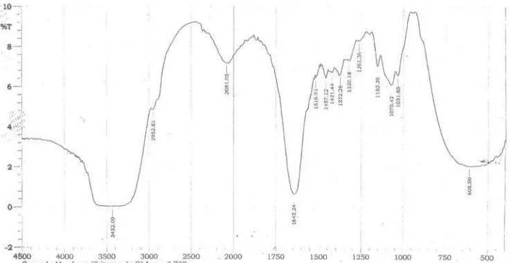
Figure 1. IR spectra of chitosan-ferrihydrite membrane with glutaraldehyde crosslinker.
Figure 2. Phosphate kinetic adsorption curve for
modified chitosan-ferrihydrite membrane
using XRD was not performed then the paste was prepared every two days to minimize changing of ferrihydrite.
After mounting and hydration, the wet chitosan-ferrihydrite membrane disk with diameter 3 cm had 0.8 ± 0.2 mm thickness and weight of 0.565 ± 0.100 g. Glutaraldehyde crosslinking process was qualitatively assessed from the IR spectra provided from the modified membrane (Fig. 1). When chitosan was crosslinked with glutaraldehyde, a reduction peak of primary amine (1100 cm-1) was observed, denoting that much amino groups were bound to glutaraldehyde molecules. A peak at 1655 cm-1 was also observed, which was related to the formation of imine bonds (C=N). A peak at 1700-1750 cm-1 indicated the free aldehydic groups which did not reach with amino groups of chitosan polymer. Free amino groups then could be protonated in acidic solution.
Kinetics of Phosphate Sorption
Fig. 2 presents the orthophosphate ion adsorption kinetic curves of crosslinked chitosan-ferrihydrite membranes. The initial concentration of phosphate was 5 mg P/L and pH 5.0. Relatively slow kinetic rates of phosphate uptake onto the chitosan-ferrihydrite membranes were observed. Approximately, 38% of initial phosphate concentration was uptake by the membrane during 5 h immersion. Up to 24 h, the amount of phosphate adsorbed was twice than the first five hours. It was observed also that the equilibrium had not been reached yet up to 24 h. Practically, the uptake equilibrium of solute by a DGT probe is reached for 24 h to uptake low concentration of phosphate (in micro units). Hence, kinetic experiment was not continued more than 24 h regarding with higher concentration of phosphate used in this research. However, the obtained kinetic rates data are promising to implement the crosslinked chitosan-ferrihydrite membrane as a binding layer for the DGT technique for phosphate uptake.
Adsorption Equilibrium
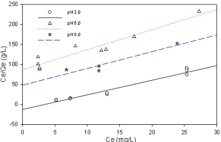
Fig. 3 depicts the Langmuir adsorption isotherms at pH 2.0, 5.0 and 8.0 while Table 1 presents Langmuir equilibrium constant. The Langmuir isotherm model assumes monolayer adsorption on a surface with a finite number of identical sites, that all sites are energetically equivalent and that there is no interaction between adsorbed molecules. The phosphate adsorption on to the modified chitosan membrane was affected by the pH of solution. pH will determine the available phosphate species, the protonation of free amino groups of chitosan and the form of ferrihydrite
Barlah Rumhayati et al.
Figure 3. Langmuir isotherm of phosphate adsorption at
pH 2.0, 5.0, and 8.0.
Table 1. Langmuir parameters for adsorption
experimental data.
Langmuir isotherm value pH
Qmax (mg/g) K (L/mg) R2
2.0 0.2748 -0.3012 0.96 5.0 0.1998 0.0578 0.91 8.0 0.2397 0.0862 0.90
surface. Phosphate are dissociated three times (pKa1 =
2.0, pKa2 = 6.8, pKa3 = 12.3) [17] while chitosan has pKa
of 6.5 [18]. The amino groups of chitosan will be protonated at pH below than pKa. Furthermore, ferrihydrite has point zero charge (pzc) between 7.8-7.9 [16]. At pH below pzc, ferrihydrite will have positive charge of FeOH2
+
and at pH above pzc, FeO- groups will be dominant on the surface of ferrihydrite.
Evaluating the effect of pH on the phosphate uptake, it was observed that the adsorption capacity of crosslinked chitosan-ferrihydrite membrane to phosphate was similar at pH 5.0 and 8.0. Even though the maximum amount of phosphate adsorbed was obtained at pH 2.0 but the adsorption energy (K) was observed negative. Errors might be occurred related to the quantitative analysis of phosphate at very high acid concentration. Ferrihydrite is diluted in acidic condition. The diluted Fe3+ could adsorb phosphate and resulted in underestimation of phosphate concentration measurement (subtractive interference) [19]. As a result, the amount of phosphate adsorbed was high but the adsorption was taken place on the diluted ferrihydrite, instead of on the membrane.
At pH 5.0, phosphate was mostly in form of H2PO4
-while free amino groups of chitosan were protonated and ferrihydrite had positive charge surface. On the other hand, at pH 8.0, HPO4
was dominant, the amino groups were not protonated and ferrihydrite surface had the same amount of FeOH2
+
and FeO- forms. It was observed that the amount of phosphate uptake by the
Figure 4. Ionic crosslinking of chitosan membrane with
sulphuric acid ions (adopted from [20])
Table 2. Desorption of phosphate ions in sulphuric
acid.
Desorption (%) H2SO4 concentration (M)
Average Sd(n-1), n=3
0.1 29.41 4.09
0.2 28.12 1.16
0.3 42.62 3.55
membranes was similar at pH 5.0 and 8.0. At these pHs, phosphate uptake was through two steps, i.e. the fast step of phosphate diffusion into active sites at surface due to electrostatic force and followed by the slow step of phosphate diffusion into microspores of ferrihydrite [16]. Theoretically, fresh ferrihydrite has 2-5 nm pore size [16]. However, the energy adsorption was higher at pH 8.0, indicating that phosphate uptake was predominantly due to electrostatic attraction to active sites of ferrihydrite surface rather than the attraction to the protonated amino groups. It means that for a binding layer of the DGT technique, the performance of the binding agent is more important than the one of the binding layer matrix. However, the use of chitosan as matrix of binding layer is a good approach regarding with the environmental friendly of material.
Desorption Performance
The binding layer of DGT technique is disposable layer for one usage. Hence, the percentage of
hosphate desorbed from a new binding layer must be determined to assess the desorption capability of the layer. Table 2 shows the percentage of desorbed phosphate using sulphuric acid eluent. The amount of phosphate desorbed was similar when using 0.1 and 0.2 M of sulphuric acid while only 42% of phosphate could be desorbed from the membrane using 0.3 M of sulphuric acid.
p
Barlah Rumhayati et al.
between the SO42- and NH3+. Hence, phosphates bound
in the protonated amino groups were released (desorbed). The presence of SO4
might crosslink two NH3
+
groups by ionic bonding (Fig. 4) during 2 h desorption. The increase of sulphuric acid content in the chitosan membrane might increase the ionic crosslinking and increased the amount of phosphate desorbed. However, not all of the absorbed sulphuric ions were crosslinking agents. Only those located between two NH3+ groups on the chains resulted in ionic crosslinking,
depending on the mobility of SO4
ions in the membrane [20].
Desorption might also occur through the dilution of ferrihydrite. Phosphates which were adsorbed by ferrihydrite could be diluted also. However, the diluted Fe3+ ions still adsorbed phosphate ions resulted in negative errors of phosphate analysis.
CONCLUSION
In this study, the performance of phosphate uptake onto the crosslinked chitosan-ferrihydrite membrane for a new binding layer of DGT technique was confirmed. The maximum adsorption capacity of phosphate on the membrane was reached at pH 5.0 and 8.0, indicating that adsorption process occurs mainly in chitosan amino groups and ferrihydrite active sites. The best desorption condition was obtained when using 0.3 M of sulphuric acid. The desorption was occurred due to the adsorption of SO42- ion to the protonated amino groups and the
ionic crosslinking. However, few amount of phosphate desorbed might due to subtractive errors of phosphate analysis. The crosslinked chitosan-ferrihydrite membrane has good performance on the phosphate uptake and, therefore, can be used as the DGT binding layer. Further investigation of other eluents solution may be conducted for increasing the desorption percentage.
ACKNOWLEDGEMENT
The authors thank Brawijaya University for DPP/SPP financial support.
REFERENCES
1. Westheimer, F.H., 1987, Science, 235, 1173-1178. 2. Ruttenberg, K.C., 2004, The Global Phosphorus
Cycle, in Tratise on Geochemistry, H.D. Holland,
K.K. Turekian, and W.H. Schlesinger, Editors, Elsevier Pergamon: Amsterdam, pp. 585.
3. Tang, H., 2000, Acta Sci. Circumstant, 20, 1. 4. Zeng, L., Li, X. and Liu, J., 2004, Water Res., 38, 5,
1318.
5. Murphy, J. and Riley, J.P., 1962, Anal. Chim. Acta, 27, 31.
6. McKelvie, I.D., Hart, B.T., Cardwell, T.J., and Cattrall, R.W., 1989, Analyst, 114, 1459.
7. McKelvie, I.D., 2004, 1. Separation, Preconcentration and Speciation of Organic Phosphorus in Environmental Samples, in Organic Phosphorus in The Environment, B.L. Turner, E. Frossard, and D.S. Balwin, Editors, CAB International, pp. 1.
8. Zhang, H., Davison, W., Gadi, R., and Kobayashi, T., 1998, Anal. Chim. Acta, 370, 1, 29.
9. Harper, M., Davison, W., Zhang, H., and Tych, W., 1998, Geochim. Cosmochim. Acta, 62, 2757. 10. Zhang, H. and W. Davison, 1995, Anal. Chem., 67,
3391.
11. Rumhayati, B, 2007, In Situ Measurement of Phosphorus Species in Overlying and Sediment
Pore Waters Using the La(OH)3-Diffusive Gradient
in Thin Films (DGT), Thesis, School of Chemistry, Monash University, Clayton.
12. Maghami, G.G. and Roberts, G.A.F., 1988, Makromol. Chem., 189, 2239.
13. Baroni, P., Vieira, R.S., Meneghetti, E., d. Silva, M.G.C., and Beppu, M.M., 2008, J. Hazard. Mater., 152, 1155.
14. Gibbs, G., Tobin, J.M., and Guibal, E., 2003, J. Appl. Polym. Sci., 90, 1073.
15. Vieira, R.S. and M.M. Beppu, 2005, Adsorpt., 11, 731.
16. Cornell, R.M. and U. Schwertmann, 1996, The Iron Oxides: Structure, Properties, Reactions, Occurrence and Uses., VCH, New York, 99.
17. Corbridge, D.E.C., 1985, Phosphate, 3rd ed. , Elsevier, Amsterdam, pp. 761.
18. Guibal, E., 2004, Sep. Purif. Technol., 38, 43. 19. Peat, D.M.W., McKelvie, I.D., Matthews, G.P.,
Haygarth, P.M.. and Worsfold, P.J., 1997, Talanta, 45, 1, 47.