www.elsevier.com/locate/jinsphys
Evidence for serine protease inhibitor activity in the ovarian calyx
fluid of the endoparasitoid Venturia canescens
Markus Beck, Ulrich Theopold, Otto Schmidt
*Department of Applied and Molecular Ecology, The University of Adelaide, Glen Osmond, SA 5064, Australia
Received 26 July 1999; received in revised form 12 January 2000; accepted 12 January 2000
Abstract
Endoparasitic wasps are able to develop inside permissive host insects due to their ability to overcome or evade the host’s immune system. In the present study, we provide experimental evidence that ovarian calyx fluid of the ichneumonid endoparasitoid
Venturia canescens has the potential to alter host haemocyte spreading and inhibit host haemolymph melanisation due to the presence
of a putative serine protease inhibitor (serpin) activity. The existance of a serpin-like activity in the calyx fluid is also supported by experiments where the synthetic protease inhibitor p-APMSF had effects on cellular and cell-free immune reactions similar to ovarian calyx fluid. In addition, based on proteolytic digestion patterns of a wasp egg surface protein, we predict an Arg-specific trypsin-like protease activity in the host haemolymph which is possibly affected by calyx fluid components as well. Our data suggest that ovarian calyx fluid, deposited into the host together with the parasitoid egg, contains serpin activity which might transiently inactivate host defence reactions until other means of protection are established on the egg surface.2000 Elsevier Science Ltd. All rights reserved.
Keywords: Insect immunity; Parasitoid–host interactions; Protease inhibitors; Serine proteases; Venturia canescens
1. Introduction
Foreign objects entering an insect are recognised as non-self leading to the induction of a variety of humoral and cellular defence reactions (Hoffmann et al., 1999). Humoral defence reactions are carried out by soluble haemolymph proteins, whereas cellular defence reac-tions are executed by haemocytes. The hallmark of humoral defence reactions is the induction of an array of antimicrobial peptides (Hultmark, 1993; Hoffmann et al., 1999). The most conspicuous cellular events are phagocytosis, encapsulation, and nodule formation (Ratcliffe, 1993). Frequently, capsule formation is accompanied by the production of the black, insoluble pigment melanin (Strand and Pech, 1995). The key enzyme responsible for melanin production is phenoloxi-dase, usually present in the haemolymph as an inactive proenzyme proteolytically activated upon non-self
rec-* Corresponding author. Tel: +61-8-8303-7269; fax: + 61-8-8379-4095.
E-mail address: [email protected] (O. Schmidt).
0022-1910/00/$ - see front matter2000 Elsevier Science Ltd. All rights reserved. PII: S 0 0 2 2 - 1 9 1 0 ( 0 0 ) 0 0 0 4 8 - 2
ognition by a serine protease cascade (Sugumaran and Kanost, 1993). Melanin seals off cellular capsules, and it is generally believed that this leads to asphyxiation of encapsulated intruders (Salt, 1970). However, consider-ing the concomitant production of a variety of cytotoxic molecules, melanogenesis may also directly inactivate invaders (Nappi and Vass, 1993).
an egg surface protein of 32 kDa has been identified that confers protection prior to the expression of a polydna-virus-encoded immune suppressor (Asgari et al. 1997, 1998). It appears that mechanisms of immune evasion are needed to cover the period between oviposition and virus-encoded host immune suppression.
For the ichneumonid wasp Venturia canescens, virus-like particles (VLPs) attached to the egg chorion have been shown to be essential for immune protection (Feddersen et al., 1986). Contrary to polydnaviruses of other wasps, V. canescens VLPs are devoid of nucleic acids (Bedwin, 1979; Feddersen et al., 1986). Analysis of the proteins constituting VLPs revealed that two of the VLP proteins are antigenically related to an Ephestia
kuehniella host protein (Feddersen et al., 1986; Schmidt
and Schuchmann-Feddersen, 1989). Since previous research indicated that the encapsulation response is not affected in E. kuehniella larvae parasitised by V.
canes-cens (Salt, 1965; Feddersen et al., 1986), Schmidt and
Schuchmann-Feddersen (1989) concluded that the egg surface is not recognised as foreign by the host immune system due to its antigenic similarity to host tissues. However, data recently obtained by Kinuthia et al. (1999) imply that components of the mature oocyte sur-face alone are probably not sufficient for immune protec-tion. They could demonstrate that shortly after ovi-position the host lipoprotein lipophorin binds to the egg surface, possibly mediated by an egg surface glyco-protein homologous to Drosophila melanogaster hemo-mucin, a glycoprotein likely to be involved in haemo-lymph coagulation (Theopold and Schmidt, 1997). Since lipophorin has been shown to prevent cell adhesion (Coodin and Caveney, 1992; Mandato et al., 1996), it was hypothesised that an immunologically inert coat for-med by wasp and host haemolymph components might protect the parasitoid egg (Kinuthia et al., 1999). Here we show that VLP-free ovarian calyx fluid has the poten-tial to inhibit humoral and cellular host defence reac-tions. It seems conceivable that ovarian calyx fluid initially confers protection until a protective layer has been formed on the egg surface.
2. Materials and methods
2.1. Insect cultures
Parthenogenetically reproducing female V. canescens Gravenhorst (Hymenoptera: Ichneumonidae) wasps were reared on final instar larvae of the Mediterranean flour moth E. kuehniella (Lepidoptera: Phycitidae). E.
kuehni-ella larvae were fed on crushed oats, and V. canescens
adult wasps, when neccessary, on a 33% honey–water solution. Both insect cultures were maintained in the lab-oratory at 25°C under a 14 h light:10 h dark photoperiod.
2.2. Collection of reservoir eggs, ovarian calyx fluid and VLPs
Prior to dissection, adult wasps were kept on honey without hosts for 7 days, leading to an accumulation of eggs and ovarian calyx fluid in the wasps’ oviducts. Thereafter, wasps were dissected in PBS (1.47 mM KH2PO4, 7.3 mM NaH2PO4, 138 mM NaCl, 2.7 mM
KCl; pH 7.5) and their reproductive tracts removed. To obtain reservoir eggs, oviducts filled with eggs were cut open. The released eggs were collected using a pipette with a 1 ml tip rinsed before in PBS containing 0.05% Tween 20 and transferred to a microfuge tube. To obtain calyx fluid and VLPs, oviducts separated from the ovar-ies were transferred into a microtiter plate well contain-ing 100 µl PBS. After collection, the volume was reduced to 50µl, and the oviducts cut into pieces using straight ‘Castroviejo’-microscissors (Probing & Struc-ture, Thuringowa Central, QLD, Australia) until the sus-pension turned whitish, indicating the release of VLPs. To remove eggs and cell debris, the suspension was cen-trifuged at 770g for 10 min in a benchtop centrifuge. The supernatant was retained and is referred to as ovarian calyx fluid. The pellet was discarded. To precipi-tate VLPs, the ovarian calyx fluid was centrifuged again at 15 400g for 10 min. The supernatant was removed from the pellet and constitutes VLP-free calyx fluid. For further use, the VLP pellet was re-suspended in 20 µl PBS. For the above samples, sample quantities are expressed as number of wasps dissected instead of pro-tein amounts.
2.3. Collection of haemolymph and haemocytes
Final instar E. kuehniella larvae were surface sterilised with 95% ethanol. To obtain cell-free haemolymph, lar-vae were held carefully between thumb and index finger wearing latex gloves, the anterior part facing the finger tips. After a pair of prolegs was cut off with microscis-sors, the larvae were gently squeezed and bled by immersing the wound into a drop of ice-cold buffer placed on a piece of Parafilm‘M’ (American National Can, Chicago, IL, USA). Usually, five larvae were bled into one 25 µl drop of PBS. To avoid melanogenesis, the sample was then immediately diluted in 100 µl ice-cold PBS and centrifuged at 760g to pellet the remaining haemocytes. The recovered supernatant constituting cell-free haemolymph was kept on ice until further use. Instead of protein amounts, the number of larvae bled for an particular sample are indicated.
2.4. Electrophoresis and immunoblotting
Herkules, CA, USA) and 15% polyacrylamide gels con-taining 0.1% sodium dodecyl sulphate (SDS). To prepare samples for SDS–PAGE, 2 parts of gel-loading buffer (100 mM Tris–HCl pH 6.8, 20% glycerol, 10% 2-Mer-captoethanol, 4% SDS, 0.2% Bromophenol blue) were added to 1 part of sample. Shortly before loading, samples were boiled for 3 min. As molecular weight standards, prestained SeeBlue markers (Novex, San Diego, CA, USA) were used according to the supplier’s instructions. The transfer of the SDS–PAGE separated proteins onto nitrocellulose filters (Amersham, Bucks, UK) and the visualisation of the proteins of interest were carried out as described previously (Hellers et al., 1996), except that the antiserum directed against the VLP pro-tein VLP1 (Beck et al., 1999) was diluted 1:3000 in the first blocking solution.
2.5. Enzymatic test for phenoloxidase activity
Phenoloxidase (PO) catalyses the initial rate-limiting steps in melanin biosynthesis (Nappi and Vass, 1993). To determine the enzymatic activity of PO, the forma-tion of pinkish-coloured dopachrome intermediates was measured spectrophotometrically according to Xylander and Bogusch (1992) with slight modifications. As sub-strate, a 20 mM solution of DL-3,4-dihydroxyphenylal-anine (DL-DOPA) in PBS was used. For the assay, 100 µl cell-free haemolymph samples were mixed with 900 µl substrate solution, and the increase in absorbance at 490 nm monitored at room temperature using a DMS100 spectrophotometer from Varian Techtron (Mulgrave, Vic, Australia).
3. Results
3.1. Proteolytic processing of a wasp VLP protein
Two allelic variants of the VLP protein VLP1, desig-nated VLP1.2 and VLP1.0, are present in a parthenogen-etically reproducing laboratory population of V.
canes-cens (Theopold et al., 1994; Hellers et al., 1996; Beck
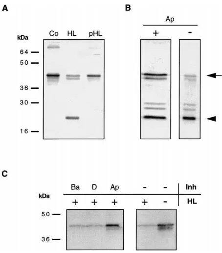
et al., 1999). Individual females of this population are homozygous for either one of the corresponding allelic genes [see Fig. 1(A), OM, OP]. Consequently, by using the allelic variance of VLP1 as a diagnostic tool, two clonal strains, addressed as RP (Repeat Plus) and RM (Repeat minus), can be distinguished (Beck et al., 1999). Even though VLP1.2 and VLP1.0 are stable in ovary samples [Fig. 1(A), OM, OP], the presence of PEST sequences common to rapidly degraded proteins indi-cated already previously that these two allelic variants might be proteolytically processed (Hellers et al., 1996). Indeed, when parasitoid eggs recovered from parasitised
E. kuehniella larvae were analysed, VLP1.2 as well as
VLP1.0 were processed [Fig. 1(A), EP, EM]. VLP1.0
and VLP1.2 differed in their respective digestion pattern. However, the major breakdown product was the same for both allelic variants displaying a molecular weight of about 22 kDa [Fig. 1(A), arrowhead].
Since VLPs cover the egg surface (Rotheram 1967, 1973; Schmidt and Schuchmann-Feddersen, 1989), they are probably exposed to host haemolymph after egg deposition. Thus, the question was asked whether host haemolymph proteases are involved in the processing of the allelic VLP1 variants. To answer this question, in vitro experiments were performed by adding cell-free host haemolymph to eggs isolated from RP wasp ovi-ducts (Fig. 2). VLP1.2 processing was observed for eggs incubated in cell-free haemolymph obtained from unparasitised larvae [Fig. 2(A), HL], but not for control eggs incubated in PBS only [Fig. 2(A), Co]. Since para-sitoid eggs are deposited together with ovarian calyx fluid, processing might be influenced by these secretions. When cell-free haemolymph obtained from highly super-parasitised E. kuehniella larvae was used, processing of the VLP protein was inhibited [Fig. 2(A), pHL].
It thus appears that the VLP protein is cleaved by a protease derived from host haemolymph and that this activity might be regulated by wasp components injected into the host during oviposition. To investigate the nat-ure of the putative host protease, the amino acid sequence of VLP1.2 and VLP1.0 was analysed employing a computer program that predicts proteolytic fragments generated by different proteases. Among these proteases, only the serine protease trypsin used under conditions with specificity for arginine residues dis-played processing patterns for both VLP1 variants simi-lar to the ones observed in vivo [Fig. 1(B), arrowheads; Fig. 1(A), EP, EM]. Firstly, this particular protease would cut VLP1.2 twice within a tandem repeated region (TR) not present in VLP1.0. This could account for the two additional bands seen with VLP1.2 compared to
Fig. 2. Inhibition of VLP1 processing in vitro and in vivo. (A) Com-parable numbers of RP reservoir eggs were incubated overnight at room temperature in various solutions and then analysed on Western blots. Eggs incubated in PBS solution (Co), cell-free haemolymph from five unparasitised larvae (HL), and cell-free haemolymph from five highly super-parasitised larvae (pHL). (B) In vivo inhibition of the VLP1 processing by p-APMSF. Prior to the exposure to wasps for 20 h, 2 µl of a 40 mM p-APMSF solution (in PBS containing 10% dimethylformamide (DMF)), or as a control 2µl PBS containing 10% DMF, were injected with a drawn-out Pasteur pipette into host larvae. (Ap+), eggs retrieved from parasitised larvae containing p-APMSF. (Ap2) a comparable number of eggs dissected from control larvae
containing buffer only. (C) Effects of synthetic protease inhibitors on VLP1 degradation in vitro. Note that in this experiment the intensity of the 40 kDa precursor band is used as a readout for proteolytic activity. Incubation of a similar number of oviduct eggs in cell-free haemo-lymph (HL, equivalent to five caterpillars), in the presence of 50 mM benzamidine hydrochloride (Ba), PBS containing 10% DMF (D), 40 mM p-APMSF (in PBS containing 10% DMF) (Ap), and PBS (2/+).
(2/2), an equivalent number of reservoir eggs from RP-females,
which were neither exposed to haemolymph nor inhibitors. All blots were incubated in anti-VLP1 antiserum and stained with alkaline phos-phatase-conjugated secondary antiserum (see Materials and methods).
VLP1.0 [Fig. 1(A), EP, dots]. Secondly, this trypsin activity would produce an 18.5 kDa fragment for both allelic variants, which resembles in size the mutual major breakdown product depicted in Fig. 1(A) (arrowhead).
To test whether a trypsin-like host enzyme might be responsible for the proteolytic cleavage of VLP1 in vivo, buffer solution with and without the synthetic serine pro-tease inhibitor p-amidinophenylmethanesulfonyl fluoride (p-APMSF) was injected into final instar E. kuehniella larvae, which subsequently were parasitised by RP-wasps. Western blot analysis of eggs retrieved from treated larvae revealed a significantly higher amount of
unprocessed VLP1.2 in the presence of p-APMSF com-pared to the control [Fig. 2(B), Ap+, Ap2, arrow].
A similar result was obtained in vitro using reservoir eggs dissected from RP wasp oviducts [Fig. 2(C)]. Since the proteolytic fragments were released from the egg sur-face under some in vitro conditions, the processing of VLP1.2 by the putative host haemolymph protease was in this case inferred from the reduction of the non-pro-cessed precursor signal compared to the control [Fig. 2(C), right blot]. When added cell-free haemolymph contained the inhibitor p-APMSF, the processing was inhibited and VLP1.2 completely retained on the egg surface [Fig. 2(C), Ap]. Interestingly, the synthetic ser-ine protease inhibitor benzamidser-ine, which is less specific than p-APMSF (Laura et al., 1980), did not prevent the degradation of VLP1 [Fig. 2(C), Ba].
3.2. Inhibition of host phenoloxidase by maternal wasps secretions
Frequently, melanisation was considerably delayed in haemolymph preparations from superparasitised E.
kuehniella larvae compared to unparasitised larvae. To
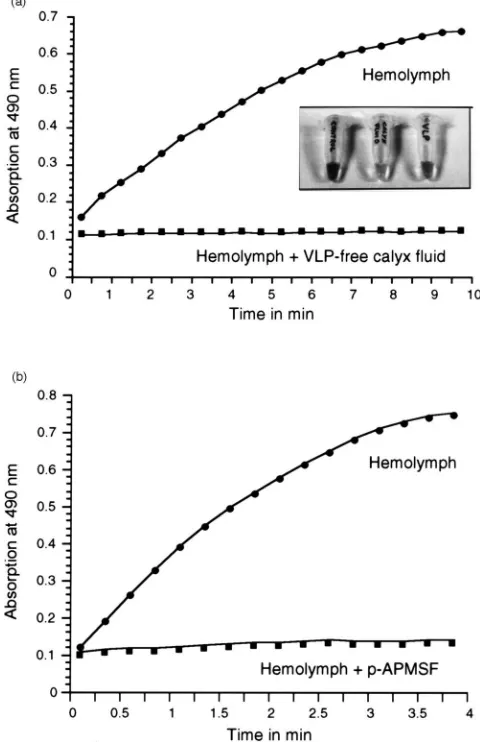
test whether ovarian wasp secretions might be respon-sible for the observed inhibition of melanogenesis, ovarian calyx fluid preparations were mixed with cell-free haemolymph [Fig. 3(A), insert]. Samples containing a high amount of VLP-free calyx fluid did not display any signs of melanogenesis [Fig. 3(A), insert, calyx fluid], in contrast to the corresponding control [Fig. 3(A), insert, control]. Purified VLPs prevented mel-anogenesis to a much lesser degree than VLP-free calyx fluid [Fig. 3(A), insert, VLP], suggesting that the observed inhibitory effect in the VLP sample might be due to the presence of contaminating soluble ovarian proteins.
Since PO is considered the key enzyme of the bio-chemical pathway leading to the production of melanin, VLP-free ovarian calyx fluid was examined regarding its ability to inhibit PO activity. When haemolymph samples with and without VLP-free calyx fluid were tested in a PO enzyme assay, samples containing VLP-free calyx fluid displayed no PO activity [Fig. 3(A), graph]. To test whether the synthetic protease inhibitor p-APMSF inhibits melanogenesis in E. kuehniella, PO enzyme activity of a haemolymph sample containing the inhibitor was measured. As seen in Fig. 3(B), the pres-ence of p-APMSF resulted in a complete inhibition of haemolymph PO activity.
3.3. Wasp induced alterations of host haemocyte behaviour
To explore whether protease inhibitors may affect cellular defence reactions, we examined the spreading of
Fig. 3. Inhibition of melanogenesis. (A) Inhibition of PO activity by VLP-free ovarian calyx fluid. Cell-free haemolymph from one E.
kuehniella larva (dots) and an equivalent sample containing VLP-free
calyx fluid from 10 wasps (squares) were assayed for PO activity using DL-DOPA as substrate. Insert: Cell-free haemolymph from four E.
kuehniella larvae (in 100µl PBS) was mixed with 50µl of PBS (left tube), VLP-free calyx fluid from 40 wasps (middle tube), and purified VLPs from 40 wasps (right tube), respectively, and inspected after overnight incubation at room temperature. (B) Inhibition of PO activity by p-APMSF. Cell-free haemolymph from two E. kuehniella larvae (dots) and an equivalent sample containing 50 mM p-APMSF (squares) were assayed for PO activity using DL-DOPA as substrate. Note that complete inhibition of melanisation is only observed in haemolymph samples from highly superparasitised host larvae, or in haemolymph samples from unparasitised hosts having a high calyx fluid content.
Haemocytes normally display attachment and spreading behaviour upon contact with a glass surface [Fig. 4(A) and (C)]. However, in the presence of p-APMSF haemo-cyte spreading, in particular of plasmatohaemo-cytes, was inhibited. Instead of spreading and expanding cellular extensions, p-APMSF treated haemocytes retained a rounded shape and small filopodia, which had already developed before the inhibitor was added [Fig. 4(A) and (B)]. Since p-APMSF and VLP-free calyx fluid had similar effects on melanization, the spreading behaviour
Fig. 4. Comparison of haemocyte behaviour in the presence of p-APMSF and VLP-free calyx fluid. E. kuehniella haemolymph was bled into a drop of PBS on a glass slide, allowing haemocytes to attach for 5 min. Then, the attached haemocytes were washed several times with PBS, and subsequently incubated for 1 h in different solutions: (A), PBS containing 10% DMF; (B) PBS containing 10 mM p-APMSF and 10% DMF; (C), PBS; (D) VLP-free calyx fluid from 25 wasps diluted in PBS (see Materials and methods). The bars represent 20µm.
of haemocytes was examined in the presence of ovarian calyx fluid. When VLP-free calyx fluid was added to haemocytes partially attached to a glass surface, spread-ing behaviour was indeed altered compared to the con-trol [Fig. 4(C) and (D)].
4. Discussion
Our data suggest an elaborate interaction between pro-teineous wasp secretions and the host defence system, involving a putative serpin activity in the calyx fluid and host protease activities associated with haemocytes and cell-free haemolymph.
proteo-lytically processed. In vitro experiments revealed that the proteolytic activity responsible for the VLP1 processing resides in the host haemolymph. A comparison of in vivo and computer-generated VLP1 processing patterns sug-gests that the host haemolymph protease is probably similar to the serine protease trypsin with specificity for arginine residues. The serine protease nature of the pro-posed enzyme was also confirmed by in vitro and in vivo experiments where VLP1 processing was inhibited by p-APMSF. This substance has been shown to cause an immediate and irreversible inhibition of plasma serine proteases such as bovine trypsin and human thrombin when applied in equimolar concentrations (Laura et al., 1980). p-APMSF was not able to completely inhibit the activity of the host haemolymph-derived protease in vivo, which could be explained in several ways: The inhibitor concentration in the host haemocoel may not have been sufficient to completely inhibit the protease activity. Alternatively, p-APMSF might have been meta-bolized or degraded over time, resulting in a processing of VLP1 derived from eggs deposited at later stages of the experiment.
Both allelic VLP1 variants were processed to an ident-ical breakdown product of around 22 kDa. The size of this fragment corresponds to the molecular weight reported for vertebrate glutathione peroxidases, includ-ing the enzyme phospholipid-hydroperoxide glutathione peroxidase from pig (Brigelius-Flohe et al., 1994), to which the N-terminal domain of VLP1 shows significant homology [see Fig. 1(B)]. Additional evidence that the 22 kDa breakdown product might indeed comprise the putative catalytic domain of VLP1 is provided by the computer simulation of the proteolytic processing gener-ating a N-terminal fragment of 18.5 kDa and the fact that this fragment was recognised by VLP1 anti-serum. The anti-VLP1 antiserum was raised against a recombinant fusion protein comprising only the putative PHGPX domain of VLP1 (Hellers et al., 1996). There-fore, every proteolytic fragment detected by the anti-serum must contain at least parts of this domain.
When VLP1 processing was examined employing haemolymph from highly superparasitised larvae, pro-teolytic cleavage of VLP1 was inhibited. This suggests that ovarian fluid injected during oviposition might con-tain protease inhibitor activity specific for trypsin-like enzymes. Further evidence for an active role of ovarian calyx fluid in host manipulation was obtained when mel-anisation in haemolymph samples from superparasitised hosts was delayed, and the delay was positively corre-lated to the degree of superparasitism. Inhibition of host phenoloxidase activity, as a consequence of parasitism, has been described previously (Sroka and Vinson, 1978; Stoltz and Cook, 1983). Yet, in most of these cases PO seems to be inhibited due to the expression of polydna-virus-encoded gene products in host tissues (Beckage et al., 1989; Lavine and Beckage, 1995; Strand and Pech,
1995). V. canescens VLPs were not able to abolish mel-anogenesis, whereas samples containing VLP-free ovarian calyx fluid prevented melanogenesis and PO activity. Since pro-phenoloxidase requires proteolytical activation (Sugumaran and Kanost, 1993), it seems con-ceivable that protease inhibitors in the ovarian calyx fluid might not only influence VLP1 processing, but also interfere with the initiation of melanogenesis.
The observation that haemocyte spreading, and there-fore possibly the host’s encapsulation response, was affected by VLP-free calyx fluid was unexpected since parasitism by V. canescens was thought to leave the cellular defence reactions of E. kuehniella intact (Salt, 1973; Schmidt and Schuchmann-Feddersen, 1989). One explanation for this apparent contradiction is that the cellular suppression is transient and locally restricted to the immediate surrounding of the egg. Since inhibition of host protease activities appears to be dependent on high ovarian calyx fluid concentrations, the necessary threshold for alterations of haemocyte properties may be reached only for a short period of time after oviposition, before the fluid around the egg becomes diluted in the surrounding haemolymph.
Effects of parasitism on host haemocyte behaviour and morphology have been documented for a number of other larval parasitoids (Stettler et al., 1998). However, little is known about the molecular mechanisms respon-sible for these effects. The fact that spreading of E.
kuehniella haemocytes was inhibited by the serine
pro-tease inhibitor p-APMSF indicates that cell adhesion is regulated by proteolytic events. During blood coagu-lation in vertebrates, the serine protease thrombin not only converts fibrinogen to fibrin, but also activates blood platelets. Upon activation, platelets change their shape, aggregate and discharge intracellular granules and lysosomes (Blockmans et al., 1995). In addition to thrombin, a variety of other physiological agents are able to activate platelets, amongst them van Willebrand factor (vWF; Blockmans et al., 1995). Recently, a lectin, desig-nated hemocytin, has been identified in the silk worm,
Bombyx mori, which shows homology to vWF and to
the coagulation factors V and VIII. Hemocytin is expressed in haemocytes and it has been speculated that this protein might be involved in encapsulation of foreign objects (Kotani et al., 1995). Such findings indi-cate that cell adhesion in insects and vertebrates share common features. Therefore, it seems conceivable that a serine protease inhibitor in the calyx fluid might influ-ence haemocyte spreading by interacting with a host enzyme that has a similar function as thrombin. Interest-ingly, Charalambidis et al. (1996) reported that serine proteases in insects are not only involved in regulatory cascades leading to the activation of humoral enzymes, but also in cellular events leading to phagocytosis.
proteases. In fact, the result that the synthetic inhibitor p-APMSF was able to mimick all effects induced by ovarian calyx fluid raises the possibility that only one serine protease inhibitor might be present. A superfamily of inhibitors referred to as serpins (serine proteinase
inhibitors) has been well described in vertebrates. The
primary function of its members seems to be the regu-lation of proteolytic events associated with a number of biochemical pathways, including blood coagulation, complement activation, and intracellular proteolysis (Potempa et al., 1994). Serine proteinase inhibitors belonging to the serpin superfamily have also been found in the haemolymph of invertebrates, among them three groups of arthropods (Jiang and Kanost, 1997). Even though their functional significance is largely unknown, it is assumed that they regulate proteases involved in immune reactions and proteolytic cascades that ulti-mately activate enzymes responsible for haemolymph clotting and melanisation (Jiang and Kanost, 1997). In the lepidopteran insect Manduca sexta, a number of ser-pin variants encoded by one gene have been character-ised (Jiang et al. 1995, 1996; Jiang and Kanost, 1997), which display highly specific inhibitory activities (Jiang and Kanost, 1997). Interestingly, one of these variants, serpin-1J, inhibited the activation of M. sexta haemo-lymph phenoloxidase (Jiang and Kanost, 1997).
In general, it appears that components derived from the egg surface, ovarian calyx fluid, and host haemo-lymph interact in a very complex manner. Only a few of these components have been identified so far, and more research is required to learn how they interact with each other in order to ensure the survival of V. canescens within permissive hosts.
Acknowledgements
We thank an anonymous referee for valuable com-ments on the manuscript. This work was supported by a grant from the Australian Research Council to OS, an ARC Research Fellowship to UT, and a Postgraduate Scholarship from The University of Adelaide to MB.
References
Asgari, S., Schmidt, O., Theopold, U., 1997. A polydnavirus-encoded protein of an endoparasitoid wasp is an immune suppressor. Journal of General Virology 78, 3061–3070.
Asgari, S., Theopold, U., Wellby, C., Schmidt, O., 1998. A protein with protective properties against the cellular defense reactions in insects. Proceedings of the National Academy of Sciences USA 95, 3690–3695.
Beck, M., Siekmann, G., Li, D., Theopold, U., Schmidt, O., 1999. A maternal gene mutation correlates with an ovary phenotype in a parthenogenetic wasp population. Insect Biochemistry and Molecu-lar Biology 29, 453–460.
Beckage, N.E., 1997. The parasitic wasp’s secret weapon. Scientific American 277, 50–55.
Beckage, N.E., 1998. Modulation of immune responses to parasitoids by polydnaviruses. Parasitology 116, S57–S64.
Beckage, N.E., Metcalf, J.S., Nesbit, D.J., Schlieffer, K.W., Zetlan, S.R., De Buron, I., 1989. Host hemolymph monophenoloxidase activity in parasitised Manduca sexta larvae and evidence of inhi-bition by wasp polydnavirus. Insect Biochemistry 20, 285–294. Bedwin, O., 1979. The particulate basis of the resistence of a parasitoid
to the defence reactions of its insect host. Proceedings of the Royal Society of London Series B 205, 267–270.
Blockmans, D., Deckmyn, H., Vermylen, J., 1995. Platelet activation. Blood Review 9, 143–156.
Brigelius-Flohe, R., Aumann, K.D., Blo¨cker, H., Gross, G., Kiess, M., Klo¨ppel, K.D., Maiorino, M., Roveri, A., Schuckelt, R., Ursini, F., Wingender, E., Flohe, L., 1994. Phospholipid-hydroperoxide glu-thatione peroxidase: genomic DNA, cDNA, and deduced amino acid sequence. The Journal of Biological Chemistry 269, 7342– 7348.
Charalambidis, N.D., Foukas, L.C., Zervas, C.G., Marmaras, V.J., 1996. Hemocyte surface phenoloxidase (PO) and immune response to lipopolysaccharide (LPS) in Ceratitis capitata. Insect Biochem-istry and Molecular Biology 26, 867–874.
Coodin, S., Caveney, S., 1992. Lipophorin inhibits the adhesion of cockroach (Periplaneta americana) haemocytes in vitro. Journal of Insect Physiology 38, 853–862.
Davies, D.H., Vinson, S.B., 1986. Passive evasion by eggs of braconid parasitoid Cardiochiles nigriceps of encapsulation in vitro by hae-mocytes of host Heliothis virescens. Possible role for fibrous layer in immunity. Journal of Insect Physiology 32, 1003–1010. Feddersen, I., Sander, K., Schmidt, O., 1986. Virus-like particles with
host protein-like antigenic determinants protect an insect parasitoid from encapsulation. Experientia 42, 1278–1281.
Hellers, M., Beck, M., Theopold, U., Kamei, M., Schmidt, O., 1996. Multiple alleles encoding a virus-like particle protein in the ichneu-monid endoparasitoid Venturia canescens. Insect Molecular Biology 5, 239–249.
Hoffmann, J.A., Kafatos, F.C., Janeway, C.A. Jr., Ezekowitz, R.A.B., 1999. Phylogenetic perspectives in innate immunity. Science 284, 1313–1318.
Hultmark, D., 1993. Immune reactions in Drosophila and other insects: a model for innate immunity. Trends in Genetics 9, 178–183. Jiang, H.B., Kanost, M.R., 1997. Characterization and functional
analysis of 12 naturally occurring reactive site variants of serpin-1 from Manduca sexta. Journal of Biological Chemistry 272, 1082–1087.
Jiang, H., Mulnix, A.B., Kanost, M.R., 1995. Expression and charac-terization of recombinant Manduca sexta serpin-1B and site-directed mutants that change its inhibitory selectivity. Insect Bio-chemistry and Molecular Biology 25, 1093–1100.
Jiang, H.B., Wang, Y., Huang, Y.L., Mulnix, A.B., Kadel, J., Cole, K., Kanost, M.R., 1996. Organization of serpin gene-1 from Manduca
sexta—evolution of a family of alternate exons encoding the
reactive site loop. Journal of Biological Chemistry 271, 28017– 28023.
Kinuthia, W., Li, D., Schmidt, O., Theopold, U., 1999. Is the surface of endoparasitic wasp eggs and larvae covered by a limited coagu-lation reaction? Journal of Insect Physiology 45, 501–506. Kotani, E., Yamakawa, M., Iwamoto, S., Tashiro, M., Mori, H.,
Sum-ida, M., Matsubara, F., Taniai, K., Kadonookuda, K., Kato, Y., Mori, H., 1995. Cloning and expression of the gene of hemocytin, an insect humoral lectin which is homologous with the mammalian von Willebrand factor. Biochimica et Biophysica Acta 1260, 245–258.
Laemmli, U.K., 1970. Cleavage of structural proteins during assembly of the head of bacteriophage T4. Nature 227, 680–685.
meth-anesulfonyl fluoride, an irreversible inhibitor of serine proteases. Biochemistry 19, 4859–4864.
Lavine, M.D., Beckage, N.E., 1995. Polydnaviruses: potent mediators of host immune dysfunction. Parasitology Today 11, 368–378. Mandato, C.A., Diehl-Jones, W.L., Downer, R.G.H., 1996. Insect
hem-ocyte adhesion in vitro: inhibition by apolipophorin I and an arti-ficial substrate. Journal of Insect Physiology 42, 143–148. Nappi, A.J., Vass, E., 1993. Melanogenesis and the generation of
cyto-toxic molecules during insect cellular immune reactions. Pigment Cell Research 6, 117–126.
Potempa, J., Korzus, E., Travis, J., 1994. The serpin superfamily of proteinase inhibitors: structure, function, and regulation. The Jour-nal of Biological Chemistry 269, 15957–15960.
Ratcliffe, N.A., 1993. Cellular defense responses of insects: unresolved problems. In: Beckage, N.E., Thompson, S.N., Federici, B.A. (Eds.), Parasites and Pathogens of Insects. Academic Press, San Diego, pp. 267–304.
Rotheram, S., 1967. Immune surface of eggs of a parasitic insect. Nat-ure 214, 700.
Rotheram, S., 1973. The surface of the egg of a parasitic insect II. The ultrastructure of the particulate coat on the egg of Nemeritis. Proceedings of the Royal Society of London Series B 183, 195– 204.
Salt, G., 1965. Experimental studies in insect parasitism XIII. The haemocytic reaction of a caterpillar to eggs of its habitual parasite. Proceedings of the Royal Society of London Series B 162, 303– 318.
Salt, G., 1970. Experimental studies in insect parasitism XV. The means of resistance of a parasitoid larva. Proceedings of the Royal Society of London Series B 176, 105–114.
Salt, G., 1973. Experimental studies in insect parasitism XVI. The mechanism of the resistance of Nemeritis to defence reactions. Pro-ceedings of the Roual Society London Series B 183, 337–350. Schmidt, O., Schuchmann-Feddersen, I., 1989. The role of virus-like
particles in parasitoid–host interaction of insects. In: Harris, J.R. (Ed.), Subcellular Biochemistry. Plenum Press, New York, pp. 91–119.
Sroka, P., Vinson, S.B., 1978. Phenoloxidase activity in the haemo-lymph of parasitised and unparasitised Heliothis virescens. Insect Biochemistry 8, 399–402.
Stettler, P., Trenczek, T., Wyler, T., Pfister-Wilhelm, R., Lanzrein, B., 1998. Overview of parasitism associated effects on host haemo-cytes in larval parasitoids and comparison with effects of the egg– larval parasitoid Chelonus inanitus on its host Spodoptera littoralis. Journal of Insect Physiology 44, 817–831.
Stoltz, D.B., 1993. The polydnavirus life cycle. In: Beckage, N.E., Thompson, S.N., Federici, B.A. (Eds.), Parasites and Pathogens of Insects. Academic Press, San Diego, pp. 167–187.
Stoltz, D.B., Cook, D.J., 1983. Inhibition of host phenoloxidase activity by parasitoid hymenoptera. Experientia 39, 1022–1024. Strand, M.R., Pech, L.L., 1995. Immunological basis for compatibility
in parasitoid–host relationships. Annual Review of Entomology 40, 31–56.
Sugumaran, M., Kanost, M.R., 1993. Regulation of insect hemolymph phenoloxidase. In: Beckage, N.E., Thompson, S.N., Federici, B.A. (Eds.), Parasites and Pathogens of Insects. Academic Press, San Diego, pp. 317–342.
Theopold, U., Schmidt, O., 1997. Helix pomatia lectin and annexin V, two molecular probes for insect microparticles: possible involve-ment in hemolymph coagulation. Journal of Insect Physiology 43, 667–674.
Theopold, U., Krause, E., Schmidt, O., 1994. Cloning of a VLP-protein coding gene from a parasitoid wasp Venturia canescens. Archives of Insect Biochemistry and Physiology 26, 137–145.