Expression of genes encoding PR10 class pathogenesis-related
proteins is inhibited in yellow lupine root nodules
Michał M. Sikorski
a,*, Jacek Biesiadka
a, Alina E. Kasperska
a, Joanna Kopcin´ska
b,
Barbara Łotocka
b, Władysław Golinowski
b, Andrzej B. Legocki
aaInstitute of Bioorganic Chemistry,Polish Academy of Sciences,Noskowskiego12/14,61-704Poznan´,Poland bDepartment of Botany,Agriculture Uni6ersity,Rakowiecka26/30,02-528Warsaw,Poland
Received 17 May 1999; received in revised form 1 July 1999; accepted 21 July 1999
Abstract
Pathogenesis-related proteins of the PR10 class have been found in many plant species, are induced under various stress conditions and act as common allergens. Here we demonstrate the presence of two PR10 proteins in yellow lupine (Lupinus luteus L. cv. Ventus). Both 17 kDa proteins, referred to as LlPR10.1A and LlPR10.1B, are composed of 156 amino acids, and have 76% parities identity (91% similarity). Identity to homologues from other plants ranges from 25 to 67% (46 – 82% similarity). Patterns of their expression in lupine organs and tissues were investigated using Western blotting and immunocytochemistry. Both proteins are constitutively expressed in roots, but expression is significantly decreased in young and mature root nodules (9 – 26 days post infection (dpi)), but not in senescent nodules (36 dpi). Immunocytochemical staining localised the proteins in the parenchymatous tissues of the root and senescent nodule, primarily in the cortex. The PR10 proteins were not detected in nodule bacteroid tissue. Expression in aerial parts of the plant is generally lower and only one of the proteins, LlPR10.1B, is expressed constitutively in the stem, leaf and petiole, while the other, LlPR10.1A, is only present in the stem and is induced in senescent leaves. © 1999 Elsevier Science Ireland Ltd. All rights reserved.
Keywords:Differential expression; Immunocytochemistry;Lupinus luteus; PR10 proteins; Protein purification; Symbiosis
www.elsevier.com/locate/plantsci
1. Introduction
One of the important features of plant response to stress is the increased expression of distinct proteins, including chitinases, glucanases, enzymes of phenylpropanoid pathway, thionins, osmotines, peroxidases, protease inhibitors, proteases, pro-line-rich glycoproteins and proteins of unknown function. The stress-induced proteins, described as ‘pathogenesis-related’ (PR) have been classified into 12 groups according to their properties or sequence homology [1]. Proteins of the PR10 class
seem to be ubiquitous in the plant kingdom, their homologues found in various species belonging to
both dicotyledonous and monocotyledonous
plants [2]. Also, based on sequence homology, common allergens present in birch pollen grains [3], celery [4] and apple [5] are included in this group. The PR10 proteins are small (around 17 kDa), slightly acidic and resistant to proteases. Although a precise cellular localisation of PR10 proteins has not been determined, the absence of an apparent signal peptide and their structural properties indicate that they are cytosolic [6,7]. Tertiary structure of the birch pollen allergen Betv1 was recently determined by X-ray crystal-lography [8]. The protein contains a long C-termi-nal a-helix, surrounded by a seven-stranded
b-sheet. Additionally, two shorter a-helices are located near the N-terminus. The amino acid
The nucleotide sequences of the full-length cDNA clones Llpr10.1a and Llpr10.1b were registered in the EMBL GenBank under accession numbers X79974 and X79975.
* Corresponding author. Tel.: +48-61-8528503; fax: + 48-61-8520532.
E-mail address:[email protected] (M. Sikorski)
quence analysis of almost 90 related PR10 proteins revealed the presence of conserved glycine residues (at positions 46, 48, 49 and 51 in Betv1) that form a so-called glycine reach loop (P-loop). This motif has been found in many nucleotide binding proteins [9]. An additional lysine residue at posi-tion 54 of Betv1 may be responsible for binding of the phosphoryl group of the nucleotide. The posi-tion of the P-loop on the surface of the PR10 protein molecule suggests that it is important for its biological function.
Several laboratories have reported that PR10 proteins accumulate around sites of pathogen in-vasion [10 – 15], wounding [16,17] and are induced by other environmental stress [6,18], suggesting their involvement in a general defence mechanism. The physiological function and any contribution of PR10 proteins to a defence mechanism remain unknown. However, high amino acid sequence homology and the similarity of the expression pattern with that of ginseng ribonuclease suggest that an RNase activity associated with these proteins may be involved in the defence reaction [19,20]. It has recently been shown that birch pollen allergen Betv1 has RNase activity in vitro [21,22]. Because of amino acid sequence similarity, PR10 proteins have been classified as ribonucle-ase-like PR proteins [1]. There are also suggestions that PR10 proteins play an important role in plant development since they have been identified in dry seeds [7,23], constitutive expression was observed in roots [18,24 – 26], and many plants have the constitutive expression of PR10 proteins in their stems [7] or various parts of the flowers [3,7,17,27 – 29]. Senescent leaves also often have elevated levels of PR10 protein expression [18]. Some PR10 homologues appeared to be induced by phytohormones like cytokinin [30], abscisic acid [31] or ethephon, an ethylene releasing com-pound [32].
Major latex proteins (MLPs), induced by wounding in bell pepper [33], also present in opium poppy, melon, strawberry, Arabidopsis and tobacco, have low (much less than 25%) homology to PR10 proteins. However, secondary structure predictions for PR10 and MLP groups are similar, suggesting that weak homology between them may be significant [34]. Interestingly, these two groups of proteins have never been found to coexist in one plant species.
The interaction between legume plants and rhi-zobia results in the formation of a new plant organ, the nitrogen-fixing root nodule, where bac-teria are present intracellularly, in so-called in-fected cells. These cells are completely packed with bacteria and obvious host defence response does not occur. The surface determinants of rhizobia (lipopolysaccharides, LPS and exopolisaccharides, EPS) as well as the host membrane, that always separates the bacteria and host cytoplasm, most likely control the avoidance of the defence re-sponse in symbiotic interactions [35 – 37].
Although, rhizobia appear to be successful in avoiding defence reactions in nodules, it is striking that even in the wild type interaction defence responses are induced [38]. Elicitation of defence mechanisms in roots by symbiotic bacteria may be a part of the mechanism by which the plant con-trols infection and, therefore, regulates nodulation (feedback control of nodulation). It has been shown by immunolocalisation assay that PR proteins associated with the defence mechanism in plants accumulated in the necrotic cells induced by rhizobia. However, a number of defence genes, enzymes of phenylpropanoid pathway, peroxi-dases, chitinases and PR proteins, were constitu-tively expressed in uninfected roots of both non legume and legume plants. Therefore, it can be proposed that the constitutive defence mechanism is a part of the root developmental program. The expression of pea RH2 protein in root epidermal cells [26] represents a good example of the gene encoding pathogenesis-related protein of PR10 class which is developmentally regulated and shows organ-specific expression.
the plant defence mechanism is observed. The ability of plants to distinguish between beneficial and parasitic interactions is an example of their genome adaptation to diverse environmental con-ditions. Since there is only a single publication available on the expression of gene encoding PR10 protein MtN13 during symbiosis development in Medicago truncatularoot nodules [42], we decided to describe in this paper the characterisation of two yellow lupine proteins of class PR10 and their corresponding cDNA clones. We also present their expression pattern and localisation studies in roots and developing nodules after inoculation with the symbiotic bacteriumBradyrhizobiumsp. (Lupinus).
2. Materials and methods
2.1. Plant growth conditions
Lupinus luteus L. cv. Ventus plants were grown in sterile perlite at 23°C with a 16 h day and 8 h night photoperiod. One day old imbibed seeds were inoculated with Bradyrhizobium sp. (Lupi-nus), strain USDA 3045.
2.2. Protein extraction and purification
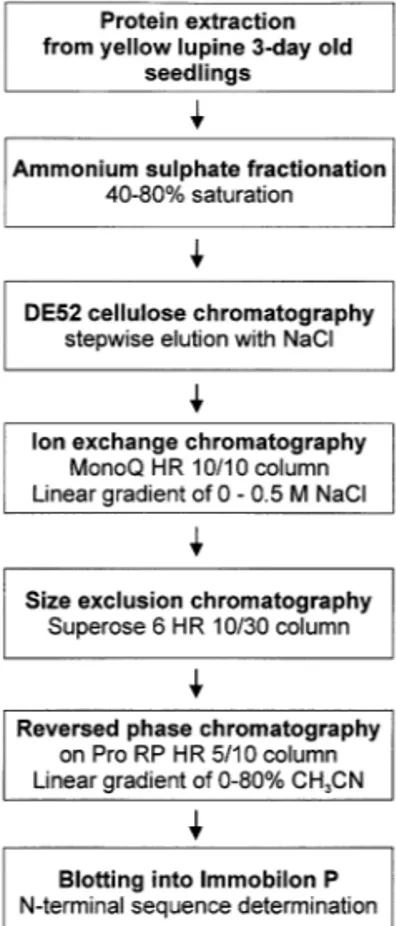
The extraction and purification of soluble proteins from plant tissues was carried out accord-ing to the procedure described by Sikorski et al. [43] with some modifications of chromatographic steps: 3-day-old lupine seedlings were frozen in liquid nitrogen and ground to a powder using a mortar and pestle and 1 g tissue was suspended in 3 ml buffer E (50 mM Tris – HCl (pH 8.0), 50 mM KCl, 5 mM MgCl2, 10 mM b-mercaptoethanol, 0.5 M sucrose). The homogenate was left on ice for 1 h and extraction was carried out by gentle stirring. The protein extract was then centrifuged twice at 15 000×g and 30 000×g for 15 min at 4°C. The supernatant was collected and fraction-ated by ammonium sulphate precipitation. The fraction of 40 – 80% saturation containing PR10 proteins was dialysed against buffer D (20 mM Tris – HCl (pH 8.0), 5% glycerol, 10 mM b -mer-captoethanol). For further purification of the PR10 proteins the 40 – 80% ammonium sulphate fraction was applied to a 30 ml DE52 cellulose column and the proteins were fractionated by stepwise elution with one column volume each of
buffer D containing 0.05, 0.1, 0.3 and 0.5 M NaCl. Fractions eluted with 0.3 M NaCl were pooled, dialysed against buffer D and submitted to a MonoQ HR 10/10 column in the linear gradient of 0 – 1 M NaCl in buffer D. Fractions eluted with 0.1 – 0.2 M NaCl, containing LlPR10.1A and LlPR10.1B proteins, were concentrated and 5.0 mg protein samples were separated by size exclu-sion chromatography on a Superose 6 HR 10/30 column (column buffer D containing 0.05 M NaCl). The PR10-containing fractions were purified to homogeneity by reversed phase chro-matography on a Pro RP HR 5/10 column in a linear gradient of 0 – 80% acetonitryle in the pres-ence of 0.1% trifluoroacetic acid (TFA) and 0.01%
b-mercaptoethanol (sample containing 1.0 mg of protein was treated with 10% HCOOH,
cen-trifuged and applied to the column). The
LlPR10.1A protein was eluted at 48.8% CH3CN and LlPR10.1B at 43.5% CH3CN.
2.3. Screening of yellow lupine cDNA library
The yellow lupine cDNA library was con-structed in UNI ZAP XR expression vector (Stratagene). The poly(A)+ RNA used for cDNA synthesis was isolated from lupine roots of non infected plants, harvested 8-, 15- and 21-days after germination. The amplified library contained 3.5×10¹⁰ pfu per 1 ml (represents 3.0×10⁶ re-combinants).Two synthetic deoxyoligonucleotide
probes: Oligo A
(TTYGCNTTYGARAAY-GARCA, 20-mer, 128× degenerated) and Oligo
B (TTYGCNTTYGARGAYGA, 17-mer, 64×
degenerated) were chemically synthesised based on the determined N-terminal amino acid sequences of LlPR10.1A and LlPR10.1B proteins. The probes were labelled with g-[32P]ATP (S.A., 5000 Ci/mmol) using T4 polynucleotide kinase as de-scribed by Sambrook et al. [44] and used for the screening of yellow lupine root cDNA.
2.4. Northern hybridisation
cDNA clones encoding either LlPR10.1A or LlPR10.1B (an internal BamHI cleavage site was used to split the cDNA to ensure the specificity of probes).
2.5. Western blot analysis of soluble protein extracts
The LlPR10.1A protein has been overexpressed in Escherichia coli expression system using vector pET-3a and purified to homogeneity [46]. The homogenous recombinant protein was used as an antigen to inject a female New Zealand rabbit. The injection was carried out with 100 mg of protein with Freund’s complete adjuvant (1:1, v/v) and repeated 2 and 4 weeks later with the same amount of protein. Antiserum was collected 2 weeks after the last boost.
Soluble proteins for Western blot were extracted from the plant tissue with buffer containing 0.1 M sodium phosphate (pH 7.5), 10% glycerol and 10 mM b-mercaptoethanol (3 ml per 1 g of tissue) using a mortar and pestle. The homogenate was centrifuged twice at 15 000×g for 15 min to remove cell debris. The protein content was mea-sured according to Bradford [47] and electrophore-sis was performed in a 15% polyacrylamide gel in the presence of sodium dodecylsulphate (SDS) ac-cording to Laemmli [48]. The samples containing 10 ng of recombinant protein or 10 mg of soluble plant protein extract were applied on the gel for Western blot analysis. The separated proteins were transferred onto an Immobilon P membrane, us-ing the MilliBlot-SDE transfer system accordus-ing to the Millipore protocol, and incubated for 2 h with anti-LlPR10.1A polyclonal antibodies. Im-munodetection was carried out with the biotin/
streptavidin-AP system of Amersham.
2.6. Immunocytochemical localisation of PR10 proteins
Plant material was sampled 9, 26 and 40 days after bacterial inoculation. Fragments of the roots
and roots with nodules were fixed in 4%
paraformaldehyde in phosphate-buffered saline (PBS). The 12 mm sections were cut from the paraffin embedded tissue. After deparaffinization and PBS soaking the sections were incubated in the solution containing rabbit anti-LlPR10A serum in PBS in the presence of 3% bovine serum
albumine for 1.5 h at 37°C. The primary antibody solution was washed out three times for 30 min at room temperature. Further steps of detection were performed using the biotin/streptavidin-AP system from Amersham, according to the standard proce-dure. Pre-immune serum or PBS was used as negative controls.
2.7. Sequence Analysis
All PR10 protein sequences retrieved from the
GeneBank database were aligned using the
CLUSTALW program [49]. Alignment of amino acid sequences has been used to construct a neigh-bour-joining tree, using programs from the PHYLIP package [50]. Percent support for each branch was estimated by analysing 1000 bootstrap
resamplings. The tree was displayed with
TREEVIEW [51]. Alignment of the amino acid sequences was displayed using GENEDOC [52].
3. Results
3.1. Identification and purification of yellow lupine PR10 proteins
Electrophoretic analysis of soluble protein ex-tracts from yellow lupine 3-day-old seedlings led to the identification of two abundant ca. 17 kDa
proteins, LlPR10.1A and LlPR10.1B. Both
proteins were purified from 3-day-old seedlings according to the flow diagram presented in Fig. 1. Sequence analysis of N-terminal 30 amino acid residues of LlPR10.1A and LlPR10.1B revealed a high level of similarity to intracellular pathogene-sis-related PR10 class proteins.
3.2. Screening of lupine cDNA library and identification of cDNA clones encoding PR10 proteins
corre-sponding sequences derived from protein sequenc-ing. The deduced amino acid sequences have sig-nificant homology to the PR10 class proteins from other plants and have the conserved amino acids characteristic for this group (Fig. 2A). The puta-tive glycosylation site NYS is located at positions 82 – 84 of each polypeptide. The conserved glycine-rich motif GXGGXGXXK between residues 46 and 54, described as the P-loop, is considered as a possible phosphate binding site [9]. The hydropa-thy profiles of the deduced polypeptides (data not shown) show no dominating hydrophobic or hy-drophilic regions, suggesting that the PR10 proteins do not contain a signal peptide. The putative polyadenylation signal AATAAA was found in the 3%untranslated region of both cDNA clones.
3.3. Northern analysis of PR10 transcripts
Total RNA was isolated from lupine root tissue at different stages of plant development (dry seeds, imbibed seeds and developing root, up to 30 days) and analysed by northern hybridisation with 3%
end fragments of cDNA clones Llpr10.1a and Llpr10.1b. The lack of cross-hybridisation of
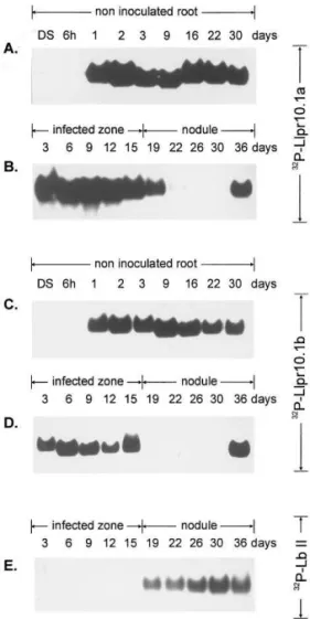
probes was confirmed by dot blot analysis (data not shown). As shown in Fig. 3A and C, transcrip-tion of both Llpr10.1a and Llpr10.1b starts be-tween 6 and 24 h after imbibition and a high level of both transcripts is detected during the root development of non inoculated plants. Fig. 3B and D present Northern analysis of the transcripts in the infected zone of the root and in the developing
nodule. Results for both LlPR10.1A and
LlPR10.1B proteins are similar: the bands corre-sponding to both the pr10.1a and pr10.1b tran-scripts appear in the lanes containing the preparations from 3- to 15-day-old infected roots and 36-day-old nodules. The pr10.1b transcript is present additionally in 19-day-old nodules. There were no detectable pr10.1aand pr10.1b transcripts in 22 – 30- or 19 – 30-day-old nodules.
3.4. Immunochemical analysis of PR10 proteins in yellow lupine: Western blotting and in situ
localisation
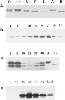
Organ specificity was determined by a series of immunoblotting of the protein extracts from the leaves, petiole, stem, root and root nodules. These analyses were performed using rabbit anti-LlPR10.1A antibody, which has approximately the same immunoreactivity with both proteins. Equal amounts of the purified LlPR10.1A and B proteins were placed in two lanes of each blot as a size reference. The general expression pattern is shown in Fig. 4A. LlPR10.1B protein is present in all the tissues tested with approximately equal intensity (slightly lower in the leaves). However the LlPR10.1A protein is present only in the roots and stems but not in the mature leaf or petiole. Details of PR10 protein expression in leaves taken from the upper, middle and lower part of the same plant, are presented in Fig. 4B. The LlPR10.1B intensity varies depending on the stage of leaf development; it is low in the preparations from young and mature leaves, but in senescent leaves it increases. Additionally, the LlPR10.1A protein is present in senescent leaves.
To detect the PR10 proteins at different stages of nodule development, equal amounts of nodule protein extracts (10 mg of total protein) were ap-plied to an electrophoresis gel and treated as above (Fig. 4C). Both LlPR10.1A and LlPR10.1B proteins were present in all stages of development, but PR10.1A is present in larger amounts in
young nodules (9 and 15 days post infection (dpi)). Both proteins were reduced to barely detectable levels in the mature nodule with PR10.1B present in slightly larger amounts, and both proteins present in approximately equal amounts in senes-cent nodule tissue (Fig. 4C). Fig. 4D shows the detection of leghaemoglobin level, a marker protein of effective symbiosis.
To determine the location of the PR10 proteins,
immunocytochemical staining of the PR10
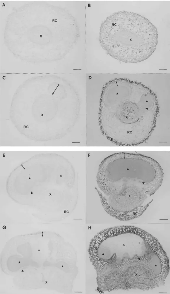
proteins was performed on root and nodule sec-tions that represent distinct developmental stages corresponding to changes in PR10 protein expres-sion during nodule development. Uninoculated, 9-day-old roots (Fig. 5B) exhibited uniform distri-bution within the primary root cortex and epiblem, but the PR10 proteins were not detected in root stele. In roots with 9-day-old nodules (Fig. 5D) the majority of the PR10 proteins was lo-calised in the root cortex, as in the uninoculated 9-day-old root. However, the root stele of the nodulated root contained PR10 proteins. No PR10 protein was observed in the young nodule cortex or bacteroid tissue. In mature 26-day-old nodules (Fig. 5F) low protein levels were de-tectable in the nodule protective tissue and inner cortex, excluding the layer of cortical cells immedi-ately adjoining the bacteroid tissue. The label was absent from the bacteroid tissue and nodule vascu-lar bundles. The PR10 proteins also accumulated in the root stele with the exception of cambium and within the remains of the root cortex. In 40-day-old nodules (Fig. 5H) the PR10 protein was observed within all the nodule cortical tissues with the exception of the innermost layer adjoin-ing the infected cells, resultadjoin-ing in an increase of the overall level of expression. The nodule vascular bundles and bacteroid tissue were free of label, except for single cells within the senescent zone. In the root stele an identical pattern of labelling was observed as in the 26-day-old material.
Fig. 3. Northern analysis ofL.luteustotal RNA of dry seeds (DS) and developing root (from 6 h after imbibition up to 30 days). The 32P-labeled BamHI/XhoI DNA fragments of
Llpr10.1a cDNA and Llpr10.1b cDNA were used as a probes. Total RNA (20 mg) was applied per lane. Total RNA was isolated from dry seeds, developing root, inoculated root and developing nodule. The RNA blots were hybridised to32
P-la-beled 3%-end fragments of cDNA encoding LlPR10.1A and LlPR10.1B proteins: (A, B) hybridisation to 32P-Llpr10.1a
cDNA; (C, D) hybridisation to 32P-Llpr10.1b cDNA; (E)
control hybridisation to 32P-LllbII; (A, C) non inoculated
root; (B, D)Bradyrhizobiumsp. (Lupinus) inoculated root and nodule. The plant material was harvested at different time points of plant development. DS-dry seeds; LllbII-L. luteus leghaemoglobin II cDNA.
Fig. 4. Western blotting analysis of PR10 proteins in different lupine organs (A – C, detection with anti-LlPR10.1A). (A) Soluble protein extracts from root (R), lateral root (Lr), stem (S), petiole (P) and leaf (L) of 4-week-old non inoculated plant; LlPR10.1A and LlPR10.1B, purified recombinant proteins. (B) Soluble protein extracts from leaves of 8-week-old lupine harvested from different parts of the plant: I, young leaf (upper); II, III, mature leaf (middle); IV, V, VI, senescent leaf (lower); L, leaf of 4-weeks old plant; LlPR10.1A and LlPR10.1B, purified proteins. (C) Soluble protein extracts from nodules. (D) Soluble protein extracts from nodules incubated with anti-LlLbI. LlPR10.1A, LlPR10.1B and LbI, purified recombinant proteins. Protein extract (10mg) from each tissue and 10 ng of purified LlPR10 and LbI was analysed by Western blotting and detected with anti-LlPR10.1A and anti-LbI antibodies (diluted 1:10 000). The biotin/streptavidin-AP system was used to develop the protein blot.
cortex. The changes in both protein levels appear to be similar, based on the results of the Northern and Western hybridisations. Since the proteins can not be distinguished in the immunocytochemical analy-sis due to their immuno-crossreactivity, it is not known whether the distribution of each homologue within the plant tissue is similar or different.
4. Discussion
The sequences of two yellow lupine PR10 proteins are 76% identical and reveal high homol-ogy with the other legume PR10 proteins (53 – 67% identity), and a 45% identity to birch pollen aller-gen BvBetv1. The most distantly related sequence, AoPR1 from a monocotyledonous plant, has only 25% identity to LlPR10. The identity values higher than 25% indicate not only a common origin of the PR10 class proteins, but also suggest a similar secondary structure for the proteins and the possi-bility that they have a common function. The blocks of highest homology correspond mainly to the regions of the well defined secondary structure (a-helices or b-strands), except for the glycine-rich loop between amino acid positions 46 and 54, which is one of the most conserved fragments and may be responsible for the protein activity (Fig. 2A).
Selected amino acid sequences were used to construct a phylogenetic tree representing genetic relations between the PR10 proteins of the subfam-ily Fabaceae (Fig. 2B). Topology of the tree is consistent with the taxonomy of plants at the level of subfamilies. The bootstrap values for the appro-priate branches are too low, indicating rather the lack of useful genetic information in the sequences. The presence of the homologous PR10 proteins within one plant species indicates the duplications of the PR10 genes. Thus, the PR10 proteins are generally encoded by gene families. The diversity of PR10 genes in legumes is more variable among the species, suggesting a lack of concerted evolution. The available data allow us to distinguish at least five paralogous groups of the PR10 genes in legumes. The most divergent group is represented by a single sequence, M. truncatula N13, the only gene expressed specifically in root nodules. Both lupine sequences fall into one group, but the pres-ence of other homologues in lupine is possible. In fact, two lupine sequences seem to be paralogous to each other due to their relatively high divergence. The results presented above support the
PR10 proteins were identified in plants in re-sponse to either biotic (pathogen infection) or abiotic stress (wounding or environmental stress like chemical pollutants, UV radiation). However, constitutive expression was also reported in differ-ent plant organs and tissues, primarily in roots, but in some cases in the leaves or various parts of the flowers. Large amounts of PR10 proteins present in fruits and vegetables or in the pollen grains of certain trees act as the common aller-gens. The biological function of PR10 proteins has not yet been determined, but their abundance, wide range of the plants, and different patterns of expression suggest that they play a significant and universal role in plant physiology, including de-fence mechanisms as well as development.
Two yellow lupine PR10 homologues are abun-dant in the roots, where their expression is consti-tutive. The level of PR10 proteins in the aerial parts of the plant is lower. Both proteins are present in the stem, but only the PR10.1B is constitutively expressed in the leaf and petiole. The increase of the PR10.1B protein, accompanied by the appearance of small amount of PR10.1A, was observed during the senescence of the leaves. The similar effect of the PR10 content increase in the lupine leaf was observed in response to infiltra-tion with the pathogenic bacteria Pseudomonas syringae (data not shown).
The major difference between the levels of PR10 proteins in the roots and aerial parts of the plant, and observation of the expression pattern in devel-oping and senescent lupine leaves suggests that the phytohormones may be involved directly or indi-rectly in the regulation of the PR10-encoding genes. This is supported by the recent findings describing the activation of PR10 gene expression by cytokinins in periwinkle callus [30], by abscisic acid in pea [31] and by ethylene in subterranean clover [32].
The main purpose of the presented work was to study the expression of pathogenesis-related proteins during the development of symbiosis with nitrogen fixing bacteria. A decrease of PR10 tran-scripts and protein accumulation in nodules be-tween 9 and 30 days after inoculation with Bradyrhizobium sp. (Lupinus) and the return to a high level in senescent nodules 36 days after infec-tion was observed. A decrease of PR10 protein content is correlated with the increase of the level of leghaemoglobin, a marker of effective
symbio-sis. The PR10 proteins are located outside of the bacteroid tissue, primarily in the parenchymatous tissue of the nodule cortex, suggesting that PR10 inhibition is not connected directly with the nitro-gen fixation process. Since it is known that PR10 proteins are induced in leaves in response to the invasion of pathogenic bacteria, it seems apparent that the suppression of a defence mechanism is part of the recognition of the bacteria as a mi-crosymbiont. However, the involvement of these proteins in some more general, developmental pro-cess, connected with the formation of nodules or other tissues might provide the most likely expla-nation, additionally supported by the reported presence of a nodule-specific PR proteins MtN1 and MtN13 in M. truncatula [42].
function of these defence proteins could be to prevent pathogenic infections of the root nodule, which is a potential carbon source for soil micro-organisms. A high homology of MtN13 to pea RH2 protein, specifically expressed in root epider-mal cells and proposed to contribute to a constitu-tive defence mechanism [26], supports the hypothesis of the protective role of MtN13 in nodules. Further similarity of MtN13 protein to Panax ginseng PR10 proteins exhibiting the ri-bonuclease activity [19,20] lead to the conclusion that MtN13 could posses such activity involved in mediating of cytotoxic effects.
The alternative hypothesis of a protective role against pathogens concerns the possible role of MtN1 as a defence protein expressed in response to inoculation of plant by Rhizobium. However, this speculation is contrary to the hypothesis that effective symbiosis development involves the sup-pression of plant defence mechanisms [40,53].
The other possibility is that MtN1 and MtN13 are involved in a developmental program not di-rectly related to the plant defence mechanism. It should be pointed out that the exact biological role of PR10 proteins during pathogenic invasion still remains unknown. The expression of some PR proteins was also observed in the absence of pathogens, e.g. during developmental processes and organogenesis [54 – 59]. Furthermore some de-fence proteins such as peroxidases and chitinases are considered to play different roles during nod-ule development and defence responces to patho-gens [60 – 62]. The PR10 proteins belong to the wide group of proteins described as ‘pathogenesis-related’. It is important to consider the relation-ship of these proteins with the plant response to pathogens. It is possible that the protein is pro-duced to act directly against the pathogen, being either cytotoxic to the infected cells or simply harmful to the pathogen, but it can be involved in a physiological mechanism that is enhanced during pathogenesis but that also occurs under other conditions. If the first possibility were true, it would allow us to describe these proteins as defen-sive; otherwise, they are only pathogenesis-in-duced. Although other authors have discussed both of these possible functions, the data available in the literature as well as the results of our own work suggest that the PR10 proteins play a more general role in plant physiology rather than that of purely defensive proteins.
Acknowledgements
This work was supported by Grant No. 6 P04B 011 10 from the Polish State Committee for Scien-tific Research. The computer work has been done in co-operation with the Poznan´ Supercomputing and Networking Centre. We thank Dr Zbyszek Michalski for his excellent editorial assistance.
References
[1] L.C. van Loon, W.S. Pierpoint, T. Boller, V. Conejero, Recommendations for naming plant pathogenesis-related proteins, Plant Mol. Biol. Rep. 12 (1994) 245 – 264. [2] M.H. Walter, J.-W. Liu, C. Grand, C.J. Lamb, D. Hess,
Bean pathogenesis-related (PR) proteins deduced from elicitor-induced transcripts are members of a ubiquitous new class of conserved PR proteins including pollen allergens, Mol. Gen. Genet. 222 (1990) 353 – 360. [3] H. Breiteneder, K. Pettenburger, et al., The gene coding
for the major birch pollen allergen Bet6 1 is highly homologous to a pea disease resistance response gene, EMBO J. 8 (1989) 1935 – 1938.
[4] H. Breiteneder, K. Hoffman-Sommergruber, G. Orior-dain, et al., Molecular characterisation of Api g 1, the major allergen of celery (Apium gra6eolens), and its immunological and structural relationships to a group of 17-kDa tree pollen allergens, Eur. J. Biochem. 223 (1995) 484 – 489.
[5] M. Vanek-Krebitz, K. Hoffman-Sommergruber, M.L.D. Machado, et al., Cloning and sequencing ofMal d1, the major allergen from apple (Malus domestica), and its immunological relationship to Betv 1, the major birch pollen allergen, Biochem. Biophys. Res. Commun. 214 (1995) 538 – 551.
[6] A. Awade, M.H. Metz-Boutique, M. Le Ret, G. Genot, I. Amiri, G. Burkard, The complete amino acid sequence of pathogenesis-related (PR2) protein induced in chemi-cally stressed bean leaves, Biochim. Biophys. Acta 1077 (1991) 241 – 244.
[7] S.A.J. Warner, A. Gill, J. Draper, The developmental expression of the asparagus intracellular PR protein (AoPR1) gene correlates with sites of phenylpropanoid biosynthesis, Plant J. 6 (1994) 31 – 43.
[8] M. Gajhede, P. Osmark, F.M. Poulsen, et al., X-ray and NMR structure of Betv 1, the origin of birch pollen allergy, Nature Str. Biol. 3 (1996) 1040 – 1045.
[9] M. Saraste, P.R. Sibbald, A. Wittinghofer, The P-loop-a common motif in ATP- and GTP-binding proteins, TIBS 15 (1990) 430 – 434.
[10] C. Breda, C. Sallaud, J. El-Turk, et al., Defence reaction in Medicago sati6a: a gene encoding a class 10 PR protein is expressed in vascular bundles, Mol. Plant-Mi-crobe Interact. 9 (1996) 713 – 719.
[12] D.P. Matton, N. Brisson, Cloning, expression, and se-quence conservation of pathogenesis-related gene tran-scripts of potato, Mol. Plant-Microbe Interact. 2 (1989) 325 – 331.
[13] P.M. Pinto, C.P.P. Ricardo,Lupinus albusL. pathogene-sis-related proteins that show similarity to PR10 proteins, Plant Physiol. 109 (1995) 1345 – 1351.
[14] E. Schmelzer, S. Kruger-Lebus, K. Hahlbrock, Temporal and spatial patterns of gene expression around sites of attempted fungal infection in parsley leaves, Plant Cell 1 (1989) 993 – 1001.
[15] I.E. Somssich, E. Schmelzer, P. Kawalleck, K. Hahlbrock, Gene structure and in situ transcript localiza-tion of pathogenesis-related protein 1 in parsley, Mol. Gen. Genet. 213 (1988) 93 – 98.
[16] S.A.J. Warner, R. Scott, J. Draper, Characterisation of a wound-induced transcript from the monocot asparagus that shares similarity with a class of intracellular patho-genesis-related (PR10) proteins, Plant Mol. Biol. 19 (1992) 555 – 561.
[17] S.A.J. Warner, R. Scott, J. Draper, Isolation of an asparagus intracellular PR gene (AoPR1) wound-respon-sive promoter by the inverse polymerase chain reaction and its characterization in transgenic tobacco, Plant J. 3 (1993) 191 – 201.
[18] D.N. Crowell, M.E. John, D. Russel, R.M. Amasino, Characterisation of stress-induced, developmentally regu-lated gene family from soybean, Plant Mol. Biol. 18 (1992) 459 – 466.
[19] G.P. Moiseyev, J.J. Beintema, L.I. Fedoreyeva, G.I. Yakovlev, High sequence similarity between a ribonucle-ase from ginseng calluses and fungus-elicited proteins from parsley indicates that intracellular pathogenesis-re-lated proteins are ribonucleases, Planta (Heidelb.) 193 (1994) 470 – 472.
[20] G.P. Moiseyev, I. Larisa, L.I. Fedoreyeva, et al., Primary structure of two ribonucleases from ginseng callus, FEBS Lett. 407 (1997) 207 – 210.
[21] A. Bufe, M.D. Spangfort, H. Kahlert, M. Schlaak, W.-M. Becker, The major birch pollen allergen, Betv 1, shows ribonuclease activity, Planta 199 (1996) 413 – 415. [22] I. Swoboda, K. Hoffman-Sommergruber, G. O’Riordain, O. Scheiner, E. Heberle-Bors, Vicente O Betv 1 proteins, the major birch pollen allergens and members of a family of conserved pathogenesis-related proteins, show ribonu-clease activity in vitro, Physiol. Plant. 96 (1996) 433 – 438. [23] D.H.P. Barratt, J.A. Clark, Proteins arising during the late stages of embryogenesis inPisum sati6umL., Planta (Heidelb.) 184 (1993) 14 – 23.
[24] A.B. Legocki, W.M. Karlowski, J. Podkowinski, M.M. Sikorski, T. Stepkowski, Advances in molecular charac-terization of the Yellow lupin-Bradyrhizibiumsp. (Lupi-nus) symbiotic model, NATO ASI Ser. 39 (1997) 263 – 266.
[25] M.M. Sikorski, A.E. Szlagowska, A.B. Legocki, cDNA sequences encoding for two homologues of Lupinus lu-teus (L.) IPR-like proteins (Accession Nos. X79974 and X79975 for LlR10A and LlR18B mRNA’s respectively) (PGR95-114), Plant Physiol. 110 (1996) 335.
[26] P. Mylona, M. Moerman, W.-C. Yang, et al., The root epidermis-specific pea gene RH2 is homologous to a
pathogenesis-related gene, Plant Mol. Biol. 26 (1994) 39 – 50.
[27] C.P. Constabel, N. Brisson, Stigma- and vascular-specific expression of the PR10a gene of potato: a novel expres-sion of a pathogenesis-related gene, Mol. Plant-Microbe Interact. 8 (1995) 104 – 113.
[28] J.-C. Huang, F.-C. Chang, C.-S. Wang, Characterization of a lily tapetal transcript that shares sequence similarity with a class of intracellular pathogenesis-related (IPR) proteins, Plant Mol. Biol. 34 (1997) 681 – 686.
[29] I. Swoboda, O. Scheiner, D. Kraft, M. Breitenbach, E. Heberle-Bors, O. Vicente, A birch gene family encoding pollen allergens and pathogenesis-related proteins, Biochim. Biophys. Acta 1219 (1994) 457 – 464.
[30] S. Carpin, S. Laffer, F. Schoentgen, R. Valenta, J.-C. Che´nieux, M. Rideau, S. Hamdi, Molecular characteriza-tion of a cytokinin-inducible periwinkle protein showing sequence homology with pathogenesis-related proteins and the Betv 1 allergen family, Plant Mol. Biol. 36 (1998) 791 – 798.
[31] E.A. Iturriaga, M.J. Leech, D.H.P. Barrat, T.L. Wang, Two ABA-responsive proteins from pea (Pisum sati6um L.) are closely related to intracellular pathogenesis-re-lated proteins, Plant Mol. Biol. 24 (1994) 235 – 240. [32] K. Broderick, C. Pittock, T. Arioli, E.H. Creaser, J.J.
Weinman, B.G. Rolfe, Pathogenesis-related proteins in Trifolium subterraneum: a general survey and subsequent characterisation of a protein inducible by ethephon and redlegged earth mite attack, Aust. J. Plant Physiol. 24 (1997) 819 – 829.
[33] P. Osmark, B. Boyle, N. Brisson, Sequential and struc-tural homology between intracellular pathogenesis-re-lated proteins and a group of latex proteins, Plant Mol. Biol. 38 (1998) 1243 – 1246.
[34] J. Pozueta-Romero, M. Klein, G. Houlne, M.-L. Schantz, B. Meyer, R. Schantz, Characterization of a family of genes encoding a fruit-specific wound-stimu-lated protein of bell pepper (Capsicum annuum): identifi-cation of a new family of transposable elements, Plant Mol. Biol. 28 (1995) 1011 – 1025.
[35] K. Niehaus, D. Kapp, A. Pu¨chler, Plant defence and delayed infection of alfalfa pseudonodules induced by an exopolysaccharide (EPS I)-deficient Rhizobium meliloti mutant, Planta 190 (1993) 415 – 425.
[36] M. Parniske, P.E. Schmidt, K. Kosch, P. Mu¨ller, Plant defense responses of host plants with determinate nod-ules induced by EPS-defective exoB mutants of Bradyrhizibium japonicum, Mol.Plant-Microbe Interact. 7 (1994) 631 – 638.
[37] S. Perotto, N.J. Brewin, E.L. Kanenberg, Cytological evidence for a host defense response that reduces cell and tissue invasion in pea nodules by lipopolysaccharide-de-fective mutants ofRhizobium leguminosarumstrain 3841, Mol Plant-Microbe Interact. 7 (1994) 99 – 112.
[38] J. Vasse, F. de Billy, G. Truchet, Abortion of infection during the Rhizobium meliloti-alfalfa symbiotic interac-tion is accompanied by a hypersensitive reacinterac-tion, Plant J. 4 (1993) 555 – 556.
[40] M.A. Djordjevic, D.W. Gabriel, B.G. Rolfe,Rhizobium — the refined parasite of legumes, Annu. Rev. Phyto-pathol. 25 (1987) 145 – 168.
[41] H.P. Spaink, The molecular basis of infection and nodu-lation by rhizobia: the ins and outs of symphatogenesis, Annu. Rev. Phytopathol. 33 (1995) 345 – 368.
[42] P. Gamas, F. de Billy, G. Truchet, Symbiosis-specific expression of two Medicago truncatula nodulin genes, MtN1 and MtN13, encoding products homologous to plant defense proteins, Mol. Plant-Microbe Interact. 11 (1998) 393 – 403.
[43] M.M. Sikorski, U. Szybiak-Strozycka, P. Strozycki, et al., Coordinated synthesis of leghemoglobin and root protein R18 in yellow lupin, Acta Biochim. Polon. 36 (1989) 63 – 72.
[44] J. Sambrook, E.F. Fritsh, T. Maniatis, Molecular Cloning: A Laboratory Manual, Cold Spring Harbor Laboratory, Cold Spring Harbor, NY, 1989.
[45] P. Chomczynski, N. Sacchi, Single-step method of RNA isolation by acid guanidine isothiocyanate – phenol – chlo-roform extraction, Anal. Biochem. 162 (1987) 156 – 159. [46] M.M. Sikorski, Expression ofLupinus luteuscDNA cod-ing for PR10 protein in Escherichia coli: purification of the recombinant protein for structural and functional studies, Acta Biochim. Pol. 44 (1997) 565 – 578.
[47] M.M. Bradford, A rapid and sensitive method for the quantitation of microgram quantities of protein utilising the principle of protein dye binding, Anal. Biochem. 72 (1976) 248 – 254.
[48] U.K. Laemmli, Cleavage of structural proteins during the assembly of the head of bacteriophage T4, Nature (Lond.) 227 (1970) 680 – 685.
[49] J.D. Thompson, D.G. Higgins, T.J. Gibson, CLUSTALW, improving the sensitivity of progressive multiple sequence alignment through sequence weighting, positions-specific gap penalties and weight matrix choice, Nucleic Acids Res. 22 (1994) 4673 – 4680.
[50] J. Felsenstein, PHYLIP (phylogeny inference package) version 3.2, Cladistics 5 (1989) 164 – 166.
[51] R.D.M. Page, TREEVIEW, An application to display phylogenetic trees on personal computers, Comput. Appl. Biosci. 12 (1996) 357 – 358.
[52] K.B. Nicholas, H.B. Nicholas Jr., GeneDoc: a tool for editing and annotating multiple sequence alignments, distributed by the author (1997).
[53] C. Baron, P. Zambryski, The plant response in patho-genesis, symbiosis, and wounding: variations on a com-mon theme?, Annu. Rev. Genet. 29 (1995) 107 – 129. [54] H.I. McKhann, A.M. Hirsch, DoesRhizobiumavoid the
host response?, in: J.A. Dangl (Ed.), Bacterial Pathogen-esis of Plant and Animals, Springer, Berlin, 1994, pp. 139 – 162.
[55] J.R. Cutt, D.F. Klessig, Genes involved in plant defense, in: T. Boller, F. Meins (Eds.), Plant Gene Research Series, Springer, Vienna, 1992, pp. 20 – 243.
[56] Q. Gu, E.E. Kawata, M.-J. Morse, H.-M. Wu, A. Che-ung, A flower-specific cDNA encoding a novel thionin in tobacco, Mol. Gen. Gen. 234 (1992) 89 – 96.
[57] T. Lotan, N. Ori, R. Fluhr, Pathogenesis-related proteins are developmentally regulated in tobacco flowers, Plant Cell 1 (1989) 881 – 887.
[58] S.B. Milligan, C.S. Gasser, Nature and regulation of pistil-expressed genes in tomato, Plant Mol. Biol. 28 (1995) 691 – 711.
[59] A.D. Neale, J.A. Wahleithner, M. Lund, et al., Chiti-nase, b-1,3-glucanase, osmotin and extensin are ex-pressed in tobacco explants during flower formation, Plant Cell 2 (1990) 673 – 684.
[60] D. Cook, D. Dreyer, D. Bonnet, M. Howell, E. Nony, K. VandenBosch, Transient induction of a peroxidase gene inMedicago truncatulaprecedes infection by Rhizo-bium meliloti, Plant Cell 7 (1995) 43 – 55.
[61] A.J. De Jong, J. Cordewener, F. Lo Schiavo, et al., A carrot somatic embryo mutant is rescued by chitinase, Plant Cell 4 (1992) 425 – 433.
[62] C. Staehelin, M. Schultze, E. Kondorosi, R.B. Mellor, T. Boller, A. Kondorosi, Structural modifications in Rhizo-bium melilotiNod factors influence their stability against hydrolysis by root chitinases, Plant J. 5 (1994) 319 – 330.