www.elsevier.com / locate / bres
Interactive report
1
The hippocampal lamella hypothesis revisited
*
Per Andersen , Anne F. Soleng, Morten Raastad
Department of Neurophysiology, Institute for Basic Medical Sciences, University of Oslo, Pb. 1104 Blindern, 0317 Oslo, Norway
Accepted 28 September 2000
Abstract
We have re-examined the hippocampal lamellar organization of the CA3-to-CA1 connection. Based on a new technique with electrophysiological quantification of Schaffer collateral density, and a review of recent literature, we conclude that the lamellar organization remains a useful concept for understanding hippocampal connectivity. Using a sheet-like hippocampal preparation, containing the whole CA1 region, we mapped the distribution of Schaffer collaterals by two procedures. First, we recorded the amplitude of the Schaffer compound action potential in various parts of CA1 after stimulation of a point in CA3. Second, we charted the CA1 positions from which we could antidromically excite individual CA3 neurones. Although the Schaffer collaterals radiated from their CA3 cells of origin within a wide, fan-shaped area, covering a large part of the septo-temporal extent of CA1, the amplitude of the compound action potential was largest in a slightly oblique, transverse band across the CA1 towards the subicular region. 2000 Elsevier Science B.V. All rights reserved.
Keywords: Hippocampus; Lamellar organization; CA3 axon; CA1; Schaffer collateral
1. A preparation for studying connectivity principles fibre tracings showed a wide, fan-shaped distribution of Schaffer collaterals. These studies were made with fibre The hippocampal formation is a useful preparation in staining of anterogradely and retrogradely transported which to study general neuroscience problems. One reason markers [1,7] or by tracing of axonal branches after is that the hippocampal formation demonstrates a number intracellular marking of individual CA3 cells [8]. These of organizational principles. First, between the narrow authors noted that the connection between CA3 neurones strip-formed cortical subdivision of the hippocampal for- and CA1 pyramidal cells, the Schaffer collaterals, were mation, there is a one-directional connectivity between the heavily branched in a markedly diverging pattern. Thus, a principal cells of each strip. Second, there is a remarkable small injection in the CA3 region led to transport of a stratification of afferent fibres such that their synapses marker substance in up to 2 / 3rds of the longitudinal extent often segregate to specific dendritic regions. of CA1. Further, retrogradely transported markers from a A third principle is the lamellar organization of intrahip- small spot in the subiculum-near part of CA1 were found pocampal fibre systems [2] to indicate that the major part in longitudinally dispersed CA3 neurones. Thus, Amaral of principal cell axons are oriented parallel to each other and Witter [1] concluded: ‘‘While the ‘lamellar hypothesis’ and course nearly transversally to the long axis of the was consistent with the known neuroanatomy, subsequent hippocampus. Andersen et al. [2] further proposed that the neuroanatomical investigations, using a variety of modern fibre orientation implied that the hippocampal cells were tracing techniques, have invariably demonstrated that all of activated in a strip-like fashion, and that the coactivation of the major hippocampal projections, except for those arising a number of cells in such a near-transverse band, called a from the granule cells of the dentate gyrus, are much more lamella, could represent a functional unit of the hippocam- divergent than would be consistent with a strict
interpreta-pus. tion of the lamellar hypothesis.’’
The lamella hypothesis was criticized, however, because A similar set of findings emerged from studies with intracellular filling of CA3 neurones with horseradish
1 peroxidase (HRP) and subsequent tracing of branches from
Published on the World Wide Web on 12 October 2000.
such single cells [8]. Axonal branches were found in *Corresponding author. Tel.:147-22-851-245; fax:147-22-851-249.
E-mail address: [email protected] (P. Andersen). numerous clusters, spread widely along the longitudinal
extension of CA1, covering up to two thirds of the entire There are some suggestions that the Schaffer axonal
length. distribution is not uniform within the longitudinal
dimen-The conclusion from such studies was that an individual sion of the axonal tree. For example, Sik et al. [13] stained CA3 neurone not only activates CA1 neurones along a a single CA3 cell which, within a set of transverse sections narrow transverse lamella, but should be able to influence a covering about 1.8 mm along the longitudinal axis, had broad sector of CA1 neurones comprising more than half more than 15 000 boutons. This high number suggest to us of the structure through the fan-like spread of its many that a major part of the total axonal tree is likely to be
Schaffer collaterals. found within this transverse band, possibly with lower
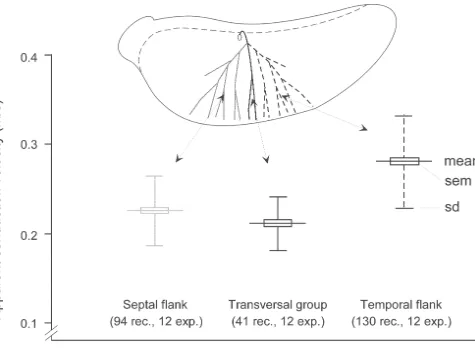
densities on either flank. A similar impression emerges from the plots of Schaffer collateral branches of Li et al. 2. Quantitative connectivity data are wanted [8]. The electron microscopical reconstruction technique gives relatively short lengths of the individual axonal Can the lamella idea survive such a set of well con- branches, so the longitudinal distribution was estimated by ducted studies? We feel that it can, focussing upon the the density of such twigs. In addition to a certain accumu-necessity to apply a numerical analysis of the relevant lation of axonal stubs along a transversally oriented region synaptic connectivity and thus offer a re-interpretation of from the stained cells, there were distinct peaks with the reported morphological results. Although these findings higher axonal branch density at various positions along the do indicate a wide distribution of the CA3-to-CA1 axonal longitudinal dimension. The number of twigs were, on the projection, they are not easily quantified. Specifically, this whole, less in the clusters found on either flank compared approach can not tell the exact proportion of axonal to those in the median region. All in all, the axonal pattern branches from a single CA3 cell which pass through the is not unlike the amplitude distribution of synaptic signals various positions of a plane oriented normal to the alvear in the original lamella paper (see Fig. 1D,C of ref. [2]). surface and along the longitudinal axis of the
hippocam-pus. This makes it difficult to estimate the density and
distribution of boutons along all axonal branches. It is this 3. A new approach synaptic density which constrains the functional efficiency
of the afferent system, and not the total width of the axonal Although we feel that the discrepancy between the
distribution. results from modern morphological studies and the lamella
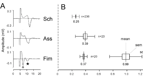
hypothesis is less than expressed by some authors, we 1A,B). Because we do not know the actual length of the appreciate that more precise data on Schaffer collateral various branches, we can only give apparent conduction fibres and their properties is desirable. Therefore, we now velocities as calculated from the surface measurements of present a set of new data with direct recording of the the recording points. Real conduction speeds must await density of Schaffer collateral branches and of the excitat- reconstruction of the fibres in question. Such studies are ory synaptic signals generated by these fibres. These feasible but time-consuming. There were two classes of observations are made in a set of horizontal slices in vitro, CA3 axon collaterals in the fimbria (Fim), having apparent covering the entire CA1 and with possibility for stimula- conduction velocities (at 238C) of 0.99 and 0.37 m / s, tion of CA3 cells giving rise to these signals. Our results respectively. Most likely, these represent fibres with and substantiate the findings of wide axonal branching of the without myelin. The mean apparent conduction velocity of Schaffer collaterals, but give quantitative data, showing longitudinal association fibres (Ass) were 0.39 m / s, that the heaviest distribution of such branches is distributed whether they were recorded in the septal or temporal along a vector running somewhat obliquely to the longi- direction or from the str. radiatum or str. oriens. The tudinal axis and with gradually falling branch densities on average conduction velocity of Schaffer collaterals was either side. Further, the Schaffer collaterals generate lowest, measuring 0.25 m / s, and with distinct variations synaptic potentials which have the same amplitude dis- between different parts of the axonal tree.
tribution as the axonal signals. In other words, both the The CA1 area from which we could record Schaffer Schaffer collaterals and the excitatory synaptic signals they collateral-associated signals had a form like a broad V with generate have the same distribution as in the original the sharp end at the point of stimulation (Fig. 2B). Within
lamella description. this sector, however, the signals differed greatly in
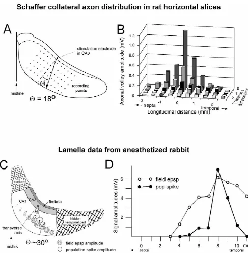
mag-In order to record the action potentials along the nitude. The largest amplitude were found in a line towards Schaffer collateral branches, we have made 500–900 mm the subiculum, deviating slightly towards the temporal thick horizontal tissue slices (parallel to the CA1 pyrami- direction from a plane transverse to the longitudinal axis dal layer) after the hippocampus was isolated and unbent (Fig. 2A,B). With increasing distance from the stimulated into a slightly curved, banana-shaped form. Such slices CA3 region, the amplitude diminished. The form of the contain the entire CA1 region from the septal nuclei till the amplitude versus position formed a ridge sloping towards temporal tip, and from the CA3 to the subicular border. A the subicular border and with a similar slope on the septal strip of CA3, containing from a third to one half of the and temporal flanks. When an average distribution was CA3 region, is attached to the CA1 part. The volume of made from a set of such experiments, the angle of this this CA3 strip varied in different preparations. ridge deviated 18 degrees from the transverse direction, These horizontal slices were mounted in a chamber in a tilted in the temporal direction (Fig. 2A). By recording at manner to secure good access of the oxygenated perfusion various distances from the fimbria, axonal action potentials fluid to all sides of the tissue and with the top close to the were recordable all the way to the subicular border. A fluid surface. The temperature was kept at 22–258C except comparison shows that the distribution of action potentials for short, 1–2 min periods at 35–378C in order to test the in the Schaffer collateral system is very close to that of signals at more physiological temperatures. After stimula- synaptic signals in the anesthetized rabbit (Fig. 2C,D). In tion of a small group of CA3 neurones, extracellular field the latter, however, the width of the band showing potentials were recorded from the CA1 region. In addition, population spikes were considerably narrower.
Fig. 2. Distribution of compound axon potentials along Schaffer collaterals. (A) Diagram of a horizontal slice with stimulated point in CA3 and recording points in CA1. The arrow indicates the direction of largest amplitudes. (B) Amplitude distribution of compound action potentials along Schaffer collaterals from a single stimulation point in CA3. (C) Diagrammatical dorsal view of a rabbit hippocampus with distribution of synaptic signals following stimulation of a CA3 point. Small dots indicate entry points of recording electrodes lowered to points of maximal responses. Triple circles give the amplitude of synaptic field potentials (epsps) while open circles give the population spike amplitude. (D) Graph of the amplitude of field epsps (open circles) and population spikes (filled circles) as a function of longitudinal recording position following stimulation of a single CA3 position. Redrawn from Fig. 6 of Andersen et al. [2].
Fig. 3. Different apparent conduction velocities within the Schaffer collaterals. The inset symbolizes three sections of the axonal tree of a single CA3 neurone. The graph shown the mean and variance of the apparent conduction velocity of the same three division s following stimulation of a small group of CA3 neurones.
m / s (Fig. 3, right side). The calculation of conduction summation of signals with the largest amplitudes along a velocities was hampered by the uncertainty about the exact line between the to ridges of maximal responses to each axonal lengths. Thus, whether these observations mean that stimulated site alone.
the temporally directed branches of the Schaffer collaterals are thicker, or less tortuous than the transversally or
oblique septally oriented subgroup is not known. No 5. Possible functional consequences of the lamellar available information allows an estimate of the possible organization
twists, bends and branching in the Schaffer axonal tree.
Whatever the explanation for the various apparent The original lamella concept was derived from studies conduction velocities, the net result is that a fibre volley of anaesthetized rabbits. The direction of the engaged emanating from a CA3 cell will not spread in a circular axons was estimated from antidromic and orthodromic fashion but with a somewhat flattened wave-front, deliver- field potentials. Characteristically, orthodromically acti-ing its simultaneous excitatory synaptic influence along a vated synaptic potentials were recorded in a nearly trans-longitudinal strip of CA1 cells. verse strip with a width of a few millimeters. Depending When synaptic field potentials were recorded, their upon the stimulus strength, the number of excited axons amplitude distribution formed a pattern similar to that of would vary and, consequently, the width of the excited the axonal signals (Fig. 2B), again repeating the slightly strip of tissue.
transversally oriented bands in both CA3 and CA1. to a band of CA1 neurones in a ridge-like fashion. The Between the two regions, CA1 cells with similar functional most effective excitation will take place along the lamellar characteristics were offset in the temporal direction by orientation with smaller effects to either side. However, 0.2–0.4 mm relative to their CA3 counterparts, roughly when two neurones, spaced some distance apart, discharge corresponding to the angled orientation of the lamellar simultaneously, the summation of the two synaptic in-main axis. This finding that cells oriented along a lamellar fluences will be largest at some intermediate position. With plane are coactivated in a physiological situation appears multiple CA3 neurones being synchronously active, sever-as a vindication of the lamellar idea sever-as having a functional al small areas of intense convergence may appear in the
role. CA1 matrix, determining the detailed pattern of discharges.
A comment may be directed at the ‘strict interpretation With asynchronous CA3 discharges, the situation becomes of the lamellar hypothesis’ used by Amaral and Witter [1]. even more complex. In such cases, the timing of the Neither the figures nor the text in [2] appear to be in various CA3 discharges will clearly determine the trans-disagreement with the results of the subsequent mor- verse location of the cells which will receive the most phological labelling experiments. Although there was a intense synaptic influence. A simple pictorial analogy definite direction along which the various afferent fibre would be to describe the spread of a single CA3 action systems showed maximal postsynaptic responses (the potential through the axonal tree as a wavefront into the lamella direction), there were also relatively broad shoul- CA1 area, somewhat similar to waves emanating from a ders on either side. This was more evident for the synaptic small opening in a stone pier. The effect of two simul-signals than for the population spike, signalling cell taneously active, but longitudinally separated CA3 neuro-discharges (open and closed circles, respectively, in Fig. nes would look like waves coming out through two
2D). neigbouring pier openings, forming an interference line.
The lamellar organization should be understood in a The direction of this interference line will be a function of population context. A strip of CA1 cells will be most the timing of various CA3 discharges. Only for syn-heavily engaged synaptically by an activated CA3 cells, chronously discharging CA3 cells would the converging but with a flank of more weakly excited cells on either excitation coincide with the lamellar direction. Remember-side. Given appropriate input to neurones at these flanks, ing the widely differing CA3 cell antidromic latencies, they could be brought to discharge. Thus, the lamella determination of the exact interference pattern requires hypothesis does not require that the excitation is confined detailed knowledge about the conduction along the axonal to a thin strip. Rather, when referring to Schaffer collater- tree of individual CA1 neurones.
als, it expresses the main excitatory synaptic influence area of a CA3 neurone.
Although there is much evidence for the proposal by References O’Keefe and Nadel [11] that the hippocampus provides the
animal with a cognitive map necessary for spatial naviga- [1] D.G. Amaral, M.P. Witter, The three-dimensional organization of the tion, we seem to be far from knowing the actual cellular hippocampal formation: a review of anatomical data, Neuroscience
31 (1989) 571–591. connectivity and discharge patterns at the basis for this
[2] P. Andersen, T.V.P. Bliss, K. Skrede, Lamellar organization of map [4,10,12]. An interesting principle emerged from
hippocampal excitatory pathways, Exp. Br. Res. 13 (1971) 222– work by Deadwyler et al. [3] that spatial retrieval seems to
238.
involve the activation of ensembles of CA1 neurones. [3] S.A. Deadwyler, T. Bunn, R.E. Hampson, Hippocampal ensemble Albeit sufficient for a simple recognition task, the activity activity during spatial delayed-nonmath-to-sample performance in
rats, J. Neurosci. 16 (1996) 354–372. of a cell ensemble in a small strip of hippocampal tissue
[4] H. Eichenbaum, Thinking about cell assemblies, Science 261 (1997) may not be sufficient for a complex task like learning a
993–994. new spatial constellation. By local inactivation of various
[5] K.M. Gothard, W.E. Skaggs, K.M. Moore, B.L. McNaughton, parts of the hippocampus, Moser and Moser [9] concluded Binding of hippocampal CA1 neural activity to multiple reference that spatial tasks probably requires neuronal ensembles frames in a landmark-based navigation task, J. Neurosci. 16 (1996)
823–835. distributed over more than half of the dorsal hippocampus.
[6] R.E. Hampson, J.D. Simeral, S.A. Deadwyler, Distribution of spatial In a landmark-based navigation task Gothard et al. [5]
and nonspatial information in dorsal hippocampus, Nature 402 found that CA1 units could be bound to multiple reference
(1999) 610–614.
frames, also suggesting the collaboration of widely spaced [7] N. Ishizuka, J. Weber, D.G. Amaral, Organization of
intrahippocam-neurones. pal projections originating from CA3 pyramidal cells in the rat, J.
The fan-like distribution of the CA3-to-CA1 axons Comp. Neurol. 295 (1990) 580–623.
[8] X.G. Li, P. Somogyi, A. Ylinen, G. Buzsaki, The hippocampal CA3 suggests that two or more CA3 neurones, separated by a
network: an in vivo intracellular labeling study, J. Comp. Neurol. certain longitudinal distance, may create a special
conver-339 (1994) 181–208.
[10] R. Muller, A quarter of a century of place cells, Neuron 17 (1996) [13] A. Sik, N. Tamamaki, T.F. Freund, Complete axon arborization of a 813–822. single CA3 pyramidal cell in the rat hippocampus, and its relation-[11] J.J.A. O’Keefe, L. Nadel, The Hippocampus as A Cognitive Map, ship with postsynaptic parvalbumin-containing interneurons, Eur. J.
Clarendon Press, Oxford, 1978. Neurosci. 5 (1993) 1719–1728. [12] M.L. Shapiro, H. Tanila, H. Eichenbaum, Cues that hippocampal