www.elsevier.com / locate / bres
Research report
Continuous intrathecal fluid infusions elevate nerve growth factor
levels and prevent functional deficits after spinal cord ischemia
a a,b ,
*
a a,bMark Bowes , Mark H. Tuszynski
, Jim Conner , Justin A. Zivin
a
Departments of Neurosciences, University of California, San Diego, La Jolla, CA 92093-0626, USA
b
Department of Neurology, Veteran’s Affairs Medical Center, San Diego, CA 92161, USA Accepted 2 August 2000
Abstract
Continuous intracerebroventricular or intrathecal infusions of neurotrophic factors have been reported to prevent neuronal degeneration, stimulate axonal sprouting and ameliorate behavioral deficits in various models of CNS injury and aging. In the present study, the ability of intrathecal infusions of recombinant human nerve growth factor (NGF) to reduce functional deficits following spinal cord ischemia was investigated. Adult rabbits underwent intrathecal cannulation and continuous infusions of either 300mg / ml recombinant human NGF or artificial CSF (vehicle) at a rate of 143ml / day for 7 days prior to induction of spinal cord ischemia. Continuous infusions were maintained after induction of ischemia. Four days later, both NGF-treated and vehicle-infused subjects showed a significant amelioration of functional motor deficits compared to lesioned, non-infused subjects (P,0.05). The average duration of tolerated ischemia increased from 23.461.8 min in lesioned, non-infused subjects to 35.563.1 min in lesioned, artificial CSF-infused subjects and 35.664.7 min in NGF-infused subjects (mean6S.E.M.). Significantly elevated NGF protein levels were attained within the spinal cords of both NGF-treated subjects and artificial CSF-infused subjects, although levels were substantially higher in NGF-treated subjects (9.863.8 ng / g in NGF-infused vs. 2.060.4 ng / g in vehicle-infused and only 0.460.2 ng / g in lesioned, non-infused animals). These findings indicate that the process of intrathecal cannulation and fluid infusion elicits alterations in the spinal cord environment that are neuroprotective, including spontaneous elevations in NGF levels. 2000 Elsevier Science B.V. All rights reserved.
Theme: Development and regeneration
Topic: Neurotropic factors: biological effects
Keywords: Nerve growth factor; Ischemia; Neurotrophin; Spinal cord; Functional recovery
1. Introduction upregulate expression of nerve growth factor (NGF),
brain-derived neurotrphic factor (BDNF) and ciliary
neuro-Neurotrophic factors prevent neuronal degeneration and trophic factor (CNTF) mRNA and increase NGF protein
promote axonal growth in responsive cell populations in production [18]. NGF levels rise in the hippocampus
various regions of the developing and adult mammalian following axotomy of cholinergic inputs, presumably as a
nervous system [7]. Neurotrophic factors such as nerve result of reduced uptake and retrograde transport by
growth factor (NGF) help to maintain normal neuronal cholinergic terminal [15]. NGF administration prevents
function and also sustain the phenotype of neurons follow- degeneration of septal cholinergic neurons after axotomy ing injury to the nervous system. Neurotrophin levels [13], and implantation into the brain of fibroblasts that are become elevated in some regions of the nervous system genetically modified to secrete NGF reduces histopatholo-after trauma, as a result of either increased production or gy following axonal transection [29] or excitotoxic injury
diminished utilization by responsive neurons [22]. For [8]. NGF infusion has also been shown to attenuate
example, Schwann cells in injured peripheral nerves cognitive impairments but not motor impairments in rats
after traumatic brain injury [26]. NGF levels are elevated following cerebral ischemia in the hippocampal CA1
*Corresponding author. Tel.: 11-858-534-8857; fax: 1
1-858-534-region and decreased in other brain 1-858-534-regions [12], and
5220.
E-mail address: [email protected] (M.H. Tuszynski). administration of exogenous NGF can reduce neuronal
necrosis following cerebral ischemia [25,27,34]. It there- days after reversible spinal ischemia in rabbits. The fore appears that the production of neurotrophic factors is a intrathecal space was cannulated and infused with NGF or
feature of several types of CNS injury, and that neuronal aCSF continuously for 3 days prior to induction of
injury may be attenuated by this response. ischemia, and continuously for 4 days thereafter.
Func-Neurotrophins have often been delivered to the CNS by tional outcomes and NGF levels in the spinal cords were
cannulating the ventricular system [13,16,25,27] or in- then assessed.
trathecal space [11,31], or by intraparenchymal infusion [26]. Although NGF appears to ameliorate the
conse-quences of neuronal injury, the infusion process itself may 2. Materials and methods
damage the CNS or elicit inflammatory processes in the
CNS, potentially resulting in upregulated production of 2.1. Experimental subjects substances such as neurotrophic factors [6,19,20]. Neuron
survival after injury has been improved by infusion of Male New Zealand White rabbits (2–3 kg) were
in-artificial cerebrospinal fluid alone [10,25,27], as well as by dividually housed and provided with food and water ad implantation of atelocollagen pellets directly into the libitum until the morning of surgery.
hippocampus [34]. This protective effect is analogous to
findings that the placement of sham grafts into the striatum 2.2. Infusion solutions can induce behavioral recovery after MPTP-induced
dopa-mine neuron lesions in primates, mimicking the beneficial Infusion vehicle consisted of a phosphate-buffered artifi-effects of fetal tissue grafts [6,20]. It has been speculated cial cerebrospinal fluid (aCSF) containing 150 mM NaCl, that a mechanism of recovery following tissue grafting to 1.8 mM CaCl , 1.2 mM MgSO , 2.0 mM K HPO , and2 4 2 4
the CNS may be elicitation of neurotrophin production in 10.0 mM glucose adjusted to pH 7.4. Recombinant NGF
the host brain resulting from limited damage of the (300 g / ml in aCSF; generously supplied by Genentech)
grafting procedure itself [6,20]. was administered to 21 animals, and 21 subjects received
In the present study, we sought to determine whether infusions of aCSF alone. Additional subjects [34] under-central infusions of nerve growth factor (NGF) or artificial went spinal ischemia / reperfusion without intrathecal
can-cerebrospinal fluid (aCSF) would ameliorate functional nulation or infusions.
deficits in a well-characterized model of adult rabbit spinal
cord ischemia [3,35]. Although NGF appears to limit 2.3. Placement of infusion tubing
hippocampal ischemic injury, the hippocampus is more
sensitive to ischemic damage than any other brain regions Rabbits were anesthetized with inhaled halothane (5%
and is therefore unrepresentative of broader neuronal induction, 2% maintenance by face mask). A [15 blade
populations. Thus, it is not clear whether NGF protection was used to incise the posterior pericervical skin layers,
also extends to other CNS cell groups. Various neuronal and muscle layers were bluntly dissected free from the
populations exhibit specificity for different neurotrophic region of the cervico-cranial junction to the level of the factors: for example, basal forebrain cholinergic neurons posterior spinal fascia. The fascia was then carefully
are primarily responsive to NGF, dopaminergic neurons to incised with a [11 blade, creating a window through
glial cell-line derived neurotrophic factor (GDNF), and which a flexible Tygon catheter (outer diameter 0.03 inch)
motor neurons to CNTF, BDNF or GDNF (see e.g. Refs. was inserted into the intrathecal space. A 15-cm length of
[10,17,23,24,30–33]). In the spinal cord, low-affinity tubing was gently advanced into the subarachnoid space,
neurotrophin receptors have been detected on injured but leaving the catheter to rest at the approximate T10 level of not intact motoneurons of the spinal cord [14], suggesting the spinal cord. Great care was taken to avoid direct
that motor neuronal responses to injury may be regulated damage to the spinal cord by halting insertion of the
by growth factors. Further, sensory projections to the catheter if any resistance was encountered during the
spinal cord express both low- and high-affinity neuro- placement procedure. If resistance was encountered, the
trophin receptors throughout life, and robustly extend new catheter was withdrawn for a distance of approximately 2 axons when provided with NGF after injury [28]. Finally, cm, then gently advanced again. Histological studies have
reactive astrocytes and microglia express neurotrophins shown that this procedure produces no tissue damage.
after injury in the CNS, suggesting that more diverse and Animals that sustained direct spinal cord damage from the widespread effects of neurotrophins may act to influence insertion procedure were detected by observing locomotor
spinal cord responses to injury [1,5,21]. function (ability to stand and walk normally) upon
re-To date, few studies that have examined the effects of covery from anesthesia; any animals with functional
NGF administration in models of CNS ischemia have also deficits were excluded from the study. Only subjects with evaluated functional outcomes, despite several reports of normal locomotor function were included in subsequent
NGF-induced neuroprotection. In the present experiment, studies of ischemia (n521 NGF-treated, n521
Upon completion of catheter insertion, the proximal end mals were classified as ‘paraplegic’ if they showed no
of the infusion tubing was connected to an Alzet Model motor response to noxious stimuli in the hindlimbs and
2ML2 mini-osmotic pump containing the experimental were completely incontinent. Rabbits were classified as
solution. The pump was placed subcutaneously in the ‘not paraplegic’ either if they were normal, or had any
infrascapular space of the animal and continuously de- motor function of the hindlimbs, including motor function livered the experimental substance of interest at a rate of that was only modest in extent. Immediate postoperative
71.4 ml / day. continence of bladder function was required for animals to
be classified in the ‘not-paraplegic’ group. In cases where
2.4. Placement of aortic ligature device the distinction between ‘paraplegic’ and ‘not paraplegic’
was uncertain, the animal was classified as paraplegic. Three days after placement of the intrathecal infusion Thus, the functional evaluation scale was conservatively
catheter, experimental animals underwent a second sur- biased.
gical procedure for placement of the descending aorta Prior to sacrifice, animals were anesthetized with
ligature device, as previously described [36]. Animals were halothane, and 3-cc CSF fluid samples were withdrawn
reanesthetized with halothane, and the abdominal aorta from the cisterna magna by transdermal C1–2 puncture.
was exposed at the level of the renal arteries using a Pumps were removed from the backs and residual fluid
paramedial incision. Small-diameter plastic tubing (outer volumes were measured to ensure adequate pump
empty-diameter 0.03 inch) was placed around the aorta just distal ing during the experimental period. Animals were then to the renal arteries. The ends of the tubing were threaded killed using Beuthansia-D (Schering-Plough, Kenilworth, through a small plastic button and then through a plastic NJ), and lower thoracic spinal cords were extruded onto
tube of larger diameter (outer diameter 0.125 inch), foil cooled on dry ice. The spinal cords were stored at
forming a snare ligature. The incision was closed around 2808C. NGF levels were assay by two-site ELISA in
the large-diameter tubing so that the free ends of the tubing residual pump fluid, CSF, and lower thoracic spinal cord were accessible externally. The animals were allowed to (T10 level), as previously described [33].
recover for at least 2 h, and all displayed normal behavior
prior to induction of spinal ischemia. 2.6. Data analysis
2.5. Induction of spinal cord ischemia Neurological damage as a function of ischemic insult
was analyzed using quantal dose–response analysis tech-The animals were restrained and aortic occlusion was niques described previously [32,37]. A computer was used performed by pulling and clamping the small tubing to fit logistic (s-shaped) curves to the fraction of abnormal around the aorta. Complete paraplegia was observed in all animals as a function of ischemic duration. The ischemic
animals within 3 min of occlusion. Occlusion durations duration necessary to produce permanent paraplegia in
encompassing all grades of neurological outcome, from 50% of a group of subjects was computed for each
complete recovery to permanent paraplegia (15–50 min of experimental group (the ET , for Effective Time). Phar-50
occlusion), were selected (see Fig. 1). At the end of the macological manipulations that improve neurological out-ischemic period, the tubing was released to restore blood come increase ET , generating a shift of the dose–re-50
flow through the aorta. The tubing was removed, the sponse curve to the right (see Fig. 1). The quantal bioassay abdominal wall closed with a suture that was placed during allows the evaluation of dose–response curves spanning a the surgery, and the skin closed with one or two wound wide degree of ischemic insult in an efficient manner and clips. Animals were returned to their home cages and using a limited number of subjects, as previously described maintained for 4 days. Rabbits that died within this period [37].
were excluded to ensure that no animals with aortic The ET s of the control, aCSF, and NGF groups were50
thrombosis were included in the data analysis. All enzyme- examined using one-way analysis of variance. A
signifi-linked immunosorbent assay (ELISA) samples were ana- cant analysis of variance was followed with Tukey’s test
lyzed simultaneously; thus, CSF and pump samples from for multiple comparisons, with P,0.05 considered signifi-NGF-treated subjects were diluted to fall within anticipated cant. NGF ELISA values amongst groups were compared NGF ranges of samples from vehicle-infused and intact using analysis of variance, and post-hoc comparisons were
subjects. made using Fisher’s least square difference.
Neurological function was evaluated at 18 h and 4 days after ischemia / reperfusion by an observer blinded to the
duration of ischemia and to the treatment group. Animals 3. Results
were classified by the presence or absence of paraplegia, as
previously described [36]. This functional outcome model 3.1. Functional studies has been validated in several previous studies as a reliable
Fig. 1. Probability of paraplegia increases with increasing duration of spinal occlusion. For each group, the ischemia duration associated with a 50% probability of paraplegia (the ET , for Effective Time), and the50
S.E.M. are calculated. Intrathecal infusion of artificial cerebrospinal fluid (aCSF) or nerve growth factor (NGF) for 3 days prior to and 4 days following ischemia / reperfusion increased the duration of ischemia
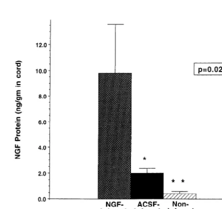
re-Fig. 2. NGF levels in spinal cord 4 days after induction of ischemia, as quired to produce permanent paraplegia. NGF administration was no
determined by ELISA. more effective than infusion of aCSF alone.
4. Discussion
infusion of aCSF or NGF improved neurological outcome
following reversible spinal ischemia and reperfusion (Fig. In the present experiment, cannulation of the intrathecal 1). At the 18-h evaluation, the ET506S.E.M. were as space and continuous infusion of either artificial CSF or follows: control non-infused, 25.761.9 min; control aCSF- human NGF resulted in significant neuroprotection after infused, 37.863.0 min; NGF-infused, 33.0564.9 min (P, transient spinal cord ischemia. Artificial CSF alone or 0.05). Post-hoc comparison using Tukey’s test at the 18-h NGF in artificial CSF were administered continuously,
evaluation indicated that the ET50 in the aCSF-infused beginning 3 days prior to induction of ischemia and
group increased significantly compared to the non-infused continuing until sacrifice 4 days later. Both groups
ex-lesion group; although the NGF group displayed a trend hibited significantly improved functional outcomes
com-toward improved outcome, it was not significant. Four pared to animals that were subjected to ischemia and did
days after ischemia / reperfusion, significant improvement not receive intrathecal instrumentation. Interestingly, levels
was observed in both the NGF-infused and aCSF-infused of NGF were elevated in the ischemic spinal cord of both
groups compared to the lesioned, non-infused group. The groups of cannulated animals, and were not elevated in the ET s were: control non-infused, 23.3750 61.8 min; control cords of animals that received lesions without instru-aCSF-infused, 35.563.1 min; NGF-infused, 35.664.7 min mentation. Thus, the process of intrathecal instrumentation
(P,0.05). itself appears to have elevated neurotrophin levels in the
ELISA measurement of NGF levels in freshly dissected ishemic spinal cord to levels sufficient to elicit neuro-spinal cords from the T10 region demonstrated signifi- trophin production, a finding that correlated with enhanced
cantly more NGF in infused subjects than in non-infused functional recovery. Although NGF levels were
approxi-controls: 9.863.8 ng NGF / g in NGF-infused subjects, mately 5-fold greater in the cords of NGF-infused subjects, 2.060.4 ng NGF / g in aCSF-infused subjects and 0.460.2 the extent of functional recovery in these animals was not
ng NGF / g in lesioned, non-infused animals (P50.02). greater than that observed in lesioned, aCSF-infused
Post hoc analysis indicated that all treatment groups subjects. These findings suggest that modest, physiological differed significantly from one another with respect to rises in neurotrophin levels may be sufficient to improve
NGF levels (Fig. 2). NGF ELISA measurements in the outcomes after spinal cord ischemia.
CSF of NGF-infused animals exceeded 600 ng /ml (upper These findings are consistent with previous studies
limits of assay condition), compared to undetectable levels demonstrating that infusion of artificial CSF improves
in aCSF-infused subjects and non-infused subjects. Simi- outcome following injury [25,27]. The mechanism by
larly, NGF levels exceeded 600 ng / ul in residual pump which neuroprotection occurs in these studies is not
fluid from NGF-infused subjects, and was undetectable in known. At least two possibilities must be considered: the
traumatize the spinal cord and the spinal nerve roots, general will be the search for substances that can stimulate potentially leading to secretion of inflammatory molecules, the activation of endogenous repair mechanisms, such as cytokines, or trophic factors that promote neuroprotection trophic factors, to mimic findings of the present study. from the subsequent ischemic deficit. Following CNS
trauma, molecules possessing neurotrophic activity are
detectable in the CNS [19]. Previous studies by Kordower Acknowledgements
[6] and Bankiewicz [20] have reported partial recovery of
motor function in hemiparkinsonian monkeys resulting This research was supported by grants from the NIH
simply from lesion placement in the brain. In these studies, (AGO0353A, NS37083, NS28121 and NS23814), the
it was hypothesized that minor trauma in the brain induced Veterans Administration, and the Hollfelder Foundation. production of neurotrophic factors and neuritic sprouting
that could have mediated partial functional recovery. The
present findings support natural growth factor elevation References
after trauma in the CNS as a potential mechanism of
generating functional recovery. Nervous system injury also [1] J.G. Assouline, P. Bosch, R. Lim, I.S. Kim, R. Jensen, N.J. Pantazis,
stimulates cytokine release that may be neuroprotective Rat astrocytes and Schwann cells in culture synthesize nerve growth factor-like neurite-promoting factors, Dev. Brain Res. 31 (1987)
[4]. Thus, the insertion of a catheter into the thecal space 3
103–118.
days before injury in the present experiment could have
[2] M.P. Bowes, E. Masliah, D.A.C. Otero, J.A. Zivin, T. Saitoh,
primed the system to express neuroprotective molecules Reduction of neurological damage by a peptide segment of the that subsequently reduced the extent of ischemic deficit. amyloid b/A4 protein precursor in a rabbit spinal cord ischemia
This finding was not observed, however, in a previous model, Exp. Neurol. 129 (1994) 112–119.
[3] U. DeGirolami, J.A. Zivin, Neuropathology of experimental spinal
study in which the intrathecal space was cannulated and
cord ischemia in the rabbit, J. Neuropathol. 41 (1982) 129–149.
drugs were administered as a single injection rather than as
[4] S.T. DeKosky, J.R. Goss, P.D. Miller, S.D. Styren, P.M. Kochanek,
a sustained infusion [2]. Thus, the presence of an ongoing D. Marion, Upregulation of nerve growth factor following cortical stimulus may be required to elicit the release of neuro- trauma, Exp. Neurol. 130 (1994) 173–177.
protective substances. The rate of fluid infusion in this [5] S. Elkabes, E.M. DiCicco-Bloom, I.B. Black, Brain microglia / macrophages express neurotrophins that selectively regulate
mi-model is sufficiently low (71.4 ml / day) compared to the
croglial proliferation and function, J. Neurosci. 16 (1996) 2508–
volume and rate of CSF formation in rabbits that fluid flow
2521.
from the catheter was unlikely in itself to have produced [6] M.S. Fiandaca, J.H. Kordower, J.T. Hansen, S.S. Jiao, D.M. Gash, damage; rather, an irritative effect of the tubing would Adrenal medullary autografts into the basal ganglia of Cebus
appear to be a more likely explanation for the observed monkeys: injury-induced regeneration, Exp. Neurol. 102 (1988) 76–91.
effect. Rabbit serum (0.1%) was added to the infusate
[7] W. Fischer, K. Wictorin, A. Bjorklund, L.R. Williams, S. Varon, F.H.
solution in both NGF-treated and control-infused animals,
Gage, Amelioration of cholinergic neuron atrophy and spatial
and it is possible that it contained a neuroprotective memory impairment in aged rats by nerve growth factor, Nature 329 substance. In vitro studies indicate improved cell viability (1987) 65–68.
in the presence of serum. However, a number of previous [8] D.M. Frim, T.A. Uhler, M.P. Short, Z.D. Ezzedine, M. Klagsbrun, X.O. Breakefield, O. Isacson, Effects of biologiclaly delivered NGF,
in vivo studies have failed to yield evidence of
mor-BDNF and bFGF on striatal excitotoxic lesions, NeuroReport 4
phological neuronal protection after infusions of artificial
(1993) 367–370.
CSF containing serum into the cerebral lateral ventricles, [9] F.H. Gage, D.M. Armstrong, L.R. Williams, S. Varon, Morphologic suggesting that serum does not mediate the protective response of axotomized septal neurons to nerve growth factor, J.
effect [9,13,16,30]. Comp. Neurol. 269 (1988) 147–155.
[10] K.M. Giehl, W. Tetzlaff, BDNF and NT-3, but niot NGF, prevent
An investigation of structural (morphological) changes
axotomy-induced death of rat corticospinal neurons in vivo, Eur. J.
underlying the observed functional effect is necessary to
Neurosci. 8 (1996) 1167–1175.
gain further insight into mechanisms underlying the pres- [11] B.G. Gold, T. Storm-Dickerson, D.R. Austin, Regulation of the ent findings. The increased functional recovery with eleva- transcription factor c-JUN by nerve growth factor in adult sensory
tion in neurotrophic factor levels suggests that examination neurons, Neurosci. Lett. 154 (1993) 129–133.
[12] Y. Hashimoto, H. Kawatsura, Y. Shiga, S. Furukawa, T. Shigeno,
of neuronal number and lesion size would be informative
Significance of nerve growth factor content levels after transient
and will be the subject of future studies.
forebrain ischemia in gerbils, Neurosci. Lett. 139 (1992) 45–46.
This study suggests that endogenous mechanisms may [13] F. Hefti, Nerve growth factor promotes survival of septal cholinergic be capable of providing significant neural protection after neurons after fimbrial transections, J. Neurosci. 6 (1986) 2155–
CNS ischemia or injury if an adequate stimulus is provided 2162.
[14] V.E. Koliatsos, D.L. Shelton, W.C. Mobley, D.L. Price, A novel
for the activation of these mechanisms. Whether such a
group of nerve growth factor receptor-immunoreactive neurons in
stimulus must be provided before the neural insult, as was
the ventral horn of the lumbar spinal cord, Brain Res. 541 (1991)
the case in the present study, or can be provided after the 121–128.
onset of injury, remains to be determined. A fruitful [15] S. Korsching, R. Heumann, H. Thoenen, F. Hefti, Cholinergic
transient accumulation of nerve growth factor (NGF) without tion or hippocampal cell loss following fluid-percussion brain injury
NGF
change in mRNA content, Neurosci. Lett. 66 (1986) 175–180. in rats, J. Neurochem. 65 (1995) 2209–2216.
[16] L.F. Kromer, Nerve growth factor treatment after brain injury [27] K. Tanaka, T. Tsukahara, N. Hashimoto, N. Ogata, Y. Yonekawa, T. prevents neuronal death, Science 235 (1987) 214–216. Kimura, T. Taniguchi, Effect of nerve growth factor on delayed [17] L.F. Lin, D.H. Doherty, J.D. Lile, S. Bektesh, F. Collins, A glial neuronal death after cerebral ischaemia, Acta Neurochir. 129 (1994)
cell-line derived neurotrophic factor for midbrain dopaminergic 64–71.
neurons, Science 260 (1993) 1130–1132. [28] M.H. Tuszynski, K. Gabriel, F.H. Gage, S. Suhr, S. Meyer, A. [18] M. Meyer, I. Matsuoka, C. Wetmore, L. Olson, H. Thoenen, Rosetti, Nerve growth factor delivery by gene transfer induces Enhanced synthesis of brain-derived neurotrophic factor in the differential outgrowth of sensory, motor and noradrenergic neurites lesioned peripheral nerve: different mechanisms are responsible for after adult spinal cord injury, Exp. Neurol. 137 (1996) 157–173. the regulation of BDNF and NGF mRNA, J. Cell Biol. 119 (1992) [29] M.H. Tuszynski, J. Roberts, M.-C. Senut, H.-S. U, F.H. Gage, Gene 45–54. thereapy in the adult primate brain: Intraparenchymal grafts of cells [19] M. Nieto-Sampedro, E.R. Lewis, C.W. Cotman et al., Brain injury genetically modified to produce nerve growth factor prevent causes a time-dependent increase in neuronotrophic activity at the cholinergic neuronal degeneration, Gene Ther. 3 (1996) 305–314. lesion site, Science 217 (1982) 860–861. [30] M.H. Tuszynski, H.S. U, D.G. Amaral, F.H. Gage, Nerve growth [20] R.J. Plunkett, K.S. Bankiewicz, A.C. Cummins, R.S. Miletich, J.P. factor infusion in the primate brain reduces lesion-induced
choliner-Schwartz, E.H. Oldfield, Long-term evaluation of hemiparkinsonian gic neuronal degeneration, J. Neurosci. 10 (1990) 3604–3614. ¨ monkeys after adrenal autografting or cavitation alone, J. Neurosurg. [31] V.M.K. Verge, P.M. Richardson, Z. Wiesenfeld-Hallin, T. Hokfelt, 73 (1990) 918–926. Differential influence of nerve growth factor on neuropeptide [21] J.S. Rudge, R.F. Aldewrson, E. Pasnikowski, J. McClain, N.Y. Ip, expression in vivo: A novel role in peptide suppression in adult
R.M. Lindsay, Expression of ciliary neurotrophic factor and the sensory neurons, J. Neurosci. 15 (1995) 2081–2096.
neurotrophins-nerve growth factor, brain-derived neurotrophic factor [32] D.R. Waud, On biological assays involving quantal responses, J. and neurotrophin 3 in cultured rat hippocampal astrocytes, Eur. J. Pharmacol. Exp. Ther. 183 (1972) 577–607.
Neurosci. 4 (1992) 459–471. [33] G. Weskamp, U. Otten, An enzyme-linked immunoassay for nerve [22] S.A. Scott, S. Liang, J.A. Weingartner, K.A. Crutcher, Increased growth factor (NGF): A tool for studying regulatory mechanisms NGF-like activity in young but not aged rat hippocampus after involved in NGF production in brain and in peripheral tissues, J. septal lesions, Neurobiol. Aging 15 (1994) 337–346. Neurochem. 48 (1987) 1779–1786.
[23] M. Sendtner, B. Holtmann, R. Kolbeck, H. Thoenen, Y.-A. Barde, [34] S. Yamamoto, T. Yoshimine, T. Fujita, R. Kuroda, T. Irie, K. Brain-derived neurotrophic factor prevents the death of motoneurons Fujioka, T. Hayakawa, Protective effect of NGF atelocollagen mini-in newborn rats after nerve section, Nature 360 (1992) 757–759. pellet on the hippocampal delayed neuronal death in gerbils, [24] M. Sendtner, G.W. Kreutzberg, H. Thoenen, Ciliary neurotrophic Neurosci. Lett. 141 (1992) 161–165.
factor prevents the degeneration of motor neurons after axotomy, [35] J.A. Zivin, J. DeGirolami, Spinal cord infarction: A highly reproduc-Nature 345 (1990) 440–440. ible stroke model, Stroke 11 (1980) 200–202.
[25] T. Shigeno, T. Mima, K. Takakura, D.I. Graham, G. Kato, Y. [36] J.A. Zivin, U. DeGirolami, E.L. Hurwitz, Spectrum of neurological Hashimoto, S. Furukawa, Amelioration of delayed neuronal death in deficits in experimental CNS ischemia, Arch. Neurol. 39 (1982) the hippocampus by nerve growth factor, J. Neurosci. 11 (1991) 408–412.
2914–2919. [37] J.A. Zivin, D.R. Waud, Quantal bioassay and stroke, Stroke 23 [26] G. Sinson, M. Voddi, T.K. McIntosh, Nerve growth factor adminis- (1992) 767–773.