SOME TEACHING TIPS ON THE MECHANISMS
OF URINARY CONCENTRATION AND DILUTION:
COUNTERCURRENT MULTIPLICATION BE DAMNED
Stephen A. Katz
Depa rtm ent of Physiology, University of Minnesota Medica l School, Minnea polis, Minnesota 55455
U
nless one is teaching the mechanisms of urinary concentration and dilution to medical students or graduate students, it is best to stay away from countercur-rent multiplication mechanisms and concentrate more on the physiological results. When one is teaching medical or graduate students, an overview of the basic countercurrent multiplication and exchange mechanisms is important, because it provides a conceptual foundation for an understanding of water balance. In order not to lose the forest for the trees, teaching aides including demonstrations, relevant clinical examples, contemporary cellular and molecular findings, and a little comparative physiology can be mixed in with traditional educational approaches. In this paper, the teaching of urinary concentration and dilution is first addressed by an educational philosophy synopsis, followed by an outline of the basic mechanisms of urinary concentration and dilution and a presentation of some useful teaching aides. Common student questions are also discussed. This material can be wonderfully fun to teach and is extremely important. The danger is in getting bogged down in explanations involving overly complex mechanisms.AM. J. PHYSIOL. 275 (ADV. PHYSIOL. EDUC. 20): S195–S205, 1998.
Key words:loop of Henle; urine; urea; water balance
I believe I am qualified to write a paper regarding the teaching of renal countercurrent mechanisms because I have never published a single paper on the subject. I suspect that most people who must teach this material do not actively engage in research involving mecha-nisms of urinary concentration and dilution, and therefore I am in the majority. In addition, I have had to teach this subject at three different levels (under-graduate, dental/pharmacy, and medical/graduate) for the last ten years. I may have questionable expertise, but I definitely have experience.
CLASSROOM IDEAS
A teaching idea that is certainlyin vogue is ‘‘active learning.’’ Break the class down into small groups and
have the students actively pursue the subjects at hand with their own resources, possibly with the aid of a group facilitator. For really difficult subjects, such as mechanisms of urinary concentration and dilution or countercurrent multiplication, I am not in favor of this approach. A few well-taught lectures can save every-one a lot of time and effort. It turns out that lectures can be fine examples of active learning. However, the lecture format is not without its downfalls. What follows are a few classroom lecture ideas for teaching difficult subjects with special regard to water balance.
In these days of co-op notes, a few poor lectures can instill a teaching philosophy in a student’s mind that is less than an educator’s ideal.
Most people tire of a lecture in ten m inutes, clever people ca n do it in five. Sensible people never go to lecture a t a ll.
—Stephen Leacock,My Discovery of Engla nd, 1922
Check out the classroom ahead of time. I wish I had a dollar for every teacher who wandered down to the front of the lecture hall in preparation for their lecture only to find it necessary to spend the first 10 minutes of class mastering the microphone controls, light switches, projection equipment, and accompanying tables and chairs.
Be organized. This is difficult because it involves effort. It is not a good idea to lecture on renal countercurrent multiplication on the basis of last year’s lecture notes that were hurriedly put together in a frantic effort to meet an unrelated grant deadline. Most students can sense instructional floundering even without knowledge of the subject at hand. They pay tuition, and they increasingly want their money’s worth. A short practice session before teaching a difficult subject can greatly enhance one’s teaching outcome while lowering student animosity.
Lectures are boring unless the lecturer tries to be entertaining. You do not have to tell jokes, but you do have to put yourself in the student’s shoes.
A 50-m inute m onologue in a m onotone is m ore tha n m onotonous, it’s m orta l.
A small witticism is potent medicine against loss of student interest. Active learning is no more important than active teaching. As soon as students get bored, the battle is lost. As long as students are actively listening, the probability for a positive educational outcome is good. If you can have fun teaching, the students will have fun learning. If one approaches teaching as an unwelcome chore, one should not expect a great deal of instruction to take place. Teaching Physiology is just like telling a story. There is an introduction (the pertinent facts), there is rising action (how the facts interact to evolve into a physi-ological system), and there is a climax (how the
system interacts with all the other systems). Students generally like stories; they do not necessarily enjoy lectures.
Remember your teachers. I remember all of my teachers’ names from kindergarten through graduate school. Perhaps only four or five were truly magnifi-cent.
The a vera ge tea cher tells. The good tea cher expla ins. The superior tea cher dem onstra tes. The grea t tea cher inspires.
To this day, I try to emulate those great teachers. I try to do what they did to capture the minds of my students, just as they captured my mind so many years ago. I had a few really atrocious teachers, too. I remember why they were bad, and if I catch myself falling into their old practices, I quickly alter my teaching tactics.
Promote questions. Even in a lecture hall with 200 students, if 5 minutes go by without a question from the class, I know I am in trouble. The class has to be involved; the students have to participate in some modest fashion or they will wither and die. Make them think. If I do not get questions, I start asking the class questions. When I get a good question, I am openly thankful. When I get a bad question, I try and expand it into a better question.
humor in my exams to break up the accompanying tension. It may be a good idea to start an exam with a few easy questions; they act as confidence boosters for the remainder of the exam. Writing exam ques-tions partly based on quesques-tions asked in class ensures fresh exams. Using old exam questions only ensures that collectors of old exams will have a field day.
Have a rudimentary knowledge of subjects to which your students have already been exposed. I was once shocked to learn that my first-year medical students had already been taught about the hormone vasopres-sin in five separate classesbefore their renal physiol-ogy sequence. (The biochemists had talked about vasopressin secretion; the neuroscientists, evidently reasoning that vasopressin was secreted by the brain, had given a complete vasopressin overview; my cardio-vascular colleagues had talked about vasopressin and blood pressure control; the histology/anatomy lec-tures mentioned vasopressin a half-dozen times; and the endocrinologists had compared oxytocin with vasopressin). None of the teachers involved probably had given much thought to the possible overlap or integration of the medical curriculum with respect to vasopressin. If you know what your students have already been exposed to, it is easy to build on the previous material. Short of sitting in on all of your students’ classes, how do you know what your stu-dents have been exposed to? The question goes curiously unanswered in many medical schools.
It is a good idea to spend a little time in class reviewing difficult concepts. A minireview of a previous, difficult concept (perhaps derived from a question asked after class) is a good way to start off a fresh lecture, and it can provide a smooth transition to new material.
Repetition, redunda ncy, a nd sa ying the sa m e thing over a ga in should be shunned, a voided, a nd es-chewed, except when tea ching.
Maintain a ‘‘new renal material file.’’ I routinely copy articles during the year that contain new information. Before teaching, I can go over the file and integrate at least some new material into the presentation. This keeps the presentations fresh from year to year and also allows contemporary topics to be presented long before they make it into textbooks.
Hand out problems on complex subjects so that the students can practice working through difficult con-cepts. This is the status quo in math, chemistry, and physics and certainly applies to problems in fluid distribution, renal clearance, acid-base balance, and perhaps water balance.
Here is the final classroom idea, and I can only recommend it for teaching fanatics. Just recently I told my class to E-mail me or call me at home with any questions before their Monday exam covering, among other things, countercurrent multiplication. The down-side to this idea was a barrage of phone calls starting Saturday afternoon and extending somewhat late into Sunday night. Also, it was not easy to respond to the
.40 separate E-mail messages (many with multiple questions) that descended over the Internet on the weekend. Another downside was that my wife be-came annoyed with all the phone calls and started harassing the students when they called. Besides doing a lot of one-on-one teaching, the only other positive outcome was that many questions were on the same topics, and these common topics presum-ably identified areas in which I had stumbled as a teacher. Next year, maybe I can try new approaches to those problems (or not give out my home phone number).
URINARY CONCENTRATION AND DILUTION MECHANISMS
The road to success when one is teaching water balance is always slippery and wet, even when com-bined with the noblest classroom strategies. This is because the molecular, cellular, and system dynamics involved in urinary concentration and dilution are so complex that students become fixated on the over-abundant and intricate mechanisms, thereby ignoring the far more important results. In general, medical or graduate students probably do require exposure to the underlying mechanisms of urinary concentration, including the countercurrent multiplier system. This is because these students intellectually need to have a mechanism to fall back on. Other students, especially those given only a handful of renal lectures, should be spared the derivation of the countercurrent multiplier system.
k now the intrica cies of countercurrent m ultiplica -tion (unfortuna tely unlik ely), he or she ca n a lwa ys consult the textbook , providing tha t it ha s not been sold (unfortuna tely lik ely).
When too few lectures prohibit the teaching of renal countercurrent multiplication, students need only be told that the medullary interstitial fluid is hyperos-motic, rising to 1,200–1,400 mosmol/kg H2O in the deepest portion of the inner medulla. This represents an exception to the basic rule that all body tissues are isosmotic (,300 mosmol/kg H2O). Collecting ducts traversing first the outer and then the inner medulla carry urine toward the calyx of the renal pelvis via papillary collecting ducts. When antidiuretic hormone (ADH) or vasopressin plasma levels are increased during negative water balance or low plasma volume, the collecting ducts become highly permeable to water due to vasopressin-induced insertion of water channels. Water moves out of the collecting duct into the hyperosmotic medullary interstitium down its chemical gradient until the collecting duct lumen and corresponding medullary interstitium have equal wa-ter concentrations. So much wawa-ter leaves by the end of the collecting duct that urine volume is low (perhaps 500 ml/day) and the urine osmolality is high (1,200–1,400 mosmol/kg H2O). The kidneys have saved volume. During positive water balance or ele-vated plasma volume, vasopressin levels are low, water is trapped in the collecting ducts because water channels are not inserted into collecting duct mem-branes, and some solute removal still occurs in the collecting ducts; therefore a very large volume of dilute urine is formed. Water balance is maintained due to variable collecting duct water permeability, which is a function of vasopressin secretion. This explanation makes use of many half-truths and omis-sions. Many teachers cannot make themselves take such a reductionist approach for fear of being dis-honest.
It is not tha t a good tea cher k nows when to lie, it is just tha t a good tea cher k nows when to sim plify.
THE RENAL COUNTERCURRENT MULTIPLIER SYSTEM
When enough lectures permit it, the establishment of the hyperosmotic gradient from outer to inner me-dulla via the countercurrent multiplier system can be
addressed. This is a medullary phenomena and primar-ily the result of juxtamedullary nephrons (with corti-cal glomeruli very close to the medullary surface, and corresponding loops of Henle extending deep into the inner medulla, close to the renal papilla). The renal medullary countercurrent multiplier system is based on four exceptions to standard physiology and transepi-thelial water and solute movement concepts. Each exception is difficult to understand, and all four combined present a formidable obstacle for any stu-dent.
Thefirst exceptionis that the descending limb of the loop of Henle does not reabsorb Na1or Cl2but does reabsorb water. Net transepithelial NaCl reabsorption is ultimately tied to the nonsymmetrical distribution of Na1-K1 pumps located almost exclusively on the basolateral membrane, usually coupled with Na1 en-try across the luminal membrane down its electro-chemical gradient. Therefore, possible reasons for little or no net transepithelial NaCl flux in the descend-ing limb would be either a symmetrical distribution of Na1-K1 pumps across both opposing membranes or the relative lack of Na1 entry across the luminal membrane. A complete deficiency of Na1-K1pumps in the descending limb is also a possible explanation, but I think that is unlikely because living cells gener-ally require Na1-K1pumps for cell volume regulation due to Gibbs-Donnan considerations. Reabsorption of water across the descending limb of the loop of Henle occurs because a hyperosmotic medullary interstitium allows water reabsorption via descending limb water channels (aquaporin-1).
lipid is cholesterol, because the greater the choles-terol fraction of membrane lipid, the lower the water permeability.
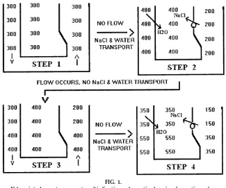
Given the above two exceptions, establishment of the third exception, namely, the hyperosmotic gradient from the outer to inner medulla via the countercur-rent multiplier system, can be addressed. The third exception is usually explained by way of simplified models, one of which is illustrated in Fig. 1, although more complex models are usually presented in text-books. Unfortunately, these types of models are nei-ther simple nor particularly accurate. Figure 1 pro-vides the basic outcome with minimal effort, and because all of these types of models are partially flawed, as explained below, it is probably a good idea not to put too much stock into these representations. The model is meant to give a student some assurance that establishment of an osmotic gradient along the longitudinal axis of the medullary loops of Henle is possible.
In Fig. 1,step 1is the initial situation before the first two exceptions take place.Step 2allows both of these exceptions to proceed toward a new equilibrium in the absence of flow through the loop of Henle. NaCl without water is pumped out of the ascending limb (exception 2), leaving the ascending limb hypoos-motic and the interstitial fluid hyperoshypoos-motic. Because water is permeable across the descending limb but no NaCl movement occurs here (exception 1), water movement from descending limb to interstitium oc-curs until the descending limb and medullary intersti-tium have the same osmolality (water concentration). Even though water is diluting the medullary interstitial osmolality, NaCl transport out of the ascending limb continues until some steady-state gradient is reached; in the case in Fig. 1, the gradient is 200 mosmol/kg H2O across any portion of the ascending limb. In proceeding to step 3, flow occurs in the nephron in the absence of solute and water movement. Thus two new 300 mosmol/kg H2O units enter the top of the descending limb, two 400 mosmol/kg H2O volume
FIG. 1.
Abbr eviated counter curr ent multiplication schematic showing for mation of an osmolality gradient in medullary loops of Henle. Numbers r efer to osmolality values in mosmol/ kg H2O. Arr ows denote dir ection of flow in steps 1and 3.
units move from descending to ascending limb, and two 200 mosmol/kg H2O units exit the thick ascend-ing limb. The medullary interstitial fluid shows no osmolality change because nephron flow is absent in this area. Now the cycle repeats itself. Step 4 again makes use of the two exceptions without flow. Ascending limb NaCl reabsorption occurs until a 200 mosmol/kg H2O gradient is again established across any portion of the ascending limb, and water leaves the descending limb to equilibrate with the corre-sponding medullary interstitium.
The key points of the medullary osmotic gradient are already evident. The osmolality increases (water con-centration or water activity decreases) from the outer to inner renal medulla (exception 3). Fluid leaving the thick ascending limb is hypoosmotic, and this is exception four, because the osmolality of body fluids is rarely less than 300 mosmol/kg H2O (sweat, saliva, and tears are normally ,300 mosmol/kg H2O). The osmolality of the luminal fluid leaving the thick ascending limb is usually stated as being between 100 and 150 mosmol/kg H2O. In the model, the final medullary osmotic gradient is a function of the degree of the NaCl gradient established across any portion of the ascending limb. In reality, nephron flow does not proceed in the absence of NaCl and water transport, rather the two occur together. By letting each one proceed independently in tiny steps, we can approxi-mate what happens when both processes occur simultaneously.
The distal tubule and cortical collecting duct are impermeable to water in the absence of vasopressin or ADH, and, coupled with some NaCl reabsorption in these units, the luminal fluid osmolality may be slightly less than 100 mosmol/kg H2O. In the presence of ADH, latter portions of the distal tubule and the cortical collecting duct are permeable to water, and approximately two-thirds of the incoming water can be reabsorbed, causing luminal osmolality to rise from 100 to 300 mosmol/kg H2O and allowing a relatively small volume of isosmotic fluid to be delivered to the medullary collecting ducts for still further water reabsorption and the production of a low volume of hyperosmotic urine.
A common student question is, ‘‘Why doesn’t the progressive osmolality gradient (or NaCl gradient)
from the outer to inner medulla disappear due to simple diffusion of NaCl down its concentration gradient?’’ The answer is that simple diffusion of NaCl cannot take place over the entire depth of the renal medulla because diffusion is not effective over such large distances (many millimeters) and because the renal countercurrent multiplication system is con-stantly generating the gradient.
Another common problem with a model such as that in Fig. 1 is that the volume of the ascending limb seems too small compared with that in the descending limb and interstitium for appreciable solute buildup in the latter two compartments at the expense of the ascending limb. Of course, Fig. 1 is only a schematic (the actual interstitial volume is negligible compared with the tubular volumes, but the countercurrent multiplication system must be able to extend a hyper-osmotic gradient through all extracellular spaces, including the vasa recta plasma, and also through some intracellular spaces). It is interesting to note that a cross section of the renal medulla would reveal five separate types of circular structures with very little relative interstitial space. The five separate tubes are the ascending and descending limbs of the loop of Henle, the ascending and descending vasa recta, and the collecting ducts.
The example used in Fig. 1 is an oversimplification and has two major inaccuracies. First, there does not appear to be active NaCl pumping in the thin ascend-ing limb, and second, the system ignores urea, a solute responsible for,50% of the osmotic activity in urine and medullary interstitial fluid when vasopressin (ADH) is present.
but none are completely adequate to explain the generation of an osmolality gradient along the thin ascending limb (4), and most theories cannot resolve how NaCl is more concentrated in the inner medulla compared with the outer medulla. Unfortunately, there is much that is unknown about how water moves out of the lower portion of the descending limb. Teaching these points to medical students is often compromised because of the inherent complex-ity as well as time constraints.
Here is a good theme to remember when teaching countercurrent multiplication or any topic that has a major unknown component:
It will a ll be different next yea r. —Horace W. Davenport
Another relatively unknown or gray area of urinary concentration and dilution is the renal handling of urea. Because urea appears to be nonionized, many students assume that urea is nonpolar and that its tubular fluid-to-plasma concentration ratio ([TF/P]urea) is equal to one throughout the nephron. This is not quite true in the proximal tubule, and not at all true in more distal nephron segments. In fact, there is some secretion of urea into portions of the loop of Henle, and the descending limb and cortical and outer medullary collecting ducts are all relatively imperme-able to urea. The inner medullary collecting duct urea permeability is variable. In the presence of ADH, carrier-mediated facilitated diffusional reabsorption of urea occurs, and this serves to load the medullary interstitium with urea so that nearly 50% of the medullary interstitial osmolality is due to urea in the presence of high ADH levels. Some of this medullary urea is evidently secreted back into the loop of Henle to yield medullary recycling of urea. Thus when plasma ADH is high, the permeability of the inner medullary collecting duct to both water and urea is high. These effects are mediated by the basolateral vasopressin-2 (V2) receptors operating with cAMP as the second messenger. The net effect is the excretion of a low volume of urine, with high osmolality (up to 1,400 mosmol/kg H2O).
A demonstration of urea facilitated transport can be helpful, and is diagrammed in Fig. 2. In Fig. 2, five small petri dishes or tissue culture dishes are each
My own view is that students will become hopelessly confused if it is correctly pointed out that, unlike NaCl, urea does not cause water to be reabsorbed from the collecting duct in the presence of ADH during the steady state. For a different view, see the paper by Vander (9) in this issue. Unlike NaCl active transport, urea transport to the medullary interstitium is via facilitated diffusion, resulting in nearly equal concentrations of urea in the collecting duct lumen and medullary interstitium in the inner medulla. Thus there is no urea osmolality difference across the collecting duct and no urea solute gradient for water reabsorption. If this comes out in class, it may be best to point out that if ADH did not increase the facilitated diffusion of urea out of the collecting duct, then all the urea would be trapped in the collecting duct lumen and the medullary interstitium osmolality maximum would be perhaps only 700 mosmol/kg H2O. In order to excrete the same amount of urea and nonurea solutes in a 700 mosmol/kg H2O urine, more water would be committed to the urine.
Another common point of confusion is the indepen-dence of urea, sodium, and water balance. During negative water balance, plasma ADH is high and urea is reabsorbed across the collecting duct via carrier-mediated facilitated diffusional transporters. This does not really cause positive urea balance because only the relatively small medullary interstitial compartment becomes loaded with urea. Urea will be a major osmotic solute in urine, but the urine volume will be small and, in the steady state, urea production will still equal urea excretion. Sodium balance and water balance are also nearly independent. Urine may con-tain as little as 10 mM Na1, and even during negative water balance, Na1can be a relatively minor contribu-tor to urine osmolality.
FREE WATER CLEARANCE
Another key point is that excretion of isosmotic (300 mosmol/kg H2O) urine does not change the water concentration in body fluid compartments, whereas
FIG. 2.
excretion of hyposmotic (,300 mosmol/kg H2O) urine will decrease the water concentration (increase the osmolality) in body fluid compartments. The appropriate response to positive water balance (water intake greater than excretion) is the excretion of hyposmotic urine, and the appropriate response to negative water balance is the excretion of a hyperos-motic urine. This concept can be treated analytically by calculating the free water clearance. Free water clearance is the volume of distilled water that must be subtracted or added to the urine to make the urine isosmotic. The free water clearance (net transport of distilled water per unit time) is greater than zero during positive water balance when the urine is hypo-smotic. Unfortunately, the free water clearance is not a true renal clearance, and this can lead to confusion.
THE RENAL COUNTERCURRENT EXCHANGE SYSTEM
Medullary capillaries called vasa recta run parallel to the loops of Henle and carry a small fraction of renal blood flow through the renal medulla. The main purpose of the vasa recta is to absorb the,25% of the filtered NaCl and the 10% of the filtered water that is reabsorbed by the loop of Henle. Just as Starling forces, capillary permeability, and rate of blood flow favor a net filtration pressure profile in glomerular capillaries, these factors combine to yield a net absorp-tive pressure profile in the vasa recta. Relaabsorp-tively low vasa recta hydraulic pressure and relatively high vasa recta oncotic or protein osmotic pressure are the main determinants of the net absorptive flux across the vasa recta.
A similar story can be told for renal peritubular capillaries in the cortex. The complicating fact for vasa recta capillaries is that they must run through the hyperosmotic medullary interstitium. If the vasa recta entered the outer medulla area, ran in a straight line parallel to the descending limb of the loop of Henle, and exited the kidney via the hyperosmotic inner medulla, vasa recta blood would leave the kidney nearly equilibrated with the hyperosmotic intersti-tium. Because the vasa recta form hairpin loops and run back to the renal cortex, vasa recta blood be-comes progressively hyperosmotic when paralleling the descending limb and then returns to near normal
osmolality (perhaps 325 mosmol/kg H2O) by the time it leaves the renal medulla. This is called countercur-rent exchange. Water may very well leave the descend-ing vasa recta down its concentration gradient (vasa recta filtration) into the hyperosmotic medullary inter-stitial fluid, but the vasa recta oncotic pressure favor-ing absorption increases still further, makfavor-ing up for any initial fluid loss by favoring still more reabsorption in the ascending vasa recta. Most importantly, counter-current exchange does not prevent vasa recta absorp-tion and in general does not interfere with countercur-rent multiplication. If vasa recta flow is too high, it is possible to ‘‘wash out’’ the medullary osmotic gradient.
URINARY CONCENTRATION AND DILUTION TEACHING TOPICS
My students tend to like these topics, many of which originated from the new renal material file mentioned previously.
Immediately after vasopressin levels increase and vasopressin binds to its renal epithelial receptor (the V2G protein-linked receptor), cAMP levels increase in the principal cells and promote apical membrane insertion of vesicles containing previously synthe-sized AQP2, without the immediate need for in-creased AQP2 gene expression. Insertion of previ-ously synthesized cell membrane proteins stored on intracellular vesicle membranes is a common phenom-ena. For instance, the same general mechanism is used by certain insulin-sensitive cells, causing membrane insertion of glucose facilitated diffusion carriers after insulin exposure.
Nephrogenic diabetes insipidus refers to the inad-equacy of the kidney to produce appropriately concen-trated urine, even in the presence of high vasopressin levels. Two general mutations, one in the V2-receptor gene and one in the AQP2 gene, cause a congenital failure to produce appropriately concentrated urine (1). In addition, V2-receptor blockers are currently being evaluated as possible diuretic agents.
Ethyl alcohol always perks student interest. Each 0.1% increase in blood alcohol produces an ,17 mos-mol/kg H2O increase in plasma osmolality. Ethyl alcohol shuts down vasopressin secretion, producing a temporary central diabetes insipidus and dehydra-tion. The hangover symptoms are related to the severe dehydration and may also involve the slow clearance of ethyl alcohol from the endolymph within the vestibular labyrinth of the inner ear, causing endo-lymph-specific gravity alterations and subsequent diz-ziness and nausea.
How can renal cells maintain their volume in the hyperosmotic inner medulla? In part, these cells synthesize and accumulate osmotically active solutes (osmolytes), including sorbitol, betaine, inositol, tau-rine, and glycerophosphocholine (2). Increasing osmo-lality causes appropriate gene expression and synthe-sis of these osmotically active intracellular solutes. Thus renal medullary cells need not shrink in an increasingly hyperosmotic extracellular fluid.
The vascular countercurrent exchange system is not unique to the kidney. Countercurrent exchange often results in the tra pping or shuntingof highly perme-able molecules or heat. For instance, blood vessels
running in a countercurrent arrangement trap heat in the vascular system of whales exposed to near-freezing ocean water. Warm blood moving to the periphery (fins) loses heat to cold blood moving away from the fin surface, effectively trapping and conserv-ing heat in these endotherms. This same system operates in the tongue of the gray whale (3). Despite the fact that the tongue is well vascularized, has little insulation, and is constantly exposed to ice-cold water in the oral cavity during filter feeding, the gray whale’s tongue loses relatively little heat to the cold ocean because the arterial and venous systems run in a countercurrent fashion, effectively trapping heat. Countercurrent trapping of CO2 and countercurrent shunting of O2 probably play a role in the renal medulla and the intestinal villus, where capillaries are arranged in a countercurrent fashion.
Finally, no countercurrent multiplication class is com-plete without mention of the kangaroo rat, a desert dweller with 100% juxtamedullary nephrons and the surprising ability to excrete urine with an osmolality of 6,000–8,000 mosmol/kg H2O. Water balance is maintained in part by excreting very little water in the urine. The rat kidneys are small, so it is not the length of the loops of Henle that gives rise to this astound-ingly concentrated urine. Perhaps the secrets of the inner medulla can best be examined by observing the renal physiology of the kangaroo rat. It is also interest-ing to compare the physiology of the kangaroo rat with that of the beaver, because the beaver has only short-loop cortical nephrons, has no countercurrent multiplication system, and cannot concentrate its urine. The beaver is resigned to this situation by always being in or near an aqueous environment and by dealing with negative water balance only by drinking more, not excreting less. This is analogous to diabetes insipidus, because the major complications of both a beaver out of water and diabetes insipidus can be alleviated by adequate water intake. As far as the beaver is concerned, countercurrent multiplica-tion be damned.
I acknowledge Dr. David Levitt for providing the ideas behind the red blood cell demonstration shown in Fig. 2.
Refer ences
1. Bichet, D. G., A. Oksche, and W. Rosenthal. Congenital nephrogenic diabetes insipidus. J. Am . Soc. Nephrol. 8: 1951– 1959, 1997.
2. Bur g, M. B. Molecular basis of osmotic regulation. Am . J. Physiol. 268 (Rena l Fluid Electrolyte Physiol. 37): F983–F996, 1995.
3. Heyning, J. E., and J. G. Mead. Thermoregulation in the mouths of feeding gray whales. Science 278: 1138–1139, 1997.
4. Knepper, M. A., and F. C. Rector, Jr.Urinary concentration and dilution. In:The Kidney(5th ed.), edited by B. M. Brenner. Philadelphia, PA: Saunders, 1996.
5. Nielsen, S., and P. Agr e. The aquaporin family of water channels in the kidney.Kidney Int.48: 1057–1068, 1995. 6. Pallone, T. L., B. K. Kishor e, S. Nielsen, P. Agr e, and M. A.
Knepper. Evidence that aquaporin-1 mediates NaCl-induced water flux across descending vasa recta. Am . J. Physiol. 272 (Rena l Physiol. 41): F587–F596, 1997.
7. Sands, J. F., R. T. Timmer, and R. B. Gunn.Urea transporters in kidney and erythrocytes.Am . J. Physiol.273 (Rena l Physiol. 42): F321–F339, 1997.
8. Schafer, J. A. Renal water and ion transport systems. Am . J. Physiol. 275 (Adv. Physiol. Educ. 20): S119–S131, 1998. 9. Vander, A. J.Some difficult topics to teach (and not to teach) in